Information Links
Related Conferences
Previous Issues Volume 7, Issue 1 - 2023
Surgical Approach for Reconstruction of a Morbus Bowen Defect in the Genital Area: A Case Report
Smilja Tudzarova-Gjorgova*, Ana Selchanec, Gloria Gjorgova, Marija Spasovska
University Clinic for Plastic and Reconstructive Surgery, Faculty of Medicine, Ss. Cyril and Methodius University, Skopje, North Macedonia
*Corresponding Author: Dr. Smilja Tudzarova-Gjorgova MD, PHD, University Clinic for Plastic and Reconstructive Surgery, Mother Teresa No. 17, 1000 Skopje, Republic North Macedonia; Email: [email protected]
Received Date: April 12, 2023
Publication Date: May 17, 2023
Citation: Tudzarova-Gjorgova S, et al. (2023). Surgical Approach for Reconstruction of a Morbus Bowen Defect in the Genital Area: A Case Report. Mathews J Dermatol. 7(1):22.
Copyright: Tudzarova-Gjorgova S, et al. © (2023)
ABSTRACT
Bowen’s disease is also known as intraepithelial squamous cell carcinoma in situ which can appear on the skin or mucous membranes (including mouth, anus and genitalia). The macroscopic appearance of Morbus Bowen is as erythematous, rough, scaly and sharply demarcated plaques, and histologically there is hyperkeratosis, parakeratosis, dyskeratosis and acanthosis present. The precursor lesion is slow-growing, and if left untreated, it can progress and transform into squamous cell carcinoma (SCC). In this particular case, our 68 year old female patient presented with a genital form of Morbus Bowen, and she had had the plaques for 15 years. The lesions were previously treated with cryotherapy and dermatological creams, but to no avail. This patient was treated in our Clinic with a surgical excision and a Thiersch skin graft (STSG) to close the defect. Consequently, we have evaluated her 3 months post-op and the results are satisfactory.
Keywords: Bowen’s disease, Surgical excision, Split-thickness skin graf
INTRODUCTION
Chief complaints
The patient is a 68 year old woman from Skopje who was admitted to the University Clinic of Plastic and Reconstructive Surgery with a history of long-lasting genital lesions, pruritus and persistent local erythema in the genital region. She informed us that the lesions were bothersome, especially when sitting and going to the bathroom. There was never any pain nor bleeding, and her OB/GYN indicated to her that the growths were likely condylomas, and recommended that they should be surgically excised [1].
History of past illness
The patient had genital lesions for more than 15 years. The growths had been treated conservatively with different creams and cryotherapy by a dermatologist. The conservative treatment consisted of 10 sessions of cryotherapy (two sessions weekly), although the patient was initially supposed to have 5 more sessions, which she didn’t finish. The lesions then disappeared for a year, but they reemerged again and more numerous than the initial presentation. Consequently, the growths as well as the pruritus vulvae, was not relieved and/or cured with that specific course of treatment.
Personal and family history
Regarding her personal history of illnesses, the patient had had two caesarian sections (35 and 33 years ago), and later had an incisional hernia on the c-section scar site. She had undergone two surgeries for the aforementioned hernia. Then, 10 years ago, the patient had a laparoscopic cholecystectomy. Also, she has been treated for herpes zoster in 2018, and currently has essential primary hypertension, for which she takes medication. She is a smoker, and has an allergy to penicillin.
Her family history was unremarkable.
Physical examination upon admission
On inspection, numerous conglomerates of round pink and dark brown papilomatous lesions with cauliflower-like masses were observed on both labia majora. There was no ulceration, exudation nor bleeding present, and clear lesion boundaries from the surrounding macroscopically normal skin tissue were noted.
On palpation, the skin growths were not painful, and easily moveable. Regarding their consistency, it was palpably firm, and each of the small warts had a diameter of 1-2 cm, while the diameter of the clusters of growths was 3-4 cm. The patient informed us that all of the growths started out as pink, and as time passed, the older growths became darker, and the new ones - light pink (Figure 1).
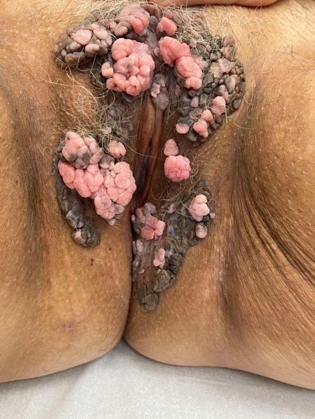
Figure 1: The patient pre-op.
Laboratory examinations
Tissue specimens were obtained by biopsy.
The skin biopsy revealed verruciform lesions with variable acanthosis, parakeratosis, hypergranulosis and hyperkeratosis. There was koilocytosis present, and in some of the tissue samples, there was evident cellular atypia throughout the whole width of the epithelium, and inflammatory infiltrate in the subepithelial connective tissue. All of these findings are consistent with Bowen’s disease. [2] The resection margins are with orderly morphology.
TREATMENT PLAN
The lesions were surgically excised while the patient was under spinal anesthesia. The safe margin for resection of the lesions is 5 mm and the depth - the full thickness of the skin. [3] A split-thickness skin graft (Thiersch) was taken from the right femoral region with an electric dermatome, and the skin graft was transplanted onto the surgical wound of the left labia majora. Vicryl 2/0 and Silk 3/0 stitches were used for the direct suture on both labias. The patient was prescribed Ciprofloxacin, and Belcura spray (spray emulsion containing microsilver) for local application, and was advised to maintain proper hygiene. No adverse events occurred.
Outcome and Follow-Up
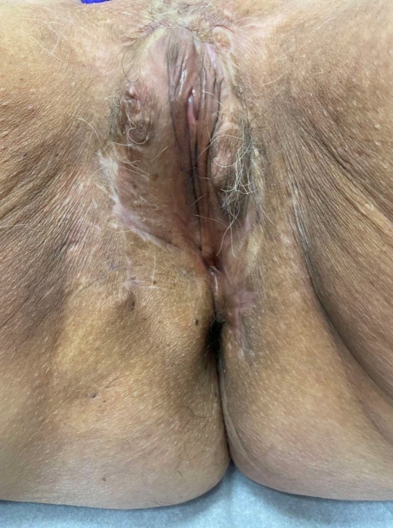
The symptoms of pruritus disappeared in the skin lesion of the patient after surgery, and no recurrence was observed at the 3 month follow-up. [4] There were no adverse effects experienced by the patient (such as hematoma or seroma formation, pain, infection or dehiscence of the wound), and the scarring is minimal, as evident on the photo.
Figure 2. The patient 3 months post-op.
 We will continue to follow this patient in order to see if there is recurrence in the following period.
DISCUSSION
Morbus Bowen is an intraepithelial squamous cell carcinoma in situ which can appear on the skin or mucous membranes. Macroscopically, Bowen’s disease presents as a pigmented, exophytic, scaly and sharply demarcated plaques. The lesions are slow-growing, and as is common with most precancerous lesions - they have a long clinical course (generally years). Microscopically, there are findings of hyperkeratosis, parakeratosis, dyskeratosis and acanthosis present within the epithelial layers. This precursor lesion, if left untreated, can progress and undergo malignant transformation into squamous cell carcinoma (SCC). [5]
The differential diagnosis for the genital lesions was condyloma acuminata - anogenital warts also caused by HPV. There is a well documented correlation between the Human Papilloma Virus and Bowen’s disease, or Bowenoid papulosis as one of the major causal agents, more precisely HPV types 16, 18, 34, 48.
There are many different treatment modalities for Bowen’s disease, such as curretage with cautery, cryotherapy, surgical excision, 5-fluorouracil, radiotherapy, laser and etc. [6] The treatment plan for each patient differs keeping some factors in mind: the patient’s age, immune status, comorbidities, medication, and location, number and size of the lesions. Each treatment option has its advantages and disadvantages - some of them are associated with scarring, ulceration, infection, hypopygmentation after cryotherapy, atrophic epidermis after extensive use of CO2 laser. All of this should be taken into consideration when selecting the treatment plan. For our patient specifically, the conservative treatment was deemed ineffective, and the surgical excision and placement of a split-thickness skin graft was our choice. Surgical excision of BD remains one of the standard treatments, with its biggest advantage being the securing of the histological free excision margins. The prognosis of the surgical approach for Bowen’s disease is excellent with minimal tumor recurrence.
CONCLUSION
Out of the many possibilities for treating BD, this case illustrates the successful curative therapy of genital Morbus Bowen with surgery. Wide excision and surgical treatment for the genital form of BD is the treatment of choice, and rightly so.
CONFLICT OF INTEREST
The authors declare no conflicts of interest.
REFERENCES
- Vukšić Polić M, Cutvarić N, Marjanović K, Mihalj M. (2022). Unrecognized Bowen's disease in previously treated condylomata acuminata: indication of a common etiology? Acta Dermatovenerol Alp Pannonica Adriat. 31(1):33-37.
- Giuffrida R, Conforti C, Resende FSS, Hamilko de Barros M, Uranitsch M, Favero F, et al. (2018). Clinical and dermoscopic features of genital pigmented Bowen disease. Clin Exp Dermatol. 43(7):813–816.
- Morton CA, Birnie AJ, Eedy DJ. (2014). British Association of Dermatologists' guidelines for the management of squamous cell carcinoma in situ (Bowen's disease) 2014. Br J Dermatol. 170(2):245-260.
- Ahmed I, Berth-Jones J, Charles-Holmes S, O'Callaghan CJ, Ilchyshyn A. (2020). Comparison of cryotherapy with curettage in the treatment of Bowen's disease: a prospective study. Br J Dermatol. 143(4):759-766.
- Bath-Hextall FJ, Matin RN, Wilkinson D, Leonardi-Bee J. (2013). Interventions for Bowen's Disease. Cochrane Database Syst Rev. 2013(6):CD007281.
- Neubert T, Lehmann P. (2008). Bowen's disease - a review of newer treatment options. Ther Clin Risk Manag. 4(5):1085-1095.