Information Links
Related Conferences
Current Issue Volume 10, Issue 1 - 2026
Lichen Striatus - Making Accurate Diagnosis in the Young and Older Adult: A Case Series
Amaewhule Mary Nnenda1, Amadi Ekechi Stella1,2,*
1Dermatology Unit, Department of Internal Medicine, Rivers State University & Rivers State University Teaching Hospital, Nigeria
2Dermatology in Clinical Practice, University of South Wales, Pontypridd, United Kingdom
*Corresponding author: Ekechi Stella Amadi, Dermatology Unit, Department of Internal Medicine, Rivers State University & Rivers State University Teaching Hospital, Nigeria and Dermatology in Clinical Practice, University of South Wales, Pontypridd, United Kingdom, Phone: 08032790394, E-mail: [email protected]
Received Date: April 07, 2026
Published Date: April 30, 2026
Citation: Amaewhule MN, et al. (2026). Lichen Striatus - Making Accurate Diagnosis in the Young and Older Adult: A Case Series. Mathews J Dermatol. 10(1):31.
Copyrights: Amaewhule MN, et al. © (2026).
ABSTRACT
Lichen striatus(LS) is an uncommon linear dermatosis that commonly affects children aged 5-15 years however case reports of adult onset disease have been documented. Lichen striatus is usually benign, occurs spontaneously and it is self-limiting, resolving without any treatment. This case series focuses on the clinical cases of lichen striatus seen in our center over a 5-year period from January 2021- December 2025 and the challenges in making accurate diagnosis using clinical features and investigations.
Keywords: Lichen Striatus, Rare, Skin Punch Biopsy, Young, Older Aldult.
INTRODUCTION
Lichen striatus (LS) is an uncommon linear dermatosis that commonly affects children aged 5-15 years, however case reports of adult onset disease have been documented [1]. The word lichen striatus is gotten from two different Greek words - ‘lichen’ which refers to symbiotic organisms (algae and fungi) coexisting together and striatus which means straight, linear, stripped or streaked [2,3]. It is so named because of its appearance not because it is a fungal lesion or it is induced by a scratch. The incidence of lichen striatus is rare. When such a skin disorder occurs we are prone not to consider it; preferring one of the other commoner linear dermatoses, such as lichen planus, linear psoriasis, nevus unius lateris (congenital linear verrucous epidermal nevus) and even herpes zoster [4].
The skin is the primary site affected by lichen striatus; however, there may be affectation of the nails. Lesions of LS follow the lines of Blaschko. Blaschko lines which are thought to be embryologic in origin resulting from the segmental growth of clones of cutaneous cells or the mutation-induced mosaicism of cutaneous cells. The exact cause of LS is unknown but it is believed that combination of genetic predisposition and environmental stimuli causes the formation of LS [1]. Atopy, autoimmune response, infections such as varicella, post therapy with drugs such as enternecept and adalimumab, post vaccination, trauma from plants and bee stings have been implicated in causing LS [1]. Currently there is no racial or gender predilection for LS.
The histopathology of lichen striatus varies depending on the stage of evolution majorly characterized by spongiosis and lichenoid dermatitis but it differs from lichen planus may result in a dense, usually perivascular, lympho histiocytic infiltrate that extends deep into the dermis and surrounds the hair follicles and eccrine sweat glands and ducts [1,5]. Epidermal changes include focal hyperkeratosis, parakeratosis, mild acanthosis, basal cell vacuolization, and apoptotic keratinocytes (Civatte bodies), reflecting damage to the basal layer. Spongiosis is seen occasionally, particularly in early lesions [5]. Additional findings may include melanophages in the upper dermis, correlating with postinflammatory hyperpigmentation [5]. The combination of lichenoid inflammation with adnexal affectation along Blaschko lines aids the diagnosis, strengthening the need for clinical and histopathological correlation [5,6].
CASE SERIES
Within the period of January 2021- December 2025, amongst the several dermatology disorders only three cases of lichen striatus were seen at the dermatology out patient clinic of our center. Two were made clinically and one via histopathology. The first clinical diagnoses of LS were made in a 17-year-old post-secondary school student with a complaint of skin coloured papules coalescing together in a linear fashion on her right upper arm. It was said to be itchy occasionally. She had chicken pox (varicella zoster) infection when she was younger. Her retroviral status was seronegative. A diagnosis of lichen striatus was made with differentials of linear verrucous epidermal nevus and herpes zoster. A skin punch biopsy was requested for to obtain samples for histopathology but the patient was lost to follow up.
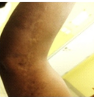
Figure 1. Skin coloured papules in a linear form on the right upper arm.
The second case was that of a 75-year-old motor mechanic who noted a gradual onset of bilateral skin rash following the lines of Blaschko of 11 months duration. It was first noticed on his right shoulder which was initially skin coloured papules and later began to coalesce to form a hyper pigmented band. The extent was from the middle contour of the right shoulder to the elbow. Similar rash was then noticed on the left shoulder, however with a shorter length. There was no history of trauma, itching, burning or crawling sensations prior to this time. The rash was not photosensitive or discharging any fluid. HIV I & 2, HbSAg and HCVAb screening were all seronegative. He was placed on daily topical 0.05% clobestasol propionate cream and the lesion regressed after four weeks of application. The patient was satisfied with the treatment and continued applying the cream at home however he was lost to follow up after the initial three visits.
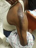
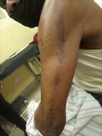
Figures 2 & 3. Lichen striatus on the right and left shoulder of an older adult.
The third case was that of an 11-year-old girl junior secondary school student referred from the pediatrics clinic on account of hypo pigmented papules appearing on the posterior aspect of the axilla and right for arm for the past 3months. The lesions were papular and occasionally pruritic. It has been increasing in size in a linear form following the lines of Blaschko. The lesions were also undergoing Koebner’s phenomenon. The patient was initially prescribed topical terbinafine by the paediatrician which was used for three weeks but the lesions did not resolve. At the initial time the outline was not very clear and Wood lamp examination done on the first visit showed enhanced hypopigmented area at the posterior forearm and axilla suggestive of the lesion being located within the epidermis. There were orangish specks noted at the axilla. A clinical diagnosis of pityriasis versicolor was made with differentials of erythrasma, guttate psoriasis and epidermodysplasia verruciformis. She was placed on 2% ketoconazole shampoo application every alternate day for three weeks and topical ticonazole cream b.d for 2 weeks. She was seronegative to HIV I &2, HbsAg and HCVAb. She had the skin punch biopsy for histopathology done a week later after the initial visit. She came for a follow-up visit after 8 months with her caregiver(mother). The result of histopathology showed mild spongiosis and mild acanthosis with foci of acantholysis. There was focal moderate infiltrate of lymphocytes and plasma cells at the dermal-epidermal junction. The papillary dermis contained few melanophages.
The lesions were now more visible as seen in Figure 6. They were now easily comparable to other clinical photographs of LS in those with darker skin tones already in existence. The patient and caregiver were counselled on the self-limiting nature of the disease, the need to avoid scratching and the side effects of drug therapy such as topical corticosteroids. She was placed on topical bethametasone cream, loratadine tablets and encouraged to continue with regular follow up at the clinic.
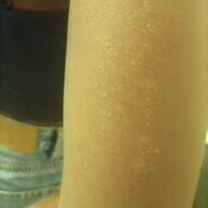
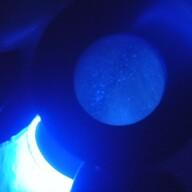
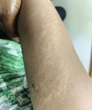
Figures 4, 5 & 6. Hypopigmented papules at first visit to the skin clinic; Wood Lamp showing enhanced hypopigmentation at the junction of posterior axilla and forearm; Post skin punch biopsy visit -lesions have become more pronounced.
DISCUSSION
In making the diagnosis of LS, a high index of suspicion is required. It is commonly seen in the younger age group between 5-15 years and has more females affected as it has been reported in medical literature [1, 5]. LS have been reported amongst adolescents with equal sex affectation in a study within the same region [7]. Cases of older adults with LS have been seen, such as the cases reported by Shepard et al of a 56-year-old Japanese female and Jones et al of a 63-year-old female with skin of colour [8,9]. When seen in adults it is called acquired blaschkoid dermatitis or blaschkitis [6]. There are different schools of thought as to LS and acquired blaschkoid dermatitis; if this is a distinct disorder from that seen in childhood or if they are both within the same spectrum. This case series has two females of adolescent age and an older adult male. It typically presents as an asymmetrical lesion in most cases but can also be symmetrical as reported by Dickman et al. [10] Koebner’s phenomenon was also reported in the case by Dańczak-Pazdrowska et al. [11] LS has been known to involve the nails as seen in other studies but this was not seen in any of the cases reported in this study [5,9,12]. Other parts of the body have been noted to have LS but the extremities are common sites [12]. Viruses such as varicella virus or hepatitis whether as clear cut diseases or as inactivated viruses used as vaccines have been implicated in the etiology of LS [1,9]. Other possible associated factors were not seen in any of the cases in this series. In making a diagnosis histopathology and dermoscopy have been established in aiding diagnosis. Despite clinical diagnosis being accurate most times if typical presentation occurs; it can also be difficult to make based on clinical features alone because the lesions may also evolve or change with treatment particularly with over counter drugs not appropriately prescribed just as it was seen in this case series. In lighter skin tones it appears hypopigmented, reddish, pinkish, violaceous or skin coloured [6,8,11]. In darker skin tones it can be skin coloured, violaceous, hypo or hyperpigmented [6,9]. Striae distensia should be clearly differentiated from LS as it is a common finding even in children and adolescents [13].
Patient education is key as LS is usually self-limiting. The treatment mostly prescribed is topical steroid. Patients usually respond to this therapy as it was seen in this case series. Treatment may also be prescribed based on accompanying symptoms and signs such as itching, dryness or pigmentation [1,6,9].
CONCLUSION
Lichen striatus is a rare, acquired, asymptomatic, self-limiting linear skin lesion which requires a high index of suspicion to make diagnosis as seen in this case series.
CONFLICTS OF INTEREST
None.
FUNDING
None.
REFERENCES
- Kim J. Nelson D, editor. (2024). Lichen Striatus. Available at: https://emedicine.medscape.com/article/1111723-overview
- Merriam -Webster Dictionary. Available at: https://www.merriam-webster.com/dictionary/lichen#word-history
- Oxford languages Dictionary. Striatus. Available at: https://www.google.com/search?q=meaning+of+striatus+in+english+oxford+dictionary&sca
- Staricco RG. (1959). Lichen Striatus: A Study of Fifteen New Cases with Special Emphasis on the Histopathological Changes and a Review of the Literature. AMA Arch Derm. 79(3):311-324.
- Syed HA, Jamil RT, Ramphul K. (2022). Lichen Striatus. [Internet]. Treasure Island (FL): StatPearls Publishing. Available at: https://www.ncbi.nlm.nih.gov/books/NBK507830/
- Coulson I, Ngan V. Lichen Sttriatus. Available at: https://dermnetnz.org/topics/lichen-striatus
- Amadi ES, Maduagwu MC, Pepple EF. (2022). Adolescents’ Skin Care Visits: A Ten Years Survey. Asian J Res Dermatol Sci. 5(1):62-67.
- Shepherd V, Lun K, Strutton G. (2005). Lichen striatus in an adult following trauma. Australas J Dermatol. 46(1):25-28.
- Jones J, Marquart JD, Logemann NF, DiBlasi DR. (2018). Lichen striatus-like eruption in an adult following hepatitis B vaccination: a case report and review of the literature. Dermatol Online J. 24(7):13030/qt1hr764kv.
- Dickman JS, Frandsen MD, Racette AJ. (2018). Asymmetric Bilateral Lichen Striatus: A Rare Presentation following Multiple Blaschko's Lines. Case Rep Dermatol Med. 2018:6905175.
- Dańczak-Pazdrowska A, Pawlaczyk-Gabriel K, Bowszyc-Dmochowska M, Polańska A, Gabriel M, Gabriel Z, et al. (2018). Reverse Kőbner response in lichen striatus: the first case described in the literature. Postepy Dermatol Alergol. 35(4):434-435.
- Leung AKC, Lam JM, Barankin B, Leong KF. (2025). Lichen Striatus: An Updated Review. Curr Pediatr Rev. 21(3):233-244.
- Abdul Kadir A, Badrin S, Hoong LL. (2021). Striae in Adolescence: A Suspicious of Bully. Electron J Gen Med. 18(5):em306.
