Information Links
Related Conferences
Previous Issues Volume 8, Issue 3 - 2023
Extra-pleural Hematoma : A Case Report
F Lamouime1,*, M Rhaouti1, I Arramach1, M Lakranbi1,2, Y Ouadnouni1,2, M Smahi1,2
1Department of Thoracic Surgery, CHU Hassan II-Fès, Morocco
2Faculty of Medicine and Pharmacy, Sidi Mohamed Ben Abdellah University, Fès, Morocco
*Corresponding Author: Lamouime fatima ezzahrae, Department of Thoracic Surgery, CHU Hassan II-Fès, Morocco; Email: [email protected]
Received Date: February 17, 2023
Publication Date: February 21, 2023
Citation: Lamouime F, et al. (2023). Extra-pleural Hematoma : A Case Report. Mathews J Case Rep. 8(3):92.
Copyright: Lamouime F, et al. © (2023)
INTRODUCTION
Post-traumatic extrapleural hematoma is a collection of blood in the space between the parietal pleura and the endothoracic fascia. It usually results from thoracic trauma. The diagnosis is suspected in front of a characteristic radiographic image. The clinical condition of the patient as well as the importance of the bleeding may lead to a thoracotomy for hemostasis. In this case, we report a case of extrapleural hematoma in a 64-year-old patient following a thoracic trauma.
OBSERVATION
The patient was a 64-year-old man admitted to the emergency room following a 3-day-old fall from a staircase with a left thoracic impact point. The clinical examination on admission found a polypneic patient with a respiratory rate of 29 cycles/minute. Physical examination revealed multiple bruises on the left hemithorax and the left clavicle.
The frontal chest X-ray showed a homogeneous opacity of the left thoracic hemifield with pulmonary collapse (Figure A). The patient underwent a body scan, which showed a clotted hemothorax on the left side, a small to moderate amount of right pleural fluid, multiple bilateral rib fractures with a left rib flap, and fracture of the outer third of the clavicle, As the patient had undergone a left pleural puncture which did not yield anything, the indication for an emergency decalcification was given, and the approach was a left posterolateral thoracotomy through the fifth left intercostal space (Figure B). Exploration revealed a left extra pleural hematoma that extended to the top of the lung with a clotted hemothorax.
Hemostasis was satisfactory, followed by removal of the pleural cavity with respect for the extrapleural hematoma, given its extensive nature (Figure C). The postoperative follow-up was simple with a control angioscanner which did not show any extravasation of the contrast product at the different times (Figure D). The patient was discharged after removing the pleural drain at 5 days postoperatively with satisfactory radiological control.
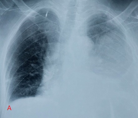
Figure A: Opacity of the left pulmonary hemichap with pulmonary collapse.

Figure B: Clotted left pleural effusion.
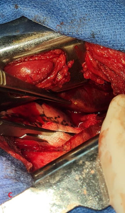
Figure C: Left extrapleural hematoma (yellow star).
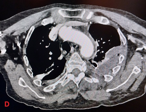
Figure D: Left extrapleural hematoma.
DISCUSSION
Extrapleural hematoma most often results from chest trauma, sometimes iatrogenic. Traumatic causes are usually secondary to rib or vertebral fractures or rupture of an aortic aneurysm [1]. Iatrogenic causes are mainly represented by the placement of a pleural drain, a venous catheter, trans-parietal biopsy or a surgical procedure [2,3]. The other etiologies are of spontaneous origin, or following coronary bypass surgery [4,6].
The diagnosis is essentially radiological and is most often made by chance. The suggestive radiological image is a D-shaped opacity with a parietal base [7,8]. Sometimes it may be an opaque hemithorax. The exact diagnosis of the lesion involved is often difficult preoperatively. Thoracic CT scan shows a collection of high spontaneous density located in the extrapleural space. This density is surrounded by a fatty border around its entire periphery, corresponding to the extrapleural fat medially and to the fat lining the external face of the endothoracic fascia laterally [1]. This fatty border sign is used to distinguish between an extrapleural hematoma, a hemothorax, and a parietal hematoma. Treatment of an extrapleural hematoma can be conservative if the patient is hemodynamically stable and thoracic drainage allows for its evacuation.
Thoracotomy is recommended in case of hemodynamic instability or in case of thoracic drainage of more than 1,500-2,000 ml, or in case of clotted extrapleural hematoma with persistent radiological image. Other authors propose treatment by thoracoscopy if the patient's hemodynamic state allows it [4] and other teams have divided extrapleural hematomas into two groups according to their CT appearance: biconvex and non-convex. Those with a biconvex shape are similar to that of an intracranial extradural hematoma. Biconvex morphology suggests hemorrhage of arterial origin and a greater likelihood of surgical intervention. The nonconvex extrapleural hematoma has lobulated margins conforming to the inner chest wall, reflecting low-pressure venous bleeding, and does not usually require surgery [5].
A case of intercostal artery embolization in the management of an extrapleural hematoma in a patient involved in a motor vehicle accident has been reported [7].
It is important to be aware of this diagnosis because its management is different from that of a hemothorax. Thus, drainage is not indicated because there is a risk of pleural injury, which can have serious consequences if the patient is put on positive pressure ventilation [5].
CONCLUSION
Extrapleural hematoma is a rarely reported clinical entity. Although thoracic computed tomography is a key diagnostic test, exploratory thoracotomy remains the only means of rapidly identifying extrapleural hematoma, determining its origin and performing hemostasis procedures. Its indication is dictated by the precarious hemodynamic state of the patient and/or in front of an active bleeding that may require emergency life-saving surgery.
LINKS OF INTEREST
The authors declare no relationship of interest.
REFERENCES
- Moulin G, Bartoli JM, Fighiera M, et al (1992). Hématome extrapleural. J Radiol 73:327–330
- Aquino SL, Chiles C, Oaks T. (1997). Displaced extrapleural fat as revealed by CT scanning: evidence of extrapleural hematoma. AJR 169:687–689
- Gilkeson RC, Abramson S. (1999) Displacement of extrapleural fat with extrapleural hematoma. AJR 173:243–234
- Crema MD, Monnier-Cholley L, Maury E, Tubiana JM,Arrive L. (2004). Quel est votre diagnostic ? J Radiol 85:419–421
- Rashid MA, Wikström T, Ortenwall P. (2000). Nomenclature, classification and signficance of traumatic extrapleural hematoma. J Trauma. 49:286-290
- Konen O, Hertz M, Klein H, et al (2002) Hematoma as an unexpected finding on a follow-up chest X-ray after coronary surgery. Eur J Radiol 44:225–227
- Seiji M, Tomoatsu T, Tomokazu F, et al (2009) Arterial embolization of an extrapleural hematoma from a dislocated fracture of the lumbar spine: a case report. Scand J Trauma Resusc Emerg Med 17:27
- Masuda R, Ikoma Y, Oiwa K, et al (2013) Delayed hemothorax superimposed on extrapleural hematoma after blunt chest Injury: a case report. Tokai J Exp Clin Med 38:97–102