Information Links
Related Conferences
Current Issue Volume 11, Issue 3 - 2026
Alzheimer’s Disease with Carotid Intraluminal Findings and Rheumatoid Arthritis–Like Symptoms: A Case Report
Takuji Shirasawa1,2,*, Luis Carlos Aguilar Cobos3
1Ochanomizu Health and Longevity Clinic, Tokyo 101-0062, Japan
2Shirasawa Anti-Aging Medical Institute, Tokyo 101-0062, Japan
3Livant Neurorecovery Center, Guadalajara, Mexico
*Corresponding Author: Takuji Shirasawa, Ochanomizu Health and Longevity Clinic, Tokyo 101-0062, Japan & Shirasawa Anti-Aging Medical Institute, Tokyo 101-0062, Japan, E-mail: [email protected]
Received Date: May 30, 2026
Published Date: June 22, 2026
Citation: Shirasawa T, et al. (2026). Alzheimer’s Disease with Carotid Intraluminal Findings and Rheumatoid Arthritis–Like Symptoms: A Case Report. Mathews J Case Rep. 11(3):228.
Copyrights: Shirasawa T, et al. © (2026).
ABSTRACT
Alzheimer’s disease (AD) is a progressive neurodegenerative disorder for which no curative therapy has been established. In this case report, a 79-year-old female patient with the APOE ε4/ε4 genotype presented at our clinic with a complaint of memory impairment. Initial evaluation, including magnetic resonance imaging (MRI) and electroencephalography (EEG), suggested AD with concomitant carotid intraluminal findings and rheumatoid arthritis–like symptoms. The patient subsequently received cytokine-based therapy in combination with a proteolytic treatment protocol. During follow-up, interval changes were observed in hippocampal morphology, carotid intraluminal findings, and rheumatoid arthritis–like symptoms. Although causal inferences cannot be drawn from a single case report, this report documents multimodal clinical and imaging changes in a patient with complex dementia-associated comorbid features.
Keywords: Alzheimer’s Disease, Case Report, Carotid Intraluminal Findings, Rheumatoid Arthritis–Like Symptoms, Cytokine-Based Therapy.
INTRODUCTION
Alzheimer’s disease is the leading cause of dementia and is characterized by progressive impairment of memory, cognition, and activities of daily living [1]. Its neuropathological hallmarks include amyloid-β deposition, tau-mediated neurodegeneration, synaptic dysfunction, and cerebral atrophy [2,3]. Although therapeutic development has advanced in recent years, effective disease-modifying interventions remain limited, underscoring the importance of early diagnosis and continued investigation of potential treatment strategies [4]. Among genetic risk factors, APOE ε4 is the strongest common determinant of late-onset Alzheimer’s disease, conferring increased risk in a dose-dependent manner and often being associated with an earlier age at onset [5,6]. The hippocampus, particularly the dentate gyrus, plays a central role in memory formation, and structural and functional change in this region has been proposed as a potentially relevant marker of disease progression and response to intervention [7,8]. Current therapeutic approaches are shifting from symptomatic management toward disease-modifying strategies, including anti-amyloid antibodies, tau-directed therapies, and biomarker-guided early intervention, with the goal of slowing disease progression and enabling more personalized care [9,10]. Prior reports of cytokine-based therapy in Alzheimer’s disease have documented both clinical and imaging changes, including structural alterations in the hippocampus and cerebral cortex [11,12].
It has also been documented that the presence of carotid intraluminal material, as indicated by imaging findings, is associated with a decrease in blood flow within the internal carotid artery. This association may be indicative of a potential cerebrovascular relevance. [13]. Autoimmune manifestations have also been reported after administration of SARS-CoV-2 mRNA vaccines, although a causal relationship has not been established [14].
In this case report, a 79-year-old female patient with the APOE ε4/ε4 genotype presented at our clinic with a complaint of memory impairment. Initial evaluation showed imaging findings consistent with Alzheimer’s disease together with intraluminal findings in both internal carotid arteries. During the subsequent clinical course, the patient also developed rheumatoid arthritis–like symptoms. The patient subsequently underwent cytokine-based therapy and a proteolytic treatment protocol, during which multimodal clinical and imaging changes were observed during follow-up.
METHODS
Cytokine-Based Therapy
The cytokine cocktail used in this study was developed by Luis Carlos Aguilar Cobos at the Livant Neurorecovery Center in Mexico, as previously described [11,12]. Five formulations were used in the present case. Gabatrof (ADPK) contains HGF, GCSF, and adiponectin. MUSS EM contains progranulin, PGF, IGF-1, and IGF-2. Neurogen EP contains exosome-derived material from young adult porcine brain, with miR-124 as the predominant component. Renotrof (23) SAL EM contains GDNF and omega-3 fatty acids. Coragen contains purine, pyrimidine, and nucleotide bases intended to support nucleic acid synthesis. All formulations were administered sublingually three times daily. Because this is a single-case report, the rationale for formulation selection is descriptive and does not permit inference regarding the contribution of any individual component.
Proteolytic Treatment Protocol
A proprietary proteolytic enzyme formulation produced by Phytomedic Labs, Houston, TX 77082, USA, was used in this case. The formulation was selected on the basis of a previous report describing imaging and clinical observations related to carotid intraluminal material [13]. In this case, the protocol consisted of one capsule administered three times daily together with aspirin 100 mg once daily. Its use in the present report should be interpreted as an observational clinical intervention rather than as evidence of efficacy for any specific mechanism.
CASE PRESENTATION
Initial Diagnosis
A 79-year-old female patient presented with gradually progressive memory impairment, with relatively preserved language comprehension, emotional control, and orientation to time and place. At the first visit to our clinic on May 22, 2023, the Mini-Mental State Examination (MMSE) score was 28/30, indicating near-normal global cognition with mild memory-related impairment. APOE genotyping showed ε4/ε4 homozygosity. Her past medical history included nephrectomy for renal tuberculosis at age 20, as well as hypertension and hyperlipidemia diagnosed in her seventies.
Cognitive function was assessed using Cognitrax and showed impairment in verbal memory. In contrast, the evaluation performed on May 22, 2023, indicated that reaction time, motor speed, sustained attention, cognitive flexibility, executive function, reasoning, and working memory were within normal limits (Figure 1).
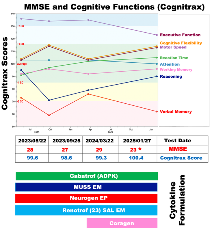
Figure 1. Cognitive function was assessed using Cognitrax and the Mini-Mental State Examination (MMSE) at four time points: May 22, 2023; September 25, 2023; March 22, 2024; and January 27, 2025. MMSE scores are shown in the upper part of the graph. Cognitrax scores are plotted chronologically for cognitive flexibility (orange), executive function (brown), working memory (magenta), attention (light blue), reasoning (dark blue), reaction time (green), motor speed (plum), and verbal memory (red). Green indicates the mean ±1 standard deviation (SD), yellow indicates -1 to -2 SD below the mean, red indicates more than -2 SD below the mean, light blue indicates +1 to +2 SD above the mean, and blue indicates more than +2 SD above the mean. A Cognitrax score of 100 represents the average score for an age-matched Japanese population. Administered cytokines and exosomes are shown below the graph.
Analysis of MRI data acquired on May 22, 2023, revealed mild cortical atrophy in the parietal and frontal lobes (Figure 2A). A sagittal section of the right hemisphere showed an indentation in the posterior corpus callosum (Figure 2C, yellow arrow). A coronal section through the prefrontal cortex demonstrated cortical volume loss, widening of the Sylvian fissures (Figure 2E, red asterisk), and atrophy of the parahippocampal cortex and hippocampus (Figure 2E, red and yellow arrows).
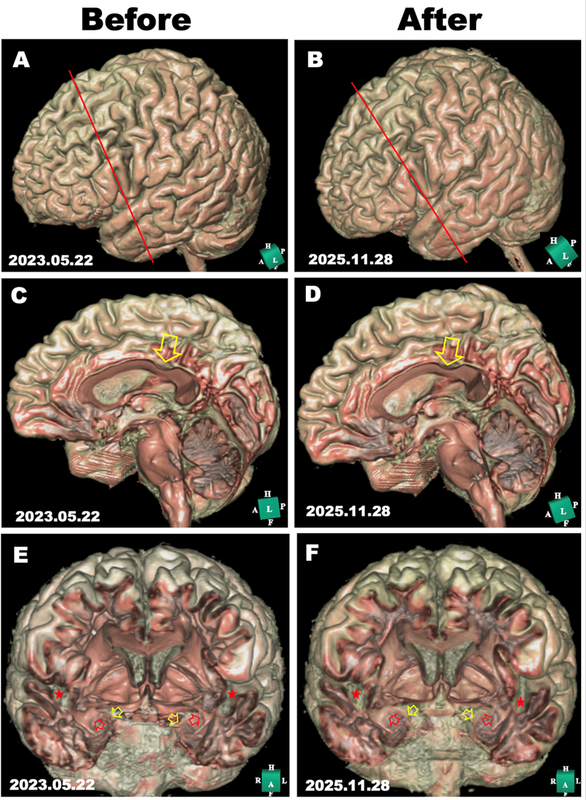
Figure 2. MRI scans were obtained on May 22, 2023, and November 28, 2025, before and during follow-up after cytokine-based therapy. Panels A and B show three-dimensional reconstructions of the cerebral cortex (lateral view of the left hemisphere) generated in silico with Expert INTAGER software from T1-weighted MRI data with 1-mm sagittal slices. Panels C and D show three-dimensional reconstructions of the right hemisphere in a sagittal view from the midsection. Yellow arrows indicate the indentation of the corpus callosum. Panels E and F show cut-surface images of the prefrontal region corresponding to the red lines in panels A and B. Red arrows indicate the hippocampus, yellow arrows the parahippocampal gyrus, and red asterisks the Sylvian fissures.
Virtual endoscopic analysis of the left and right hippocampus, viewed from within the anterior temporal cortex, revealed moderate atrophy involving the medial neck and head regions (Figures 3A and 3B). As illustrated in Figure 5, the virtual endoscopic images also demonstrated intraluminal material within the right and left internal carotid arteries, with apparent attachment to the endothelial surface on the left side (Figure 5A, 5C, and 5E, yellow arrows). On the basis of morphology and vascular location, these lesions were considered compatible with thrombotic material, as previously described [13]. The patient received the Pfizer mRNA vaccine on five occasions in June 2021, July 2021, February 2022, July 2022, and October 2023.
Electroencephalography performed on May 5, 2023, showed slow-wave activity at the frontopolar, frontal, central, and parietal leads at rest (data not shown). Analysis of P300 EEG data using Neuroscan software showed hyperexcitable responses with delayed peaks at the frontal, central, left temporal, parietal, and occipital leads after target stimulation with a high-pitched sound (Figure 4A, red line). P300 responses to the frequent low-pitched stimulus showed asymmetry at the frontopolar leads (Figure 4A, black line). The attention test demonstrated an increased response to the cue compared with the control group (Figure 4C, red vs blue lines). The Visual Space Memory test likewise showed a heightened response compared with the control group (Figure 4D, red vs blue lines). Coherence analysis of the P300 indicated elevated frontal connectivity values with limited variability, findings that may be compatible with altered inhibitory network function in the frontal leads (Figure 4, left panel, blue arrows).
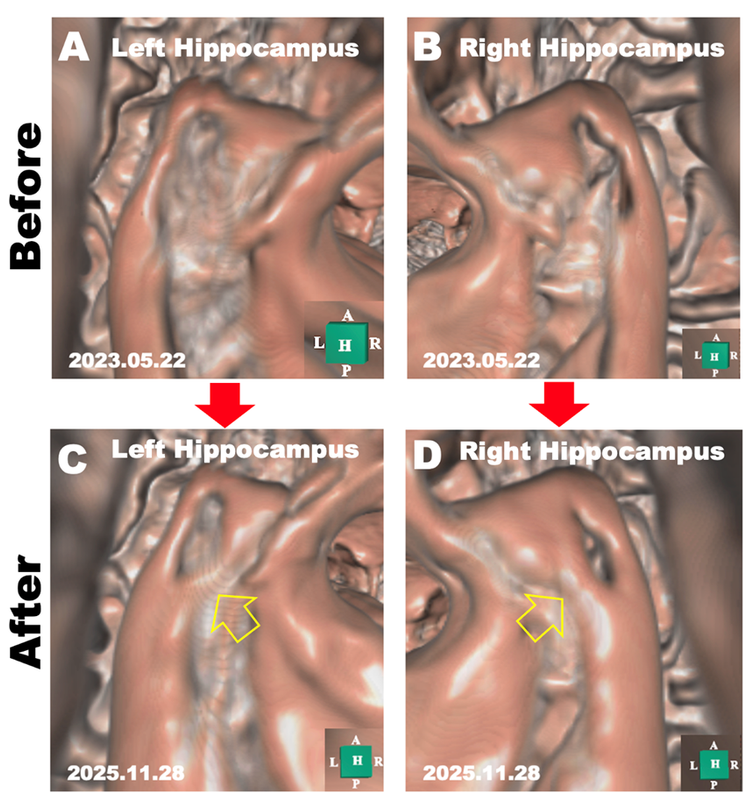
Figure 3. Morphological evaluation of the hippocampus before and during follow-up after cytokine treatment. Panels A and B show in silico endoscopic images of the left (A) and right (B) hippocampus, demonstrating atrophy at the neck of the hippocampus. Panels C and D show corresponding in silico endoscopic images of the left (C) and right (D) hippocampus obtained at follow-up, demonstrating interval morphological changes at the hippocampal neck. Arrows indicate the regions of structural change.
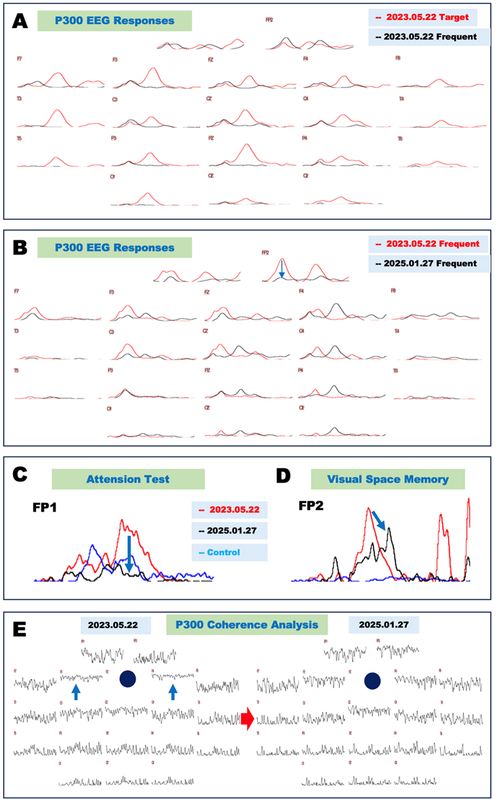
Figure 4. Neurophysiological evaluations obtained before and during follow-up after cytokine treatment.
A. P300 electroencephalogram (EEG) responses obtained at baseline and follow-up during cytokine-based therapy. Responses to the frequent stimulus (low-pitched sound) at baseline on May 22, 2023, are shown in black, and responses to the target stimulus (high-pitched sound) are shown in red.
B. Comparison of P300 EEG responses to the frequent stimulus (low-pitched sound) at baseline and follow-up during cytokine-based therapy. Baseline responses recorded on May 22, 2023, are shown in red, and follow-up responses recorded on January 27, 2025, are shown in black.
C. Electrocardiographic (ECG) and electromyographic (EMG) recordings during the attention test at baseline and follow-up during cytokine-based therapy. The heightened responses observed on May 22, 2023 (red) were reduced on January 27, 2025 (black). Blue indicates the control.
D. Electrophysiological data from the Visual Space Memory test at baseline and follow-up during cytokine-based therapy. The heightened responses observed on May 22, 2023 (red) were reduced on January 27, 2025 (black). Blue indicates the control.
E. Coherence analysis of P300 at baseline and follow-up during cytokine-based therapy. Analysis of neural network connectivity in the frontal electrodes (F3 and F4, indicated by red arrows) showed interval changes by January 27, 2025, compared with May 22, 2023.
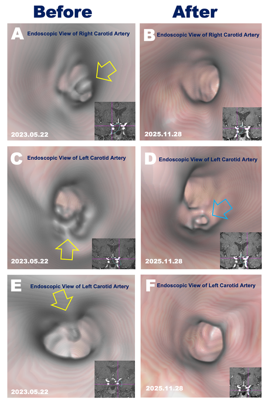
Figure 5. Three-dimensional (3D) computer-generated images show carotid intraluminal findings in the right and left carotid arteries (yellow arrows in A, C, and E) obtained on May 22, 2023, before initiation of the proteolytic treatment protocol. Corresponding follow-up images are shown in panels B, D, and F. The left ophthalmic artery is indicated by a blue arrow. Lower right insets show the position and viewing window of the in silico camera within the carotid artery.
A comprehensive evaluation of blood chemistry, complete blood count (CBC), thyroid function, and glycated hemoglobin (HbA1c) levels did not reveal evidence of common metabolic or endocrine contributors to cognitive impairment. As demonstrated in Figure 6, analysis of the IgG fraction showed a decreased IgG3/IgG4 ratio together with elevated rheumatoid factor (RF). These findings were accompanied by rheumatoid arthritis–like symptoms, although the underlying mechanism remains uncertain. The D-dimer level was within the reference range on May 22, 2023 (Figure 6). Taken together, the clinical, imaging, laboratory, and electrophysiological findings were considered consistent with Alzheimer’s disease in an APOE ε4/ε4 carrier, accompanied by carotid intraluminal findings and rheumatoid arthritis–like symptoms. Given the observational nature of this case, any etiologic relationship among these findings should be interpreted cautiously.
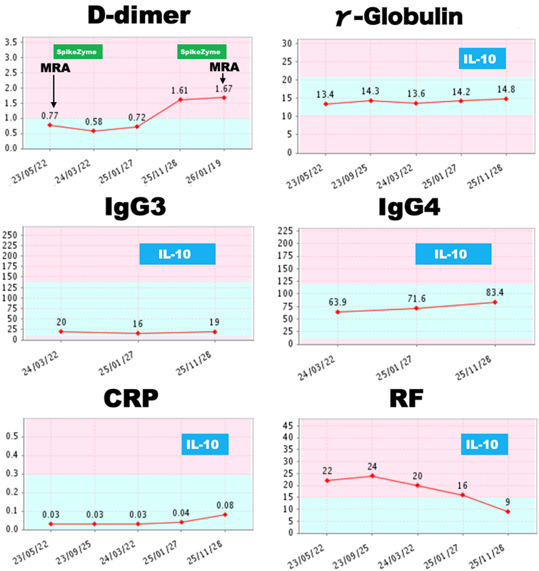
Figure 6. Clinical course of biomarkers. Graphical representation of D-dimer (µg/mL FEU), γ-globulin (g/dL), IgG3 (mg/dL), IgG4 (mg/dL), C-reactive protein (CRP; mg/dL), and rheumatoid factor (IU/mL) measured during clinical follow-up from May 22, 2023, to November 26, 2025, including periods of cytokine-based and proteolytic treatment.
Clinical Course During Cytokine-Based Therapy
The cytokine-based therapy protocol consisted of Gabatrof (ADPK), threefold concentration, 3.0 mL; MUSS EM, threefold concentration, 4.0 mL; Neurogen EP, threefold concentration, 4.0 mL; and Renotrof (23) SAL EM, threefold concentration, 3.0 mL, each administered three times daily from May 22, 2023, to January 27, 2025. Coragen, threefold concentration, 3.0 mL three times daily, was added from March 22, 2024, to January 27, 2025, as shown in Figure 1. The clinical protocol and formulations were developed at the Livant Neurorecovery Center in Mexico, as previously described [12].
As shown in Figure 1, approximately 20 months had elapsed between initiation of cytokine treatment and the assessment on January 27, 2025. Over this interval, the overall Cognitrax profile did not show marked deterioration, with a score of 100.4 at follow-up compared with 99.6 at baseline. Reaction time improved across serial assessments, whereas other domains—including cognitive flexibility, motor speed, attention, working memory, reasoning, and verbal memory—remained broadly stable. Although the MMSE score increased to 29/30 on March 22, 2024, it was 23/30 on January 27, 2025. Review of the record suggested that reduced orientation performance at the later assessment may have reflected transient inattention rather than definite progression of global impairment, but this interpretation remains uncertain.
A follow-up electroencephalogram (EEG) was obtained on January 27, 2025, and showed reduced slow-wave activity compared with the previous study (data not shown). P300 electroencephalogram (EEG) responses also changed over time: responses at the frontopolar leads were reduced (Figure 4B, blue arrow), whereas earlier P300 responses were observed at the frontal, central, right parietal, and right occipital leads (Figure 4B, black lines). These findings may be compatible with changes in inhibitory network function; however, the underlying mechanism cannot be determined from a single case.
In the attention test, the heightened response observed on May 22, 2023 (Figure 4C, red line) was reduced on January 27, 2025 (Figure 4C, black line). In the Visual Space Memory test, the heightened response observed on May 22, 2023 (Figure 4D, red line) was also reduced on January 27, 2025 (Figure 4D, black line), although it remained above the control response. These findings are compatible with interval change in attention-related and visuospatial electrophysiological measures during follow-up, but they do not permit firm conclusions regarding treatment effect.
Coherence analysis of the P300 signals recorded on January 27, 2025, showed interval changes in frontal network connectivity compared with baseline (Figure 4E). Although these findings may be consistent with altered inhibitory network function, any inference regarding GABAergic neurogenesis or synaptic restoration remains speculative in a single-case report.
On November 28, 2025, MRI showed a similar degree of atrophy in the frontal and parietal cortex compared with the prior study (Figures 2A and 2B). The sagittal reconstruction of the right hemisphere also showed comparable atrophy of the cingulate and prefrontal cortex, together with a similar indentation of the corpus callosum (Figure 2D, yellow arrows), relative to the earlier MRI (Figure 2C). A coronal section through the prefrontal, hippocampal, and parahippocampal regions likewise showed persistent cortical and hippocampal atrophy, ventricular dilatation, and widening of the Sylvian fissures (Figure 2F), similar to the initial examination (Figure 2E). In contrast, in silico endoscopy of the temporal lobe demonstrated interval morphological changes in the left and right hippocampus compared with the baseline images (Figures 3A–3D). These findings may be compatible with localized structural remodeling; however, they do not establish hippocampal regeneration, and any relationship to cognitive performance should be interpreted cautiously.
Clinical Course of Carotid Intraluminal Findings During Proteolytic Treatment
For the carotid intraluminal findings, the proteolytic treatment protocol was administered from May 22, 2023, to March 22, 2024, and again from November 26, 2025, to January 19, 2026, as described in the Method section. On November 28, 2025, virtual endoscopy showed that the previously observed intraluminal material was no longer visible in either internal carotid artery (Figures 5B, 5D, and 5F). These imaging findings are compatible with interval resolution of the lesions; however, their precise composition cannot be determined from imaging alone. The lesions were interpreted as thrombotic on the basis of their morphology and vascular location, as previously described [13], despite a D-dimer level within the reference range on May 22, 2023. On November 26, 2025, the D-dimer level increased to 1.61 µg/mL, and the proteolytic protocol was resumed. On January 19, 2026, follow-up virtual endoscopy again showed no visible intraluminal material, although treatment was continued because the D-dimer level remained elevated at 1.67 µg/mL.
IgG4 is generally considered to have anti-inflammatory or functionally suppressive properties. Repeated SARS-CoV-2 mRNA vaccination has been reported to increase spike-specific IgG4 responses, although the clinical significance of this shift remains uncertain [15]. The patient began experiencing swelling in the wrists and other joints in 2023. On May 22, 2023, the patient had an elevated rheumatoid factor (RF 22 IU/mL), bilateral wrist swelling, and an increased IgG4/IgG3 ratio. These findings occurred in parallel with rheumatoid arthritis–like symptoms; however, their relationship remains uncertain. A cytokine-based immunoregulatory approach was implemented. This approach encompassed the administration of Renagen Spleen, a formulation comprising porcine IL-10 that has been purified from porcine spleen tissue, from January 27, 2025, to November 25, 2025, as delineated in the Method section. As shown in Figure 6, C-reactive protein (CRP) showed a slight increase, whereas rheumatoid factor decreased to within the reference range by November 25, 2025, accompanied by improvement in joint symptoms. Although these observations may be compatible with a potential immunoregulatory effect of the treatment approach, no causal inference can be drawn from a single case.
DISCUSSION
This case report describes a patient with Alzheimer’s disease, carotid intraluminal findings, and rheumatoid arthritis–like symptoms who underwent cytokine-based therapy and a proteolytic treatment protocol. During follow-up, interval changes were observed in hippocampal morphology, electrophysiological measures including P300 EEG and coherence analysis, carotid intraluminal findings, and rheumatoid arthritis–like symptoms. Because the report is observational and limited to a single patient, these findings should be regarded as hypothesis-generating rather than confirmatory. The temporal clustering of multimodal changes may support future investigation in complex dementia-associated presentations.
APOE ε4 and Electrophysiological Findings
Previous studies have suggested that patients with Alzheimer’s disease (AD) who carry the APOE ε4 allele may show increased electrophysiological excitability on P300 electroencephalogram (EEG), possibly reflecting altered inhibitory network function [11]. In the present case, hyperexcitable and temporally altered P300 responses were observed following both frequent and target auditory stimuli. These findings are compatible with prior reports suggesting relative vulnerability of GABAergic circuits in Alzheimer’s disease [11,16]. Coherence analysis also showed changes in frontal network connectivity after treatment (Figure 4E). Although the mechanism cannot be determined from a single case, these observations may be consistent with partial restoration of inhibitory neuronal function. Any contribution of specific cytokine components, including HGF and GCSF in Gabatrof (ADPK), remains hypothetical and requires further investigation.
Hippocampal Morphological Findings During Follow-up
In this case report, although MRI showed no overt evidence of cortical structural recovery, virtual endoscopic imaging demonstrated interval morphological changes in both hippocampi. As shown in Figure 3, tissue filling was observed between the head and body of the hippocampus, involving the previously eroded medial neck region and producing a central opening-like morphology in the hippocampal head (Figure 3C, 3D). These imaging findings are qualitatively similar to our previous observations in Alzheimer’s disease [11]. As previously discussed, this pattern may be compatible with the known vulnerability of the medial hippocampal head and dentate gyrus in Alzheimer’s disease [11]. In this context, relative preservation of CA1–CA3 regions with greater involvement of the dentate gyrus has been described in prior studies [17,18]. As illustrated in Figure 7, the principal input to the hippocampus arises from the entorhinal cortex, whereas major outputs are conveyed through CA1 and the subiculum [19]. Although the findings in Figure 3C and 3D may be consistent with intrahippocampal structural change, no apparent recovery was observed in the entorhinal cortex or subiculum in the coronal temporal lobe section obtained on November 28, 2025 (Figure 2F). Accordingly, any inference that these changes represent hippocampal neurogenesis remains tentative. One possible interpretation is that the observed changes reflect a localized or early-stage remodeling process within the hippocampus rather than restoration of broader afferent or efferent pathways. In this context, persistent impairment of memory function would not be unexpected [20]. Given that adult-born hippocampal neurons have been implicated not only in memory but also in cognitive flexibility, pattern separation, stress regulation, emotional control, and social cognition [7,8,20], the improvements in cognitive flexibility (Figure 1), attention-related hyperreactivity (Figure 4C), and visuospatial memory testing (Figure 4D) observed in this case may be compatible with functional improvement, although they cannot be directly attributed to neurogenesis. These findings may also be compatible with broader hippocampal contributions to emotional regulation in addition to memory-related processing [21,22].
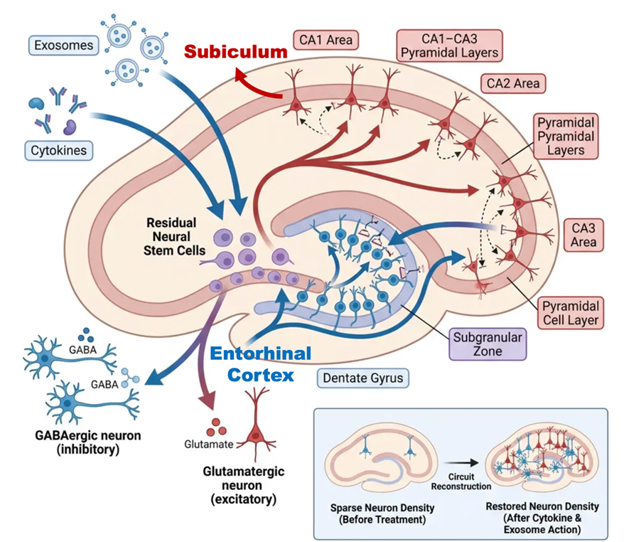
Figure 7. Illustrative schematic of possible pathways by which cytokines and exosome-derived components may relate to hippocampal structural change and neuronal circuit remodeling. The figure was created using Figurelabs.ai.
Carotid Intraluminal Findings During Follow-up
In this case, virtual intravascular endoscopy demonstrated interval disappearance of previously observed intraluminal material along both carotid arteries after treatment with the proteolytic enzyme complex SpikeZyme™. These imaging findings are compatible with interval resolution of the lesions; however, the precise composition of the material cannot be determined from imaging alone. The interpretation in this report is based on morphological features including protrusion into the vascular lumen, apparent attachment to the endothelial surface (Figures 5A and 5E), and localization within the internal carotid artery in the region shown in Figure 5. Despite a negative D-dimer result on May 22, 2023, the lesions were interpreted as thrombotic on the basis of these imaging characteristics. On November 26, 2025, the D-dimer result became positive, and SpikeZyme™ was readministered, as shown in Figure 6.
It has been hypothesized that such intraluminal material may form through aggregation and polymerization of macromolecules, potentially including fibrin and other proteinaceous components [13]. SpikeZyme™ is a plant-derived combination of proteolytic enzymes developed with the intended purpose of promoting proteolysis of proteinaceous intraluminal material (Figure 8). The formulation contains nattokinase, lumbrokinase, serratiopeptidase, fungal proteases, bromelain, and papain. Although the temporal association between treatment and disappearance of the intraluminal findings is notable in this case, the mechanism of lesion formation and resolution remains uncertain and cannot be established from a single case report.
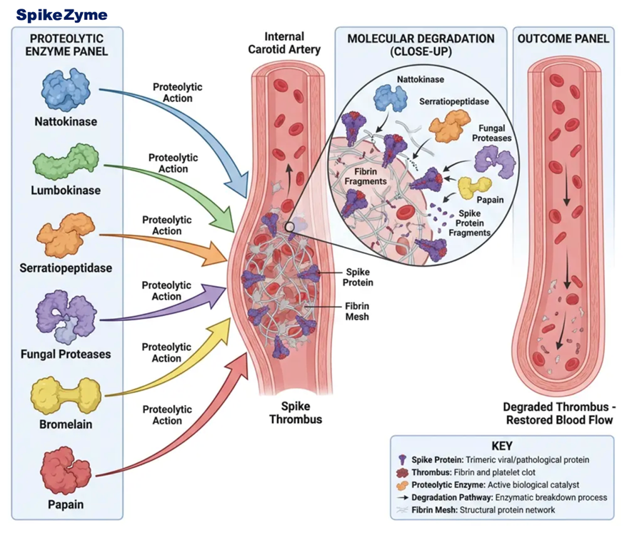
Figure 8. Illustrative schematic of a proposed relationship between proteolytic enzyme activity and carotid intraluminal material. The figure was created using Figurelabs.ai.
Rheumatoid Arthritis–Like Symptoms and Immunological Findings
As illustrated in Figure 9, the present case developed rheumatoid-like symptoms during the clinical course, together with elevation of rheumatoid factor (Figure 6). Case reports and reviews have described new-onset arthritis, rheumatoid arthritis, and disease flares after SARS-CoV-2 vaccination; however, a causal relationship has not been established [23]. Therefore, the present findings should be interpreted as a temporal association rather than evidence of vaccine-induced rheumatoid disease. Regulatory T cells (Tregs) play a central role in maintaining immune tolerance [24,25]. In rheumatoid arthritis, abnormalities in Treg number and/or function have been reported, and such dysregulation may contribute to persistent synovial inflammation and impaired self-tolerance. In particular, disruption of the Th17/Treg balance is thought to promote chronic inflammatory responses in RA [26,27]. Repeated SARS-CoV-2 mRNA vaccination has been reported to alter spike-specific IgG subclass responses, including increased IgG4 responses, although the clinical significance of this shift remains uncertain [15,28]. In this case, an increase in the IgG4/IgG3 subclass ratio was observed during follow-up (Figure 6). This serological change occurred in parallel with the development of rheumatoid-like symptoms and rheumatoid factor positivity; however, the relationship between these findings remains uncertain. Although IL-10-related immune regulation and Treg-based therapeutic strategies are of considerable interest, their clinical significance remains to be fully established [26]. In this case, porcine IL-10 purified from porcine spleen was administered, and improvement in clinical symptoms of arthritis, including joint swelling, was observed together with normalization of rheumatoid factor (Figure 6). Although this observation may suggest a potential immunoregulatory effect, the finding should be interpreted cautiously in light of the single-case design.
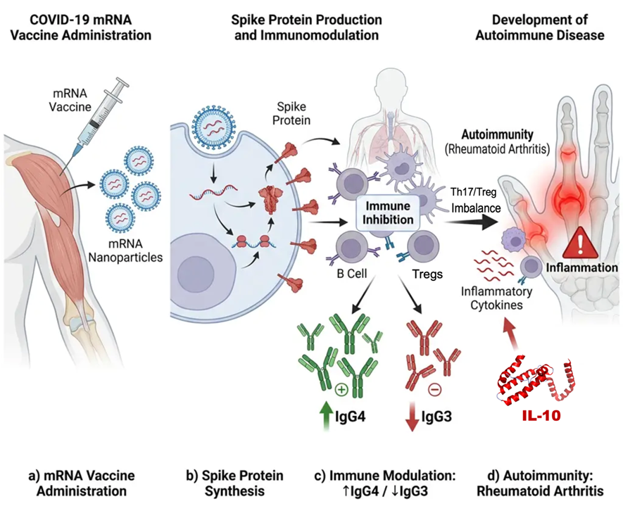
Figure 9. Illustrative schematic of immune dysregulation and rheumatoid arthritis–like manifestations discussed in this case, shown in the context of temporal association with repeated COVID-19 mRNA vaccination. The figure was created using Figurelabs.ai.
Integrated Synergistic Regenerative and Immunomodulatory Strategy
This report describes a complex translational clinical observation involving a patient with Alzheimer’s disease, rheumatoid arthritis–like manifestations, and thrombotic vascular pathology who received an integrated regenerative and immunomodulatory treatment strategy including HGF, progranulin, IL-10, miR-124, miR-146a, IGF-1/IGF-2, irisin, and juvenile porcine cardiac-derived regenerative signals (Figure 10). Collectively, these cytokines and exosome-derived components may contribute to a multifaceted biological response involving neuroprotective, immunoregulatory, and vascular reparative processes. Based on their known or proposed biological activities, HGF, progranulin, miR-124, IGF-1/IGF-2, and irisin may have contributed to mechanisms relevant to neurodegeneration in Alzheimer’s disease [29-33]. Similarly, HGF, progranulin, IL-10, miR-124, miR-146a, and irisin may have supported immunomodulatory processes relevant to rheumatoid arthritis–like manifestations [34-37]. In addition, HGF, IL-10, and SDF-1 may have contributed to biological processes relevant to vascular repair, endothelial protection, and thrombo-inflammatory pathology [38,39]. However, these interpretations remain hypothetical and should be regarded as exploratory in the context of a single-case observation.
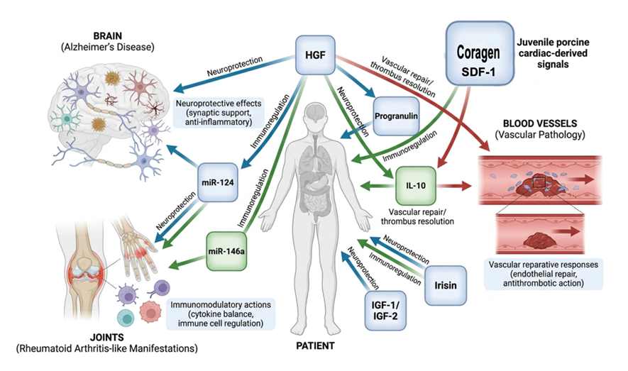
Figure 10. Conceptual schematic summarizing the combined regenerative and immunomodulatory framework discussed in this case report. The figure was created using Figurelabs.ai.
CONCLUSION
In this case report, a 79-year-old female patient with the APOE ε4/ε4 genotype was presented with cognitive impairment and diagnosed with Alzheimer's disease. The patient also exhibited carotid intraluminal findings and symptoms reminiscent of rheumatoid arthritis. During follow-up after cytokine-based therapy and a proteolytic treatment protocol, interval changes were observed in hippocampal morphology, carotid intraluminal findings, and rheumatoid arthritis–like symptoms. Although a single case does not permit causal inference, this report provides descriptive multimodal observations that may inform future hypothesis-driven investigation in complex dementia-associated presentations.
ABBREVIATIONS
AD: Alzheimer’s Disease; APOE: Apolipoprotein E; CBC: Complete Blood Count; CRP: C-Reactive Protein; ECG: Electrocardiogram; EEG: Electroencephalogram; EMG: Electromyography; GCSF: Granulocyte Colony-Stimulating Factor; GDNF: Glial Cell Line-Derived Neurotrophic Factor; HbA1c: Glycated Hemoglobin; HGF: Hepatocyte Growth Factor; IgG: Immunoglobulin G; IL-10: Interleukin-10; MMSE: Mini-Mental State Examination; MRI: Magnetic Resonance Imaging; RF: Rheumatoid Factor; SD: Standard Deviation; Tregs: Regulatory T Cells.
ACKNOWLEDGEMENTS
The authors thank Ms. Sayuri Sato and Ms. Fernanda Diaz for assistance with manuscript preparation.
ETHICAL APPROVAL AND INFORMED CONSENT
Written informed consent for publication was obtained from the patient.
CONFLICT OF INTEREST
The authors have no conflicts of interest.
REFERENCES
- Safiri S, Ghaffari Jolfayi A, Fazlollahi A, Morsali S, Sarkesh A, Daei Sorkhabi A, et al. (2024). Alzheimer's disease: a comprehensive review of epidemiology, risk factors, symptoms, diagnosis, management, caregiving, advanced treatments, and associated challenges. Front Med. 11:1474043.
- Liu E, Zhang Y, Wang JZ. (2024). Updates in Alzheimer's disease: from basic research to diagnosis and therapies. Transl Neurodegener. 13(1):45.
- Abdulkhaliq AA, Kim B, Almoghrabi YM, Khan J, Ajoolabady A, Ren J, et al. (2026). Amyloid-β and Tau in Alzheimer's disease: pathogenesis, mechanisms, and interplay. Cell Death Dis. 17(1):21.
- Peng L, Zhang Z, Li Q, Song Z, Yan C, Ling H. (2024). Unveiling the multifaceted pathogenesis and therapeutic drugs of Alzheimer's disease: A comprehensive review. Heliyon. 10(20):e39217.
- Belloy ME, Andrews SJ, Le Guen Y, Cuccaro M, Farrer LA, Napolioni V, et al. (2023). APOE Genotype and Alzheimer Disease Risk Across Age, Sex, and Population Ancestry. JAMA Neurol. 80(12):1284-1294.
- Wan Z, Ma T. (2025). The impact of apolipoprotein E, type ∊4 allele on Alzheimer's disease pathological biomarkers: a comprehensive post-mortem pilot-analysis. PLoS One. 20(2):e0303486.
- Berdugo-Vega G, Dhingra S, Calegari F. (20230. Sharpening the blades of the dentate gyrus: how adult-born neurons differentially modulate diverse aspects of hippocampal learning and memory. EMBO J. 42(22):e113524.
- Tuncdemir SN, Grosmark AD, Chung H, Luna VM, Lacefield CO, Losonczy A, et al. (2023). Adult-born granule cells facilitate remapping of spatial and non-spatial representations in the dentate gyrus. Neuron. 111(24):4024-4039.e7.
- Beshir SA, Hussain N, Menon VB, Al Haddad AHI, Al Zeer RAK, Elnour AA. (2024). Advancements and Challenges in Antiamyloid Therapy for Alzheimer's Disease: A Comprehensive Review. Int J Alzheimers Dis. 2024:2052142.
- Wang Q, Chen S, Wang J, Shang H, Chen X. (2024). Advancements in pharmacological treatment of Alzheimer's disease: the advent of disease-modifying therapies (DMTs). Brain Sci. 14(10):1-13.
- Shirasawa T, et al. (2023). Cytokine-induced Neurogenesis Can Reverse Cognitive Decline in Alzheimer’s Disease. Mathews J Case Rep. 8(3):97.
- Shirasawa T, et al. (2024). Cytokine-induced Neurogenesis for the Prevention and Treatment of Alzheimer’s Disease. Mathews J Case Rep. 9(9):187.
- Shirasawa T. (2025). Imaging Diagnosis of and Treatment for Spike Protein-Induced Thrombosis after COVID-19 mRNA Vaccination. Mathews J Case Rep. 10(1):195.
- Kim S, Bea S, Choe SA, Choi NK, Shin JY. (2024). Autoimmune disorders reported following COVID-19 vaccination: a disproportionality analysis using the WHO database. Eur J Clin Pharmacol. 80(3):445-453.
- Marchese AM, Fries L, Beyhaghi H, Vadivale M, Zhu M, Cloney-Clark S, et al. (2024). Mechanisms and implications of IgG4 responses to SARS-CoV-2 and other repeatedly administered vaccines. J Infect. 89(6):106317.
- Carello-Collar G, Bellaver B, Ferreira PCL, Ferrari-Souza JP, Ramos VG, Therriault J, et al. (2023). The GABAergic system in Alzheimer’s disease: a systematic review with meta-analysis. Mol Psychiatry. 28(1):5025-5036.
- Shirasawa T, Hirano K, Namihira M, Cobos LCA, Tomoki T. (2023). Bangle rhizome extract promotes hippocampal neurogenesis associated with improved cognitive functions: a case study. Biomed J Sci Tech Res. 52(5):44116-44126.
- Kawles A, Minogue G, Zouridakis A, Keszycki R, Gill N, Nassif C, et al. (2023). Differential vulnerability of the dentate gyrus to tauopathies in dementias. Acta Neuropathol Commun. 11(1):1.
- Hernandez-Frausto M, Vivar C. (2024). Entorhinal cortex-hippocampal circuit connectivity in health and disease. Front Hum Neurosci. 18:1448791.
- Kim TA, Syty MD, Wu K, Ge S. (2022). Adult hippocampal neurogenesis and its impairment in Alzheimer's disease. Zool Res. 43(3):481-496.
- Surget A, Belzung C. (2022). Adult hippocampal neurogenesis shapes adaptation and improves stress response: a mechanistic and integrative perspective. Mol Psychiatry. 27(1):403-421.
- Sun D, Mei L, Xiong WC. (2023). Dorsal dentate gyrus, a key regulator for mood and psychiatric disorders. Biol Psychiatry. 93(12):1071-1080.
- Hoxha A, Striani G, Lovisotto M, Simioni P, Doria A, Ramonda R. (2024). COVID-19 vaccine and the risk of flares in inflammatory arthritis: a systematic literature review and meta-analysis. Front Immunol. 15:1503895.
- Dikiy S, Rudensky AY. (2023). Principles of regulatory T cell function. Immunity. 56(2):240-255.
- Georgiev P, Benamar M, Han S, Haigis MC, Sharpe AH, Chatila TA. (2024). Regulatory T cells in dominant immunologic tolerance. J Allergy Clin Immunol. 153(1):28-41.
- Zhang J, Liu H, Chen Y, Liu H, Zhang S, Yin G, et al. (2024). Augmenting regulatory T cells: new therapeutic strategy for rheumatoid arthritis. Front Immunol. 15:1312919.
- Alunno A, Manetti M, Caterbi S, Ibba-Manneschi L, Bistoni O, Bartoloni E, et al. (2015). Altered immunoregulation in rheumatoid arthritis: the role of regulatory T cells and proinflammatory Th17 cells and therapeutic implications. Mediators Inflamm. 2015:751793.
- Kiszel P, Sík P, Miklós J, Kajdácsi E, Sinkovits G, Cervenak L, et al. (20230. Class switch towards spike protein-specific IgG4 antibodies after SARS-CoV-2 mRNA vaccination depends on prior infection history. Sci Rep. 13(1):13166.
- Sharma S. (2010). Hepatocyte growth factor in synaptic plasticity and Alzheimer's disease. ScientificWorldJournal. 10(1):457-461.
- Moebius HJ, Church KJ. (2023). The Case for a Novel Therapeutic Approach to Dementia: Small Molecule Hepatocyte Growth Factor (HGF/MET) Positive Modulators. J Alzheimers Dis. 92(1):1-12.
- Boylan MA, Pincetic A, Romano G, Tatton N, Kenkare-Mitra S, Rosenthal A. (2023). Targeting Progranulin as an Immuno-Neurology Therapeutic Approach. Int J Mol Sci. 24(21):15946.
- Han D, Dong X, Zheng D, Nao J. (2019). MiR-124 and the underlying therapeutic promise of neurodegenerative disorders. Front Pharmacol. 10(1):1555-1564.
- Zegarra-Valdivia J, Arana-Nombera H, Perez-Fernandez L, Del Rocío Casimiro M, Gallegos-Manayay V, Del Rosario Oliva-Piscoya M, et al. (2025). Insulin-like growth factor 1 impact on Alzheimer's disease: role in inflammation, stress, and cognition. Curr Issues Mol Biol. 47(4):233.
- Reynolds G, Cooles FA, Isaacs JD, Hilkens CM. (2014). Emerging immunotherapies for rheumatoid arthritis. Hum Vaccin Immunother. 10(4):822-837.
- Katsikis PD, Chu CQ, Brennan FM, Maini RN, Feldmann M. (1994). Immunoregulatory role of interleukin 10 in rheumatoid arthritis. J Exp Med. 179(5):1517-1527.
- Zhang Y, Yang M, Xie H, Hong F, Yang S. (2023). Role of miRNAs in rheumatoid arthritis therapy. Cells. 12(13):1-24.
- Wang Z, Xu J, Mo L, Zhan R, Zhang J, Liu L, et al. (2024). The application potential of the regulation of Tregs function by irisin in the prevention and treatment of immune-related diseases. Drug Des Devel Ther. 18(1):3005-3023.
- Chen Y, Shen J, Nilsson AH, Goncalves I, Edsfeldt A, Engström G, et al. (2022). Circulating hepatocyte growth factor reflects activation of vascular repair in response to stress. JACC Basic Transl Sci. 7(8):747-762.
- Yi L, Weng T, Nie P, Zhu L, Gao M, Jia H, et al. (2022). Overexpression of interleukin-10 in engineered macrophages protects endothelial cells against LPS-induced injury in vitro. FEBS Open Bio. 12(3):605-615.
