Previous Issues Volume 4, Issue 3 - 2019
Metachronic Cerebral Metastasis of Hepatocellular Carcinoma on Cirrhotic Liver Carcinoma on Cirrhotic Liver
Sabbah M, Trad D, Ouakaa A, Bellil N, Gargouri D
Department of Gastroenterology. Habib Thameur Hospital, Tunisia
Received: Jul 20, 2019
Published: Aug 7, 2019
*Corresponding Author: Sabbah Meriam, Habib Thameur Hospital, 8 Rue El Messelekh, Montfleury, 1008, Tunis, Tunisia.
Copyright © 2019 Meriam S.
Citation: Meriam S. (2019). Metachronic Cerebral Metastasis of Hepatocellular Carcinoma on Cirrhotic Liver . Mathews J Case Rep 4(3): 56
CASE REPORT
A 60 years old patient was initially hospitalized for digestive bleeding due to gastric variceal rupture successfully treated by biological glue injection. Diagnosis of cirrhosis was performed and etiological assessment found hepatitis B with positive HBsAg. Patient received antiviral therapy as well as prophylactic B blockers. The Child-Pugh score calculated was Class B (9 points). Abdominal ultrasound completed with hepatic CT angioscan showed a three centimeters mass of the hepatic dome with a typical vascular kinetic of hepatocellular carcinoma (Figure 1). Patient underwent chemoembolization with complete initial response. One year later, he was hospitalized for headache, dizziness, right hemiparesia and loss of hearing. Cerebral CT scan objectified a hypodense centimetric lesion in the frontal wall in contact with the posterior horn of the lateral ventricle raised after injection of contrast medium associated with perilesional edema (Figure 2). Neurosurgical approach was recused because of underlying cirrhosis with high risk anesthesia. Patient died due to cerebral engagement despite corticosteroids injections and mannitol infusions.
.png)
Figure 1: Hepatic CT angioscan showing a three centimeters mass of the hepatic dome with a typical vascular kinetic of hepatocellular carcinoma (wash in and wash out).
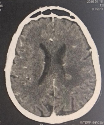
Figure 2: Cerebral CT scan objectifying hypodense centimetric lesion in the frontal wall in contact with the posterior horn of the lateral ventricle raised after injection of contrast medium associated with perilesional edema.