Information Links
Related Conferences
Previous Issues Volume 8, Issue 4 - 2023
Endometriosis with Massive Ascites: A Rare Entity
Pramod Kumar1, Pawandeep Kaur2, Heena Kauser3,*
1Professor, Obstetrics and Gynaecology, MGIMS, Sevagram, Wardha, Maharashtra, India
2Senior Resident, Obstetrics and Gynaecology, MGIMS, Sevagram, Wardha, Maharashtra, India
3Junior Resident, Obstetrics and Gynaecology, MGIMS, Sevagram, Wardha, Maharashtra, India
*Corresponding Author: Heena Kauser, Junior Resident, Obstetrics and Gynaecology, MGIMS, Sevagram, Wardha, Maharashtra, India; Email: [email protected]
Received Date: January 16, 2023
Publication Date: April 15, 2023
Citation: Kumar P, et al. (2023). Endometriosis with Massive Ascites: A Rare Entity. Mathews J Case Rep. 8(4):103.
Copyright: Kumar P, et al. © (2023)
ABSTRACT
Endometriosis associated with massive haemorrhagic ascites is an uncommon finding. In most cases, the presence of massive haemorrhagic ascites is associated with malignancies, hepatoma, tuberculosis & perforated viscera. A 23-year-old unmarried female was admitted to the gynae ward with massive ascites, leading to breathlessness, and abdominal distension, associated with cachexia and respiratory and abdominal discomfort. After, while investigating & managing her, we found that the ascitic fluid is haemorrhagic type, which led to further investigation to rule out other possible causes. Finally, the decision was made to take her for exploratory laparotomy, and 22 litres of ascitic fluid was drained. Ascitic fluid cytology and histopathology of tissue were suggestive of endometriosis.
Keywords: Endometriosis, Haemorrhagic Ascites
INTRODUCTION
Endometriosis is a benign, estrogen-dependent disease characterized by endometrial tissue outside the uterine cavity [1,2]. Endometrial implantation can be in either the pelvic or extra pelvic region with the former being more prevalent [3,4]. Clinical presentations vary by endometrial implantation site. Classical symptoms include chronic pelvic pain, dysmenorrhea, dyspareunia, and infertility [5]. Endometriosis is prevalent among 10% to 15% of women between 25 to 44 years of age group and in infertile women incidence of endometriosis is almost 25% to 40% [1]. However, massive haemorrhagic ascites mimicking ovarian malignancy in signs & symptoms is a very rare type of presentation with endometriosis with ascites & pleural effusion found.
CASE PRESENTATION
A 23-year-old unmarried female admitted was to the gynae ward from the outpatient department with chief complaints of abdominal distension, amenorrhea of 6 months, respiratory discomfort and loss of appetite. Her bowel bladder habits were normal. Her menarche was at 11 years of age with regular cycles till 6 months back, and then she developed amenorrhea. On examination, she was poorly nourished and poorly built. Tachycardia was present, pallor was present. On abdominal examination, abdomen was fully distended up to xiphisternum with fullness of flanks. Shifting dullness was present. Dilated veins seen over abdomen, dull note was present over abdomen. X ray chest was suggestive of left sided pleural effusion. X ray abdomen had diffuse increase in density. In view of worsening respiratory embracement and for diagnosis, ascitic tapping was done. 1 litre of fluid was removed which was haemorrhagic and sent for AFB (ZN Smear), CB-NAAT, LJ media culture, culture and sensitivity, fluid glucose, proteins, albumin, ADA & LDH, cytological examination& cell block studies. Blood samples for tumour marker like Ca 125, B-HCG, and LDH were sent and were found to be within normal range. While CT abdomen & pelvis was done, suggestive of heterogeneously enhancing large solid cystic mass of 9.1 * 6 * 4.1 cm in pelvis, involving bilateral adnexa with loss of fat and uterus inferiorly. Posteriorly the lesion was abutting internal iliac vessel & bilateral lower ureter? bilateral ovarian neoplastic masses with gross ascites. Non enhancing massive fluid density collection in abdomen & pelvis displacing & compressing abdominal content medially & bilateral diaphragm superiorly. Herniation of fluid through a rent of approximate size 8*7 mm in right inguinal region? Right inguinal hernia. Herniation of fluid through a rent of approx. 4.4 * 3.9 cm through anterior abdominal wall in umbilical region? Umbilical hernia. Ascitic fluid cytology showed occasional group of mesothelial cells in a background of large number of hemosiderin laden macrophages and fragmented red blood cells. No evidence of malignancy seen in present smear. Cell block study also showed same picture i.e., hemosiderin laden macrophage & lymphocyte in haemorrhagic background. No malignant cells seen. Biochemical analysis of ascitic fluid was within normal limits. Then the decision was made to take her for exploratory laparotomy. Pre anaesthetic work up was done. Therapeutic ascitic tap was done before operation and 1 litre was removed. Operation was performed under general anaesthesia. Intraoperatively 20 litres of ascitic fluid drained. Abdominal cavity
carefully examined. Uterus was normal size & shape. Bilateral ovaries adhered to each other (kissing ovaries) covered entirely by colon. Massive adhesion present. Clinical picture suggestive of grade IV endometriosis, leading to chocolate cyst leading to massive ascites. Thickened matted bowel loop present. necrotic debris present over peritoneum. Multiple biopsies taken from affected areas along with peritoneum & omentum, along with rent for histopathology. Histopathological study suggestive of interspread endometrial glands & stoma with hemosiderin laden macrophage. Post-operative period was uneventful. Injection leuprolide 11.25mg subcutaneously given & advised her to take every 3 monthly. Patient is under follow up.
DISCUSSION
An estimated 10% to 15% of woman of child bearing age group have endometriosis. Presentation of endometriosis ovaries and it may present as infertility, dysmenorrhoea, chronic pelvic pain, deep dyspareunia or bleeding at external sites like the umbilicus [2]. The term ascites describes the pathologic accumulation of fluid within the peritoneal cavity [6]. Cirrhosis of the liver is the most common cause of ascites, however, other conditions, such as heart failure, kidney failure, infection (such as tuberculosis), cancer, or gynecological malignancy can also cause this condition [7]. When a woman presents with ascites, exclusion of certain gynecological disorders, including ovarian cancer, primary peritoneal cancer, fallopian tube cancer, and pelvic tuberculosis, is necessary.
Endometriosis presenting as a recurrent haemorrhagic ascites is an unusual occurrence, more than 60 cases have been reported since 1954 [3,4], and these patients are usually noncritical with minimal significant complications [5,8]. Predominant theories regarding the pathophysiology of haemorrhagic ascites include peritoneal irritation from ruptured endometriotic implants, subdiaphragmatic obstruction of lymphatics and retrograde menstruation [9]. Here we are reporting an additional case, in order to draw attention towards this rare condition.
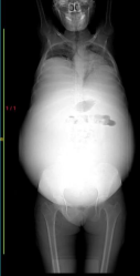
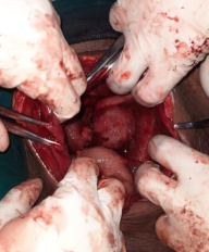
Figure 1. Figure 2.
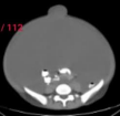
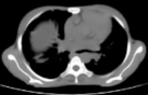

Figure 3. Figure 4. Figure 5.


Figure 6. Figure 7. Figure 8.
Diagnosis of endometriosis requires that the clinician should differentiate it from neoplastic or other inflammatory conditions [10]. In general, asymptomatic lesions of endometriosis which are found incidentally at laparotomy done for other conditions require no treatment. Hemorrhagic ascites is characterized by bloody appearance of ascitic fluid and is defined as an ascitic fluid with an erythrocyte count of more than 50,000/μL. Hemorrhagic ascites should be considered a complication of endometriosis, especially in nulliparous women of childbearing age group with abdominal distention, a pelvic mass, dysmenorrhea, abdominal pain, weight loss and eventual pleural effusion, suggesting a diagnosis of ovarian malignancy [6]. Similar to this patient, a recent review and analysis of previous cases found that 63% of women were of African ancestry, and 82% were nulliparous [10]. 38% of patients previously described in the literature also had a pleural effusion [5]. Although massive ascites has been reported as a manifestation of endometriosis, however, it can be seen in patients with acute abdomen with shock also [7]. Other known causes of bloody ascites, especially a malignant process, must be excluded. In addition to endometriosis involving various gastrointestinal organs, other findings such as adhesions and fibrosis are frequently encountered at laparotomy [11]. The diagnosis must be confirmed histologically. Recurrences have been seen commonly with severe complications and usually required multiple laparotomies and thoracotomies for associated pulmonary involvement [12]. Definitive management consists of surgical resection of endometriotic tissue combined with complete surgery or long-term hormonal suppression therapy [13]. Long-term hormonal suppression therapy is generally reserved for young nulliparous patients because it usually obviates the need for complete surgery and it can preserve fertility in some cases. The response of hormonal therapy including GnRH agonists in patients of endometriosis with ascites is often unsatisfactory according to the case studies. Oral contraceptive pills or leuprolide injection used for long term management reduces the amount of follicle stimulating hormone and luteinizing hormone, results in hypoestrogenism suppressing the growth of native and ectopic endometrial tissues. Ruptured ectopic pregnancy is the most common gynaecological cause of hemoperitoneum in a reproductive-age woman, endometriosis should also be considered as a cause, especially after excluding pregnancy [14]. Long term follow up is advised to patients who underwent conservative treatment because of the high risk of recurrence of the disease.
CONCLUSION
Presentation of endometriosis varies and it can present with significant internal bleeding. Endometriosis should be considered in the differential diagnosis when bloody ascites and a pelvic mass are present, particularly in young female patients, for which there is no obvious explanation. A very high level of clinical suspicion is required if the diagnosis of severe endometriosis is not to be missed. The surgeon should therefore be very vigilant and alert to localize and diagnose rare sites of internal bleeding due to endometriosis.
REFERENCES
- Giudice LC. (2010). Clinical practice. Endometriosis. N Engl J Med. 362(25):2389–2398.
- Young VJ, Ahmad SF, Duncan WC, Horne AW. (2017). The role of TGF-β in the pathophysiology of peritoneal endometriosis. Hum Reprod Update. 23(5):548–559.
- Davis AC, Goldberg JM. (2017). Extrapelvic endometriosis. Semin Reprod Med. 35(1):98–101.
- Machairiotis N, Stylianaki A, Dryllis G, Zarogoulidis P, Kouroutou P, Tsiamis N, et al. (2013). Extrapelvic endometriosis: a rare entity or an under diagnosed condition? Diagn Pathol. 8(1):194.
- Dunselman GA, Vermeulen N, Becker C, Calhaz-Jorge C, D'Hooghe T, De Bie B, et al. (2014). ESHRE guideline: management of women with endometriosis. Hum Reprod. 29(3):400–412.
- Hou W, Sanyal AJ. (2009). Ascites: diagnosis and management. Med Clin North Am. 93(4):801–817.
- Trent LM, Eric BT, Jason DH. (2013). Endometriosis presenting with hemorrhagic ascites, severe anaemia, and shock: Case report. Am J Emerg Med. 31:e1272-e3 272.
- Talbert LM, Kauma SM. (1990). Endometriosis. In: Danforth DN, Scott JR (Eds). Danforth's obstetrics and gynecology. (5thedn), Philadelphia: JB Lippincott.
- Otolorin EO, Ojengbede O, Falase AO (19987) Laparoscopic evaluation of the tubo- peritoneal factor in infertile Nigerian women. Int J Gynecol Obstet 25: 47-52.
- Kietpeerakool C, Rattanakanokchai S, Jampathong N, Srisomboon J, Lumbiganon P. (2019). Management of drainage for malignant ascites in gynaecological cancer. Cochrane Database Syst Rev. 12:CD007794.
- Ekoukou D, Guilherme R, Desligneres S, Rotten D. (2005). Endometriosis with massive hemorrhagic ascites: A case report and review of the literature. J Gynecol Obstet Biol Reprod (Paris). 34:351-359.
- Ussia A, Betsas G, Corona R, De Cicco C, Koninckx PR. (2008). Pathophysiology of cyclic hemorrhagic ascites and endometriosis. J Minim Invasive Gynecol. 15:677-681.
- Gungor T, Kanat-Pektas M, Ozat M, Zayifoglu Karaca M. (2011). A systematic review: Endometriosis presenting with ascites. Arch Gynecol Obstet. 283: 513-518.
- Palayekar M, Jenci J, Carlson Jr JA. (2007). Recurrent hemorrhagic ascites: A rare presentation of endometriosis. Obstet Gynecol. 110:521-522.