Previous Issues Volume 2, Issue 2 - 2017
Diagnostic Challenge of Renal Angiomyolomas
Sukhchain Singh, Dhauna Karan, Imtiaz Ismail
Rosalind Franklin University of Medicine and Science, North Chicago, Illinois.
Corresponding Author: Sukhchain Singh, Rosalind Franklin University of Medicine and Science, North Chicago, Illinois, E-Mail: [email protected]
Received Date: 26 Jul 2017 Accepted Date: 16 Aug 2017 Published Date: 17 Aug 2017
Copyright © 2017 Singh S
Citation: Singh S, Karan D and Ismail I. (2017). Diagnostic Challenge of Renal Angiomyolomas. Mathews J Case Rep 2(2): 033.
ABSTRACT
Abdominal pain in a young woman can be due to multiple causes ranging from dysmenorrhea to rare abdominal tumors. It is pertinent to evaluate for rare causes in patients with obscure diagnosis to ensure that we do not miss fatal etiologies. Here we present a case of a young healthy woman who came in with abdominal pain secondary to bleeding from renal angiomyolipoma. An appropriate history, as well as an adequate physical examination and diagnosis tests helped to diagnose a rare malignancy and hence save the life of our patient.
KEYWORDS
Renal Malignancy; Angiomyolipoma; Angioembolization; Retroperitoneal Bleeding; Nephrectomy; Nephron Sparing Surgery; m-TOR Inhibitors.
INTRODUCTION
Renal angiomyolipomas (AML) are benign neoplasms, arising from perivascular epithelioid cells [1]. They are very rare tumors and are strongly associated with tuberous sclerosis. Their incidence in the general population is very low, approximately 0.3-2.4%, and were described as first time in 1880 by Bourneville and Brissard. Most patients with renal AML are asymptomatic [2]. The dreaded complication is spontaneous retroperitoneal hemorrhage. Due to that, prophylactic surgery is recommended to prevent hemorrhage in patients with tumor size larger than 8 cm in diameter [3, 4]. Here we describe a young woman who presented with severe abdominal pain and was found to have underlying renal angiomyolipoma complicated with active hemorrhage.
CASE REPORT
A 21-year-old woman presented with sudden onset right flank and right lower quadrant abdominal pain that began when she woke up that morning. She described the pain as sharp, knife like, non-radiating, 7/10 in intensity, aggravated by minimal movement and relieved by lying still, associated with nausea and 1 episode of non-bloody vomiting. She also reported an episode of syncope earlier that day, when she lost consciousness for few seconds. She denied hitting her head, seizure like activity or post syncopal confusion. Patient denied abdominal injury, recent illness, foreign travel, fever, chills, change in bowel or bladder habits and vaginal discharge. She had no significant past medical or surgical history. She was not taking any medications. She was never married or pregnant. Her menstrual cycles were irregular. Last menstrual period was 20 days prior to admission. She reported being sexually active and used condoms for protection. She denied any significant family history. On physical examination, patient was afebrile and vitally stable. She had severe tenderness to palpation in the right upper and lower abdomen and right flank. Normoactive bowel sounds were present. Attending to blood tests, complete blood count and comprehensive metabolic panel were within normal limits except for a normochromic normocytic anemia with hemoglobin of 11.6 g/dl and minimal leukocytosis (WBC 11.1 u/kL). Urine analysis and urine pregnancy test were negative. Chest x ray was unremarkable. CT scan of abdomen and pelvis with contrast (Figure I) was obtained, which revealed a 17.5 x 13.8 x 13.1 cm retroperitoneal mass arising from the right kidney.
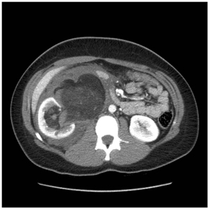
Figure 1: CT scan of abdomen revealed renal angiomyolipoma.
The mass was composed of fatty and soft tissue components along with blood products. A differential diagnosis of ruptured angiomyolipoma and malignant tumors such as liposarcoma were considered. Considering patient's young age, acuity of symptom onset and the fact that the mass arised from the kidney, the diagnosis of renal angiomyolipoma was determined to be more likely. The patient had no stigmata of tuberous sclerosis on physical examination. Initially, we decided to proceed with conservative management but within a time span of 12 hours, patient's hemoglobin decreased to 9.5 g/dl. Interventional radiologist was consulted for the management of ongoing bleeding. Emergency arteriogram with super selective embolization of AML with alcohol and coil was performed through the lower pole of right renal artery. A CT guided biopsy of right renal lesion was also performed. Histological examination confirmed our diagnosis of renal angiomyolipoma. The patient was monitored in Intensive care unit for few more days and was subsequently discharged as she improved. She is planned to undergo elective resection of angiomyolipoma at a future date.
DISCUSSION
Renal angiomyolipomas are mesenchymal tumors, occurring in association with tuberous sclerosis or sporadically in the general population. They were initially classified as hamartomas but now are considered to be benign tumors. AMLs occurring sporadically are typically large and unilateral tumors, whereas when in association with tuberous sclerosis, AML lesions tend to be smaller involving multiple foci. Sporadic AMLs occur more often in the right kidney but can occur in extra renal sites such as liver and are common in middle aged women. They can also occur in association with renal malignancies [5, 6]. Our patient had the most common classic variant of renal AML. This variant is less aggressive compared to the epithelioid variant or malignant epithelial angiomyolipoma. AML patients are asymptomatic and the tumor is found incidentally on imaging studies. The complications include retroperitoneal hemorrhage, hematuria and impingement of normal structures like normal kidney, ureter that can lead to various symptoms. “Wunderlich syndrome” is the term used to describe retroperitoneal hemorrhage occurring in AML that can cause sudden hemodynamic compromise and lead to death [7]. Diagnosis of AML is usually made based on radiological and pathological evidence of three components in tumor: fat cells, smooth muscle tissue and blood vessel components. Ultrasonography and CT scanning are the commonly used imaging modalities. In ultrasonography, the tumor is characterized by high reflectivity because of high fat content. CT scans reveal all the three components in AML and the tumor itself is usually seen as a well encapsulated mass [8, 9]. Immunohistochemical studies are rarely done and are usually positive for HMB45, MART1, HHF35, calponin, NKI-C3, and CD117 [10]. The treatment options differ based on the size of the tumor. For small (< 4cm tumors), watchful waiting is preferred. For larger tumors (> 8cm), prophylactic excision is suggested because of the high risk of retroperitoneal hemorrhage. Even smaller tumors with high vascularity are preferred to be excised [11]. mTOR (Mechanistic target of Rapamycin) inhibitors like sirolimus and everolimus have been tried with reduction in tumor size. Recent evidences suggest that though there is considerable reduction in tumor size by volume reduction of all 3 components, there is a significant rebound growth after discontinuation of m-TOR therapy [12]. Interventional Radiology guided radiographic embolization may play a role in tackling retroperitoneal hemorrhages in AML [13]. For patients selected for intervention, nephron sparing surgery is preferred if possible rather than complete nephrectomy even for larger (7 to 10cm) and/or multiple AMLs. Selective renal artery embolization may be used when the size or location precludes nephron sparing surgery.
CONCLUSION
Renal angiomyolipomas are benign tumors with natural history of slow growth. They are incidentally found in most cases except the ones presenting with complications. Retroperitoneal hemorrhage associated with AMLs can be fatal and require emergent surgeries or selective angio embolization. Prophylactic surgery is preferred for highly vascular tumors or large tumors >8cm. m-TOR inhibitors do decrease tumor size but with the disadvantage of rebound growth, they are being tried in AML associated with tuberous sclerosis as they are usually bilateral and multicentric and in metastatic malignant epithelial AML. Our case highlights the importance of considering rare etiologies for seemingly simple presentations that can help reduce mortality and morbidity.
REFERENCES
- Fletcher CD. (2014). The evolving classification of soft tissue tumours - an update based on the new 2013 WHO classification. Histopathology. 64: 2-11.
- Vasko JS, Brockman SK and Bomar RL. (1965). "Renal Angiomyolipoma A Rare Cause of Spontaneous Massive Retroperitoneal Hemorrhage. Annals of Surgery. 161: 577-581.
- Prakash G, Sankhwar S, Jhanwar A and Singh K. (2016). "Bilateral renal angiomyolipoma presenting as tuberous sclerosis syndrome. BMJ case Case reportsRep.
- Gopalakrishnan N, Dhanapriya J, Sakthirajan R, Dineshkumar T, et al. (2016). Angiomyolipoma of donor kidney: Successful transplantation and 5-yearfollow-up. Indian J Nephrol. 26: 131-133.
- Venyo AK. (2016). A Review of the Literature on Extrarenal Retroperitoneal Angiomyolipoma. Int J Surg Oncol.
- Farrell C, Noyes SL, Tourojman M and Lane BR. (2015). Renal angiomyolipoma: preoperative identification of atypical fat-poor AML. Curr Urol Rep. 16(3): 12.
- Bhatt JR, Richard PO, Kim NS, Finelli A, et al. (2016). Natural History of Renal Angiomyolipoma (AML): Most Patients with Large AMLs > 4cm Can Be Offered Active Surveillance as an Initial Management Strategy. Eur Urol. 70(1): 85-90.
- Bosniak MA, Megibow AJ, Hulnick DH, Horii S, et al. (1988). CT diagnosis of renal angiomyolipoma: the importance of detecting small amounts of fat. AJR Am J Roentgenol. 151(3): 497-501.
- Hansen GC, Hoffman RB, Sample WF and Becker R. (1978). Computed tomography diagnosis of renal angiomyolipoma. Radiology. 128(3): 789-791.
- L'Hostis H, Deminiere C, Ferriere JM and Coindre JM. (1999). Renal angiomyolipoma: a clinicopathologic, immunohistochemical, and follow-up study of 46 cases. Am J Surg Pathol. 23(9): 1011-1020.
- Harabayashi T, Shinohara N, Katano H, Nonomura K, et al. (2004). Management of renal angiomyolipomas associated with tuberous sclerosis complex. J Urol. 171: 102-105.
- Bissler JJ, McCormack FX, Young LR, Elwing JM, et al. (2008). Sirolimus for angiomyolipoma in tuberous sclerosis complex or lymphangioleiomyomatosis. N Engl J Med. 358(2): 140-151.
- Sheth RA, Feldman AS, Paul E, Thiele EA, et al. (2016). Angiographic and volumetric effects of mammalian target of rapamycin inhibitors on angiomyolipomas in tuberous sclerosis. World J Radiol. 8(3): 308-315.