Information Links
Related Conferences
Previous Issues Volume 8, Issue 2 - 2023
Development of Artificial Skin Scaffold Using Electrospun Nano Fibres for Wound Healing
N. Gokarneshan*, D. Mariya Jenita
Department of Fashion Design and Arts, Hindustan Institute of Technology and Science, Chennai, India
*Corresponding Author: Dr. N. Gokarneshan, Department of Fashion Design and Arts, Hindustan Institute of Technology and Science, Chennai, India; Email: [email protected]
Received Date: January 16, 2023
Publication Date: February 2, 2023
Citation: Gokarneshan N, et al. (2023). Children Under 5 Years Old Mortality and Associated Factors. Mathews J Case Rep. 8(2):86.
Copyright: Gokarneshan N, et al. © (2023)
ABSTRACT
Native skin consists mainly of epidermal and dermal layers. An artificial skin scaffold has been constructed mimicking the bilayered structure of the native skin using electrospinning technique for wound healing. Polyurethane (PU) and Gelatin (Ge) were used for developing the epidermal layer and the dermal layer respectively. Ciprofloxacin HCl (Cip. HCl) a fluoroquinolone antibiotic was incorporated in both layers for rapid wound healing. Morphology of the skin scaffold was studied using scanning electron microscopy (SEM) analysis and the chemical characterization was performed using FTIR spectroscopy. Water vapor transmission rate test and oxygen transmission rate test was conducted to evaluate the barrier properties of the scaffold. Thermal stability of the skin scaffold was evaluated using DSC and TGA while an understanding of the exudate absorbing capacity and degradation behavior of the scaffold was obtained from water absorption studies and in vitro degradation studies respectively. In vitro drug release study and drug release kinetics was explored to understand the release mechanism of Cip. HCl from the scaffold. Both the layers showed nano and micropores when analyzed using SEM. The dermal layer showed comparatively more water absorption capacity and degradation, hence providing a moist environment for the wound. The skin scaffold was permeable to water vapor and oxygen, and hence will speed up the process of wound healing. In vitro release for Cip. HCl showed a non-Fickian swelling type release with zero-order kinetics. Disk diffusion test conducted on the bilayers proved the antibacterial activity of the membrane. Hence the electrospun PU-Ge skin scaffold containing Cip. HCl is a promising candidate among modern day wound healing materials.
Keywords: Artificial Skin Scaffold; Polyurethane; Ciprofloxacin; Gelatin; Electrospinning
INTRODUCTION
Clinical management of deep and extensive wounds is challenging [1-3]. Any wound having a depth of more than 1 cm cannot heal by itself and requires grafts containing dermis and epidermis harvested from other parts of the body [4]. Skin tissue engineering is therefore a good option for deep wound healing as there is a limitation in getting large autografts [5-7]. For deep wounds such as second- degree burn wounds, almost all the parts of the dermis and whole of epidermis is lost. Hence the artificial skin scaffold developed for such cases should have a bilayer of dermis and epidermis mimicking the natural skin properties [8]. Bilayered collagen based skin substitutes such as Integra and Biobrane have greatly improved the long-term function and appearance of the wound, with lower wound contraction and less pigmentation [9,10]. But these collagen-based skin substitutes are usually associated with low mechanical strength, slow angiogenesis and lacks antibacterial properties as compared to the autograft. There are different types of artificial skin scaffolds made of chitosan/polylactic acid, chitosan/polycaprolactone, PEG grafted HA and collagen/chitosan hyaluronic acid [11-14]. Different Techniques such as electrospinning layer by layer assembly, solvent casting and lyophilization are some of the widely used techniques for developing wound healing membranes. Electrospinning technique has been opted to produce an artificial skin scaffold consisting of electrospun PU and Ge. Electrospinning is one of the easiest methods used for producing membranes having nano and micron-sized pores [15]. Flow rate, voltage, time, temperature, distance from the collector, density, viscosity, conductivity, surface tension, etc. are the different variables that can be optimized to get fibers of desired properties [16]. This technique has been widely used for almost all types of soluble synthetic and biopolymers. Ge has been chosen as a dermal substitute because it is the partially denatured derivative of the fibrous insoluble protein collagen, and does not express any antigenicity, is cost-effective as compared to collagen, shows good hemostatic effects, is completely bio absorbable and can be cross-linked using appropriate chemistry to improve the mechanical strength [17]. PU is an excellent medical elastomer material with good mechanical strength, oxygen permeability, barrier properties and biocompatibility and hence can be used as an epidermal substitute [18,19]. Most of these bilayered membranes do not show any antibacterial property but it is possible to add antibiotics and other growth factors so that rapid wound healing and angiogenesis can be achieved. In this case, Ciprofloxacin HCl (Cip. HCl) has been added to both the Ge and PU layers so that antibacterial property is imparted to the scaffold. The prepared bilayered membrane has been characterized and important parameters such as water vapor transmission rate, oxygen transmission rate, mechanical parameters, thermal properties, in vitro degradation, water absorption, in vitro release, drug release kinetics and antibacterial studies have been conducted so that it can be used as an effective antibacterial artificial skin scaffold.
TECHNICAL DETAILS
The following chemicals have been considered in the investigation.
Type B Gelatin conforming to USP standard
Hydrochloric acid and analytical reagents used in the preparation of pH7.4 Buffer including sodium chloride and sodium dihydrogen phosphate have been used
2,2,2- trifluoroethanol and N,N- Dimethylformamide.
The following steps have been involved in the investigation
Preparation of skin scaffold by electrospinning
Characterization of the membranes has been done using Scanning electron microscopy analysis, Fourier transform infrared spectroscopy, thermogravimetric analysis, Differential scanning calorimetry, water vapour transmission rate, and Oxygen transmission rate.
- Water absorption of the film
- In vitro degradation of the film
- Mechanical properties of films
- In vitro drug release study
Mathematical modelling for controlled release of drugs Cip. Hydrochloric acid from the electrospun fibre.
Antibacterial activity by cup diffusion method.
THE FINDINGS
The skin scaffold consists of a bilayered structure such as to mimic the native skin structure and consists of Cip. HCl incorporated electrospun gelatin layer and Cip. HCl incorporated polyurethane layer. The pH of the spinning solutions was found to be 6.15 for PU with Cip. HCl and 5.87 for gelatin solution with Cip. HCl, which is close to the skin pH ]20]. The conductivity and viscosity values for the polymer solutions were optimized for electrospinning. The continuous and smooth fibers cannot be obtained in very low viscous solutions as it may result in interruption of polymeric filaments and droplet formation whereas very high viscosity solutions make the polymers difficult to extrude [21]. According to Angammana et al., conductivity of a solution can greatly influence the average fiber diameter as well as the average fiber morphology of electrospun fibers [22]. The average diameter of electrospun fibers varies with an increase in the conductivity of the solution. The diameter of the fibers decreases with an increase in the conductivity of the solution. Here, the conductivity and the viscosity of the polymer solutions were fixed based on literature values [23]. The conductivity of Ge with Cip. HCl and PU with Cip. HCl were found to be 119.5 and 12.76 mS, respectively while the viscosity of the solutions were found to be 142.6 cps at 1000 rotations per minute (rpm) and 179.6 cps at 500 rpm respectively. Bilayer artificial skin scaffold has Cip. HCl incorporated polyurethane as the epidermal layer and Cip. HCl incorporated Ge as the dermal layer. The PU layer was white in color and the Ge layer was pale yellow. The pale yellow color of Ge layer is due to glutaraldehyde crosslinking. The appropriate thickness and weight of individual layers were found to be 0.141 mm with 0.6408 g and 0.300 mm with 0.5499 g, respectively for epidermal and dermal layers. The fabricated skin scaffold was light in weight as the approximate thickness is 0.406 mm with an average weight is 1.1895 g.
SCANNING ELECTRON MICROSCOPY
When artificial skin scaffolds have to be developed, it is useful to design them with a dense top layer which can protect the wound from getting infected and a porous lower layer having hydrophilic nature; so that it can absorb the wound exudate resulting in better cell adhesion and proliferation [24]. The SEM images of the dermal and epidermal layers are shown in Figure 1(A and B), respectively. The SEM images showed interconnected structures forming a non-woven mat-like structure. The SEM images of the Ge dermal layer under 1 kx and 3 kx showed nanofibers in the size range of 190–1500 nm. This porous structure of the bilayer membrane helps in absorbing the wound exudate and also enables cell adhesion and proliferation. The SEM images of the PU layer under 1 kx and 3 kx shows smooth, bead-free continuous nanofibers with a diameter in the range of 200–4300 nm. The mean diameter of the Ge dermal layer nanofibers and PU epidermal layer nanofibers were found to be 552 nm with most of the nanofibers in the range of 400–600 nm and 2365 nm with most of the nanofibers in the range of 2000–2500 nm respectively. This dense top layer of PU provides suitable mechanical properties and waterproof capacity to the bi-layered membrane; avoids bacterial infection and controls the gas exchange between the wound and the surrounding environment.
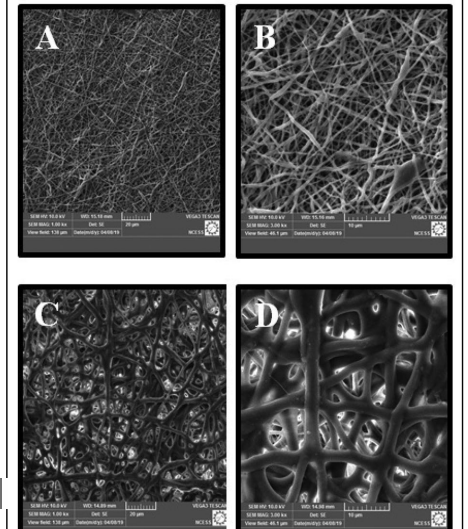
Figure 1: SEM images of the dermal and epidermal layers.
FTIR SPECTROSCOPY
FTIR spectrum of Cip. HCl and both layers of the bilayer PUGe skin scaffold containing Cip. HCl have been determined. Choi et al. conducted FTIR spectroscopy experiments for determining the structural configuration of PU, PU/Cip. HCl, and PU/Cip. HCl/Hydroxyapatite. They found the characteristic peaks of PU nanofibers as 2950 cm-1 (CH2 asymmetric vibration); 1750 cm-1 (free C–O); 1650 cm-1 (C=O bond); 1530 cm-1 (urethane amide II); 1081 cm-1 (C(O)–O–C stretching of the hard segment) and 821 cm-1 (bending vibration in benzene ring). The absorption band at 1375 cm-1 was due to the protonation of the amine group of the piperazine moiety. In the FTIR spectra of Cip. HCl blended PU nanofibers the absorption band at 1375 cm-1 was shifted to 1393 cm-1. A broad band at 3500 cm-1 is seen corresponding to OH stretching vibration. The peaks at 2950, 1650, 1530, 1081 and 821 cm-1 in PU film were shifted to 2871.27, 1699.44, 1528.94, 1072.1 and 816.9 cm-1 in drug incorporated PU film. In the case of electro spun Ge film, the peaks at 1650, 1540 and 3289 cm-1 were shifted to 1628.56 1535.08and 3277.11 cm-1, respectively. Comparing the FTIR spectra of pure Cip. HCl with that of PU film with Cip. HCl and Ge film with Cip. HCl, the O-H stretching of pure Cip. HCl at 3288.21 cm-1, -COOH stretching at 1710 cm-1, C-H stretching at 1446.65 cm-1, C= F stretching at 1266.42 cm-1 and aromatic C= C at 1383.15 cm-1 were shifted to 3319.05, 1699.44, 1471.61, 1221.43 and 1413.47 cm-1, respectively for PU film with Cip. HCl and 3277.11, 1628.56, 1450.15, 1239.58 and 1400.09 cm-1, respectively for Ge film with Cip. HCl.
DIFFERENTIAL SCANNING CALORIMETRY
The differential scanning calorimetry (DSC) technique gives an idea regarding the melting and phase transition behavior of materials and is widely used for analyzing the thermal stability of polymeric materials. DSC thermograms of electrospun PU, Ge and PU-Ge skin scaffolds have been determined. The melting peak of Cip. HCl loaded PU film was found to be at 186.21 C. The DSC thermogram was shifted to 108.5°C for the Ge layer with Cip. HCl and for PU-Ge skin scaffold with Cip. HCl, the denaturation temperature was found to be at 115.9°C. Zhang et al., performed similar studies and the results were found to be comparable [25]. TGA It was observed that the PU-Ge skin scaffold was decomposed in 4 steps. It had a slight weight loss of about 1–2% at 30 C due to the removal of residual moisture. A distinct weight loss was observed in the range of 240°C to 431°C. About 64.39% weight loss was observed due to the degradation of polymers - PU and Ge. From the DSC thermogram, it was clear that these polymers degrade after 186.21 C and 108.5 C respectively. Only 0.322% of the initial weight remained to degrade at 728 C. From the TGA curve, it was concluded that the obtained bilayer membrane was thermally stable (Kim et al., 2009). WVTR Normal skin has a WVTR value of 204 g/m2 /day while for injured skin, the value can be as high as 5138 g/m2 /day [26]. A wound healing material must have adequate WVTR so that a moist environment is maintained. A moist wound healing membrane heals the wound faster than a dry membrane. A low exudate wound can be covered with a low WVTR membrane and a high exuding wound can be healed using a high WVTR membrane. It is found that for the chitosan-alginate polyelectrolyte complex; WVTR ranges from 565 to 764 g/m2 /day (Wang et al., 2002). Huang et al. have reported a value of 2027.4 to 2069.4 g/m2 /day WVTR for konjac glucomannan films [27]. Similarly, in a recent work of wound dressings based on bacterial cellulose, WVTR was reported to be in the range of 1992 to 2688 g/m2 /day [28]. In this study, PU-Ge skin scaffold displayed a WVTR of 1172 g/m2 per day.
Oxygen transmission rate: An ideal wound dressing provides gaseous exchange especially oxygen to the wound site for rapid wound healing. Larry et al., investigated oxygen transmission rate in three kinds of film dressing’s viz. polyvinylidene chloride, a low gas permeability film; PU, a medium gas permeability film and poly (dimethyl silicone), a high gas permeability film (Sirvio & Grussing, 1989). They found that the oxygen transmission rate of polyvinylidene chloride as 0.028 (L/M’/ 24 h), PU [29] as 2.74 (L/M’/24 h) and silicone dressing as > greater than 20 (L/M’/24 h). PU-Ge skin scaffold showed an oxygen transmission rate of 43.54 (cm3 /m2 /24 h) at 0.1 MPa pressure. The high value of oxygen transmission rate will promote rapid epithelialization and enhance the process of wound healing [30].
Water absorption study: Water absorption study of PU-Ge skin scaffold and individual layers was done to further evaluate the capacity of the membranes to absorb wound exudate. Due to the hydrophilicity of the Ge, it showed 185.2% water absorption. The water absorption of the PU layer and the bilayer was found to be 165.39% and 153.86% respectively. It has been found that both PU and Ge and the skin scaffold show reasonably good water-absorbing properties.
In vitro degradation study: The degradation profile of the PU-Ge skin scaffold and individual layers was studied in PBS 7.4 at 37 C for 15 days. It has been found that the Ge layer had a significant decrease in weight as compared to PU layer. The skin scaffold consisting of PU and Ge layers showed 85% weight loss during a test period of 15 days while the PU layer and Ge layer independently showed a weight loss of 11.56% and 41.74% respectively. This is because Ge is a naturally degradable biopolymer derived from collagen, which is the main component of the extracellular matrix. During the degradation of Ge layer, the PU layer will remain intact and provide mechanical support and avoid the invasion of microorganisms.
Mechanical properties: The mechanical properties of PU, Ge and PU-Ge skin scaffold were evaluated using a universal testing machine. The maximum force at break for dry and wet PU sheet was found to be 24.9 N and 9.71 N, while it was 12.82 N and 9.90 N for dry and wet Ge layer and 11.52 N and 12.86 N for the skin scaffold. The PU nano fibrous layer exhibited high flexibility and good elastic properties with a tensile strength of 4.71 N/ mm2 and a high elongation at break of 380.27% in the wet condition as compared to 8.56 N/mm2 and 357.98 % in the dry state. The Ge nanofibers also had different results for dry and wet conditions. Tensile strength decreased from 7.37 N/mm2 to 5.15 N/mm2 and elongation at break also decreased from 24.11 % to 14.79% for wet Ge fiber. In the case of skin scaffold, tensile strength had no significant difference between dry and wet films but elongation at break was high in wet conditions. Comparing the tensile properties of skin scaffold under dry and wet conditions, the hydrated scaffolds were always significantly more flexible than the dehydrated scaffolds since Ge has better water uptake than PU. These results indicate that the mechanical properties of PU-Ge nano fibrous skin scaffolds are sufficient to allow them to serve as a support for cell adhesion and proliferation. The tensile strength of human skin is in the range of 2–16 N/mm2 and has an elongation at break in the range of 70%–77% (Silver & Shah, 2016). In vitro drug release study The in vitro drug release profile of Cip. HCl loaded Ge film, PU film and PU-Ge skin scaffold was investigated in PBS 7.4 at 37 C for 8 h. The graph of cumulative drug release of drug-loaded Ge sheet, PU sheet and PU-Ge skin scaffold have been plotted. % Cumulative release of all the membranes showed the same release of Cip HCl from one to three hours. After three hours a slight increase in drug release is seen in the case PU-Ge skin scaffold. Both PU and Ge layer released the same amount of Cip. HCl whereas PU-Ge skin scaffold showed a high % cumulative release of about 37.91%. At the end of eight hours, a cumulative release of 50 to 60% was seen. It has been found that the release of the drug is sustained for a long period of seven hours. To understand the release mechanism, the in vitro release data was analyzed using various mathematical models and the fitted parameters determined. Zero-order and first-order models are the most common kinetics models, whereas other sophisticated models such as Higuchi, Korsmeyer-Peppas add further information into the active mass transfer phenomenon. It has been found be seen that the release kinetics can be satisfactorily described by zero-order kinetics for all samples. The Korsmeyer-Peppas equation is often used to explain the mechanism of drug release from the matrix. In this case “n” explains the release mechanism of the drug. In the case of thin films, 0.5 n corresponds to a Fickian diffusion mechanism, 0.5< n < 1.0 to anomalous non-Fickian transport, and n > 1 to super case II transport. The values of n for Korsmeyer-Peppas model were observed and found to be in the range n > 1 and thus follows super case II transport (Bajpai et al., 2016). Hence it can be suggested that the drug release by diffusion rate and relaxation rate of polymer chains are comparable and the drug release follows non fickian swelling type release and follows zero-order kinetics. Antibacterial activity by cup-diffusion method the bactericidal effect of Ge, PU and PU-Ge skin scaffold in the MHA culture media was studied by the cup-diffusion method containing S. aureus and E. coli bacterial strains separately. The gelatin membrane showed a zone of inhibition of 19.75 mm and 19.13 mm in S. aureus and E. coli strains respectively, while the PU fibrous mat and PU-Ge bilayer fibrous mat showed a zone of inhibition of 18.99 mm and 19.92 mm respectively in S. aureus strains and 18.85 and 19.17 mm in E. coli strains. The interconnected PU-Ge skin scaffold provides perfect blocks and pores in the fibrous membrane such that it helps in avoiding exogenous infections effectively by preventing any bacterial penetration. As per the results, the PU-Ge skin scaffold shows good antibacterial activity and therefore can be used as a perfect wound healing material. The strong bactericidal activity for both gram-positive S. aureus and Gram-negative E. coli can be attributed to the ready availability of plenty of drug particles on the periphery and pores of the fibrous sheet [31].
CONCLUSION
Electrospinning technique was used to produce a bilayered PU-Ge skin scaffold wound dressing membrane composed of PU, Ge and Cip. HCl. The bi-layered structure of this wound dressing was designed, so as to mimic the skin native structure, with the PU layer representing the epidermal layer and the Ge layer representing the dermal layer. The nano porous interconnected structure of the bilayered scaffolds, as confirmed by SEM, is a desirable factor for burn wound healing since it helps in wound exudate absorption, better cell adhesion and proliferation. The thermal stability of the PU-Ge skin scaffolds is evident from the DSC and TGA studies while water absorption studies indicate the fluid absorption capacity of the bi-layered scaffold. The bi-layered PU-Ge skin scaffolds were found to be having optimal water vapor and oxygen transmission rates. An optimal WVTR enhances wound healing by improving the proliferation and function of epidermal cells and fibroblasts, while oxygen-permeable dressings help in faster wound healing. The PU-Ge scaffolds showed good mechanical strength in both wet and dry conditions as well as controlled biodegradation rates, which may be synchronized with the rate of epithelialization. The drug release kinetics follows zero-order controlled drug release, which can be considered ideal for wound dressing materials. The antibacterial property of the bilayered skin scaffold as well as individual layers were evaluated and was found to be adequate, such that any chances of infections can be ruled out. Hence the bilayered Pu-Ge skin scaffolds can act as effective antimicrobial wound dressing materials and can contribute towards enhanced wound healing.
REFERENCES
- Bhowmick S, Thanusha AV, Kumar A, Scharnweber D, Rother S, Koul V. (2018). Nanofibrous artificial skin substitute composed of mPEG–PCL grafted gelatin/hyaluronan/chondroitin sulfate/sericin for 2 nd degree burn care: In vitro and in vivo study. RSC Advances. 8(30):16420–16432.
- Lazarus GS, Cooper DM, Knighton DR, Margolis DJ, Pecoraro RE, Rodeheaver G, et al. (1994). Definitions and guidelines for assessment of wounds and evaluation of healing. Arch Dermatol. 130(4):489–493.
- Schulz JT, Tompkins RG, Burk JF. (2000). Artificial skin. An Rev Med. 51:231–244.
- Balasubramani M, Kumar TR, Babu M. (2001). Skin substitutes: A review. Burns. 27(5):534–544.
- Li H, Chen C, Zhang S, Jiang J, Tao H, Xu J, et al. (2012). The use of layer by layer self-assembled coatings of hyaluronic acid and cationized gelatin to improve the biocompatibility of poly (ethylene terephthalate) artificial ligaments for reconstruction of the anterior cruciate ligament. Acta Biomater. 8(11):4007–4019.
- Pedram Rad Z, Mokhtari J, Abbasi M. (2018). Fabrication and characterization of PCL/zein/gum arabic electrospun nanocomposite scaffold for skin tissue engineering. Mater Sci Eng C Mater Biol Appl. 93:356–366.
- Wang F, Wang M, She Z, Fan K, Xu C, Chu B, et al. (2015). Collagen/chitosan based two-compartment and bi-functional dermal scaffolds for skin regeneration. Mater Sci Eng C Mater Biol Appl. 52:155–162.
- Trinca RB, Westin CB, da Silva JAF, Moraes AM. (2017). Electrospun multilayer chitosan scaffolds as potential wound dressings for skin lesions. Eur Polymer J. 88:161–170.
- Ma L, Gao C, Mao Z, Zhou J, Shen J, Hu X, Han C. (2003). Collagen/chitosan porous scaffolds with improved biostability for skin tissue engineering. Biomater. 24(26):4833–4841.
- Soller EC, Tzeranis DS, Miu K, So PT, Yannas IV. (2012). Common features of optimal collagen scaffolds that disrupt wound contraction and enhance regeneration both in peripheral nerves and in skin. Biomaterials. 33(19):4783–4791.
- Chen SH, Chang Y, Lee KR, Lai JY. (2014). A three-dimensional dual-layer nano/microfibrous structure of electrospun chitosan/ poly(d,l-lactide) membrane for the improvement of cytocompatibility. J Membrane Sci. 450:224–234.
- Erencia M, Cano F, Tornero JA, Fernandes MM, Tzanov T, Macanas J, et al. (2015). Electrospinning of gelatin fibers using solutions with low acetic acid concentration: Effect of solvent composition on both diameter of electrospun fibers and cytotoxicity. J Appl Poly Sci. 132(25):42115.
- Greiner A, Wendorff JH. (2007). Electrospinning: A fascinating method for the preparation of ultrathin fibers. Angewandte Chemie. 46(30):5670–5703.
- Mao J, Zhao L, de Yao K, Shang Q, Yang G, Cao Y. (2003). Study of novel chitosan-gelatin artificial skin in vitro. J Biomed Mater Res. Part A. 64(2):301–308.
- Kim SE, Heo DN, Lee JB, Kim JR, Park SH, Jeon SH, et al. (2009). Electrospun gelatin/polyurethane blended nanofibers for wound healing. Biomed Mater. 4(4):044106.
- Tu Y, Zhou M, Guo Z, Li Y, Hou Y, Wang D, et al. (2015). Preparation and characterization of thermosensitive artificial skin with a Sandwich structure. Materials Letters. 147:4–7.
- Ali S, Yosipovitch G. (2013). Skin pH: From basic science to basic skin care. Acta Derm Venereol. 93(3):261–267.
- Amariei N, Manea LR, Bertea AP, Bertea A, Popa A. (2017). The influence of polymer solution on the properties of electrospun 3D nanostructures. IOP Conference Series: Mater Sci Engineer. 209:012092.
- Angammana CJ, Jayaram SH. (2011). Analysis of the effects of solution conductivity on electrospinning process and fiber morphology. IEEE Trans Indust Appl. 47(3):1109–1117.
- Fu SZ, Meng XH, Fan J, Yang LL, Wen QL, Ye SJ, et al. (2014). Acceleration of dermal wound healing by using electrospun curcumin-loaded poly(e-caprolactone)-poly(ethylene glycol)- poly(e-caprolactone) fibrous mats. J Biomed Mater Res B Appl Biomater. 102(3):533–542.
- Trinca RB, Westin CB, da Silva JAF, Moraes AM. (2017). Electrospun multilayer chitosan scaffolds as potential wound dressings for skin lesions. Eu Polymer J. 88:161–170.
- Zhang YZ, Venugopal J, Huang ZM, Lim CT, Ramakrishna S. (2006). Crosslinking of the electrospun gelatin nanofibers. Polymer. 47(8):2911–2917.
- Kim SE, Heo DN, Lee JB, Kim JR, Park SH, Jeon SH, et al. (2009). Electrospun gelatin/polyurethane blended nanofibers for wound healing. Biomed Mater. 4(4):044106.
- Wang L, Khor E, Wee A, Lim LY. (2002). Chitosan-alginate PEC membrane as a wound dressing: Assessment of incisional wound healing. J Biomed Mater Res. 63(5):610–618.
- Kwak MH, Kim JE, Go J, Koh EK, Song SH, Son HJ, et al. (2015). Bacterial cellulose membrane produced by Acetobacter sp. A10 for burn wound dressing applications. Carbohydr Polym. 122:387–398.
- Sirvio LM, Grussing DM. (1989). The effect of gas permeability of film dressings on wound environment and healing. J Investigat Dermatol. 93(4):528–531.
- Zhang Y, Lim CT, Ramakrishna S, Huang ZM. (2005). Recent development of polymer nanofibers for biomedical and biotechnological applications. J Mater Sci Mater Med. 16(10):933–946.
- Silver FH, Shah R. (2016). Measurement of mechanical properties of natural and engineered implants. Adv Tissue Engineer Regenerat MedOpen Access. 1:20-25.
- Bajpai M, Bajpai SK, Jyotishi P. (2016). Water absorption and moisture permeation properties of chitosan/poly(acrylamide-co-itaconic acid) IPC films. Int J Biol Macromol. 84:1–9.
- Dong C, Ye Y, Qian L, Zhao G, He B, Xiao H. (2014). Antibacterial modification of cellulose fibers by grafting b-cyclodextrin and inclusion with ciprofloxacin. Cellulose. 21(3):1921–1932.
- Letha SS, Kumar AS, Nisha U, Rosemary MJ. (2022). Electrospun polyurethane-gelatin artificial skin scaffold for wound healing. J Textile Institute. 113:378-387.