Information Links
Related Conferences
Previous Issues Volume 8, Issue 2 - 2023
Upper Jaw Cancer: A Case Report of Surgical Intervention and Defect Repair in an Upper Alveolar Squamous Cell Carcinoma
Pankaj Goyal*, Kishan Kumawat
Apollo E.N.T. Hospital, pal road, Jodhpur, Rajasthan, India
*Corresponding Author: Pankaj Goyal, Apollo E.N.T. Hospital, pal road, Jodhpur, Rajasthan, India; Email: [email protected]
Received Date: March 2, 2023
Publication Date: March 17, 2023
Citation: Goyal P, et al. (2023). Upper Jaw Cancer: A Case Report of Surgical Intervention and Defect Repair in an Upper Alveolar Squamous Cell Carcinoma. Mathews J Cancer Sci. 8(2):38.
Copyright: Goyal P, et al. © (2023)
ABSTRACT
Oral cancer is the sixth most prevalent cancer worldwide, and it is the third most prevalent in Southeast Asia. Oral squamous cell carcinoma (OSCC) of the upper alveolus is rare. There is a high frequency of gingival cancer, and it is frequently discovered in an advanced stage. It may be important to better patient education and early medical consultation because gingival cancer's clinical presentation is extremely variable. There are few English-language data have been published on their biological behaviour. Adequate surgical resection and adjuvant treatment, in the first instance, offer the best chance of disease control. Upper jaw cancer is more aggressive usually. The following is a case report on SCC of the upper alveolus in a seventy five-year-old female who developed a non-healing ulcer after tooth extraction.
Keywords: Squamous cell carcinoma, upper jaw, surgery, defect, reconstruction, buccal fat pad
INTRODUCTION
Oral squamous cell carcinoma (OSCC) confined to the upper alveolus is extremely rare, accounting for only 3.5 to 10% of all OSCC [1,2]. According to reports, the third-most frequent subsite for OSCC, the gingiva, is a prevalent site for oral cancer [3]. There could be a number of effects depending on where the OSCC is located in the gingiva. Cancer of the upper alveolar mucosa may spread to the nearby spaces due to the anatomical proximity of the upper alveolar mucosa with the maxillary gingivobuccal sulcus, making it challenging to pinpoint the disease's true origin [4]. It exhibits a wide range of clinical symptoms, such as bleeding, discomfort, swelling, ulceration, and movable teeth. These also show up in frequently occurring, non-cancerous diseases of the mouth such periodontal disease and tooth abscesses. As a result, it can be difficult to identify a lesion that is suspected of being an oral SCC because it shares traits with several other common inflammatory and infectious disorders. Therefore, a physician may fail to recognise the possibility of an oral SCC involving dentition, especially in its early stages. Suspicion for additional diseases, such as an oral SCC, should be taken into consideration if recovery following dental treatment does not proceed as anticipated. In our patient, she had a history of tooth extraction, and gradually, a non-healing ulcer developed at the same location, revealing moderately differentiated squamous cell carcinoma (MDSCC) on the biopsy report. Delayed diagnosis of an oral SCC may result in more advanced disease at the time of therapy, necessitating more time-consuming and expensive care, more morbidity, and a lower chance of survival than could be the case with an earlier diagnosis. In terms of oral cancer, a number of prognostic indicators have been put forth that may have an impact on patients' overall survival and survival specific to their disease. The tumour nodes metastases (TNM) stage, histological grade, and tumour thickness are among the established prognostic variables that are widely acknowledged. Extracapsular dissemination, lympho-vascular invasion (LVI), peri-neural invasion (PNI), involvement of the surgical margin by tumour cells, and other prognostic markers are also known to contribute to recurrence and an overall poor prognosis [5-8]. Typically, cancer of the upper jaw is more aggressive. Their delayed manifestation and early invasion of the infratemporal fossa may have contributed to their aggressive behaviour [9]. The biology of mouth cancer differs depending on where it manifests. Lower gingival-buccal malignancies, which have a greater disease-free survival rate even in advanced stages, are less aggressive than their upper gingival-buccal counterparts [10,11]. Cervical lymph node metastases developed late in the course of the illness, but it had a bad prognosis. This case report describes SCC of the upper alveolus in a 75-year-old female who developed a non-healing ulcer after tooth extraction.
CASE REPORT
A 75-year-old woman presented to our Apollo E.N.T. hospital with a non-healing ulcer on her left upper jaw that had been present for three to five months. It grew gradually until it reached its current size. Eight months prior, she attended the dentist due to a prior toothache, and a tooth extraction was performed at that time. Following that, the wound did not heal, and an ulcer developed there. She took medicine for it, but it did not get better. She denied any substance abuse. Her blood sugar level was under control despite her known diabetes. Her overall health was good when examined. Upon intraoral inspection, it was discovered that there was a sufficient mouth opening and a 3x2 cm ulcerative growth at the left side of the upper alveolus, which corresponds to the left upper lateral incisor and second molar teeth. No cervical lymphadenopathy existed. For histological diagnosis and radiological assessment, the patient had been advised to undergo punch biopsy and computed tomography (CT) scan. According to the CT scan, the soft tissue was 3.2x1.8 cm and was located in the upper alveolus on the left. The alveolar ridge had barely eroded. Therefore, it was a cT2N0M0 stage. The procedure for obtaining consent for surgery had been explained. The patient was scheduled for surgery after completing a general anaesthesia fitness test. The upper alveolus, the principal location in this case, was marked with a sufficient margin. Left infrastructural maxillectomy was done. Haemostasis has been attained. The buccal fad pad was used to reconstruct the defect. A level I–IV selective neck dissection was carried out. Sending all surgical samples for histopathology analysis Patient was sent to recovery after surgery, where she was extubated. She had intravenous antibiotics for five days following surgery, and Ryle’s tube feeding was initiated. On the sixth day following surgery, the patient had been discharged. Histopathologically, it was a squamous cell carcinoma with free margins and a moderate degree of differentiation; all of the cervical lymph nodes were tumour-free, and it was staged as pT2N0M0. The underlying bone was not involved. Patient visited our hospital on a regular basis for a year, taking three-month breaks, and is doing well with no recurrence.
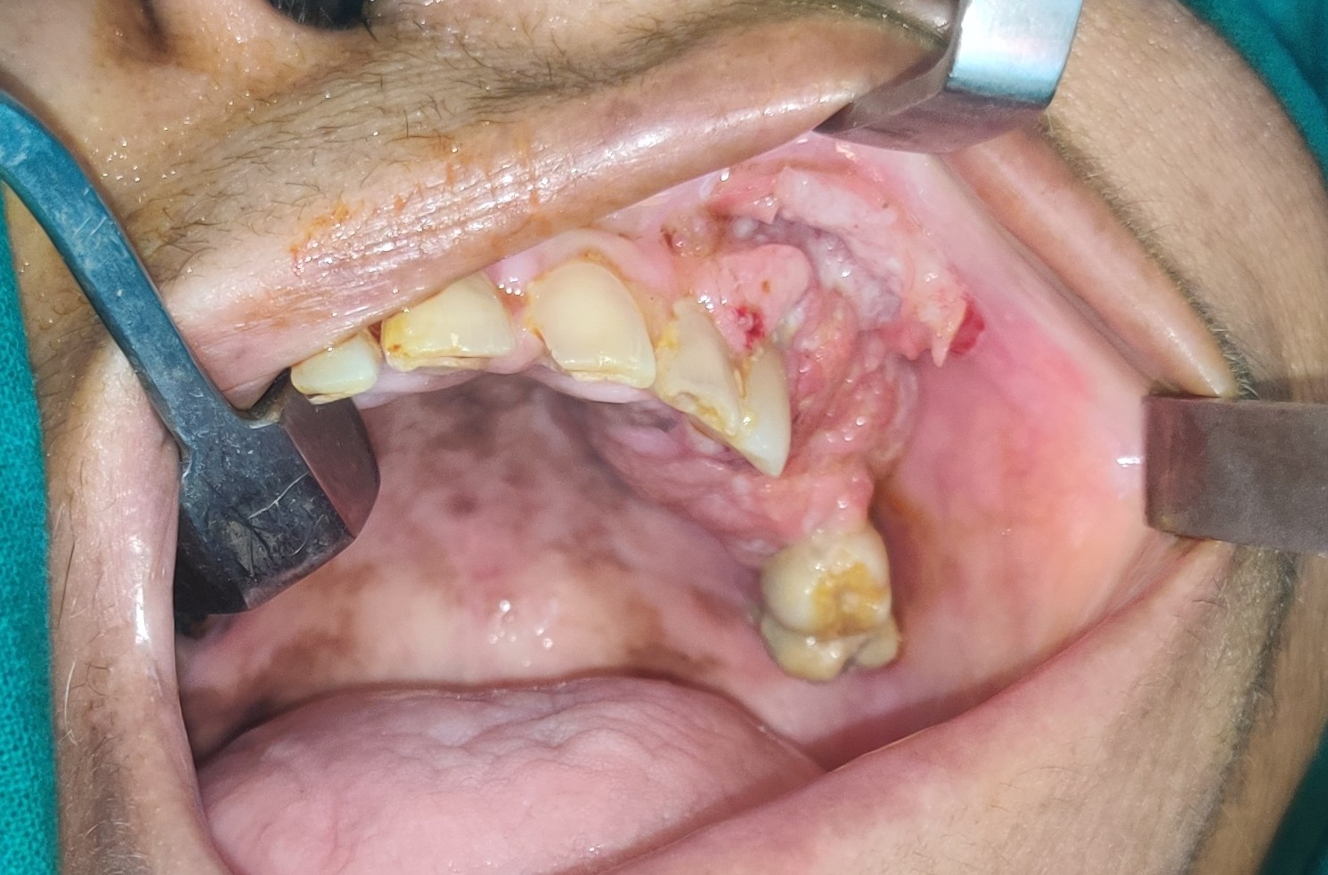
Figure 1: Clinical picture showing ulcero-proliferative growth at left sided upper alveolus with involving gingivobuccal complex. (cT2N0M0)
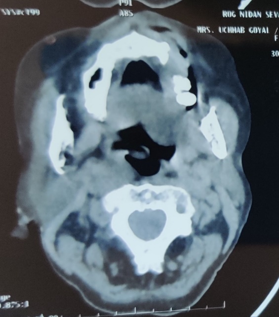
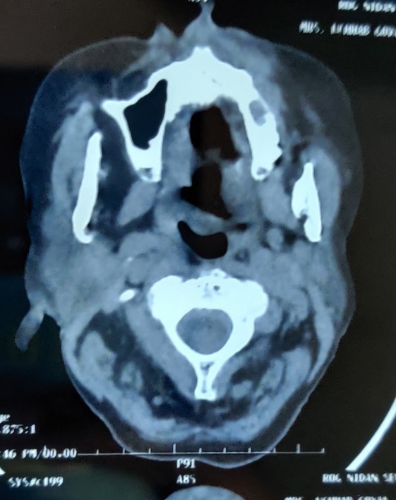
Figure 2: Computed tomography scan (axial view) showing soft tissue adjacent to left upper alveolar ridge with minimal erosion of alveolar surface of maxilla.
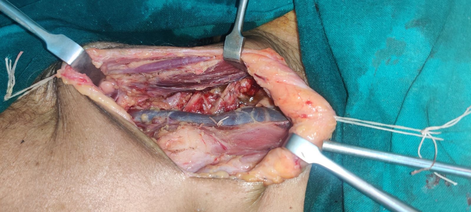
Figure 3: Intra-opeartive picture after selective neck dissection(I-IV).
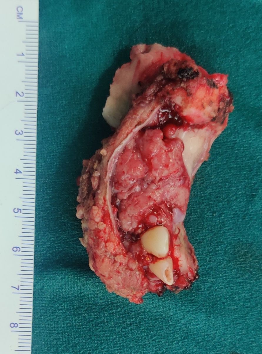
Figure 4: Main specimen: Infrastructure maxillectomy with adequate margins in all dimensions.
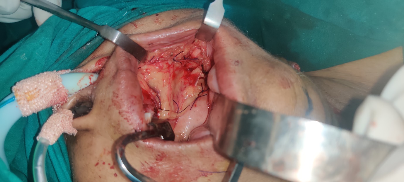
Figure 5: Repair of defect with buccal fat pad grafting after removal of primary site.
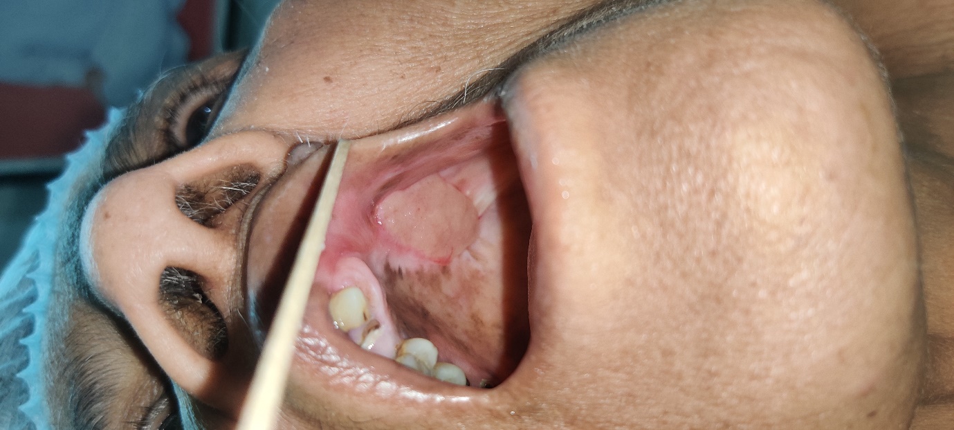
Figure 6: Picture of intra oral cavity showing healing well after three months.
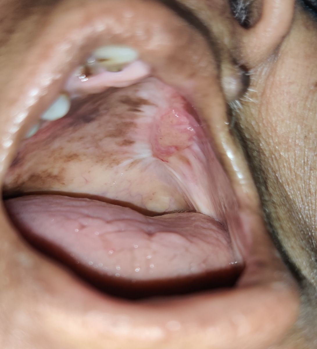
Figure 7: Picture of intra oral cavity showing well healed operated site after six months.
DISCUSSION
The alveolar processes of the maxilla and the mucosal layer that lies above them are both part of the upper alveolar ridge. The names "upper alveolus" and "upper gingiva" have been used mostly interchangeably in the field of oral oncology, and they are collectively classified as C03.0 in the International Classification of Diseases (ICD-10) system. Although the upper alveolus and overlying gingiva, upper sulcus, and adjacent parts of the upper buccal mucosa are all in close proximity to one another, the upper gingival-buccal complex is not a distinct clinical entity in ICD-10 and results in patterns of tumour spread that are similar to these patterns. Alveolar growth and trismus are frequent signs and symptoms of upper gingival-buccal malignancies, making it difficult to properly assess the area. Early cancer identification is frequently challenging because of this. Morris et al. had noted that females were more likely to develop upper alveolus cancers [12]. The patient in our situation was also female. Although there are significant age and regional variations, OSCC in gingival sites accounts for roughly 10% [13] to a quarter [14] of all oral malignancies [15]. Tumours spread from the upper alveolus and upper sulcus to the infratemporal fossa in the dentate maxilla, whereas in the edentulous jaw, with low alveolar height and a relatively thin layer of mucosa covering the alveolar process, invasion of the maxillary sinus and nose is easier. Because of the erosion of the floor of the maxillary antrum, these tumours are frequently misdiagnosed as maxillary carcinoma. A tumour originating in the floor of the maxillary sinus, on the other hand, will spread to the palate via the path of least resistance along the palatine canals rather than through the alveolar process to the superior sulcus [16]. According to Araki’s et al. research, a computed tomography scan may accurately predict the invasion of the buccal and maxillary sinuses by 75% and 86%, respectively [17]. The growth of carcinomas next to teeth results in gingival swelling, which is accompanied by discomfort and tooth sensitivity. Erroneously removing the teeth due to presumed pericoronitis or periodontitis is possible. Surgical removal could spread tumour cells and speed the metastatic process [18]. Surgical extraction of tooth without knowledge of carcinoma may result in a delay in establishing a correct diagnosis and appropriate treatment. Because the extension of the tumour-bearing period increases the rate of metastasis [19], this delay could be one of the factors that facilitated tumour dissemination. The lymphatics from the superior alveolar ridge's buccal aspect flow to the submental and submandibular nodes (level I). The infratemporal fossa, palate, maxilla, and lingual portions of the upper alveolar ridge are all affected by the disease, which also extends through the lymphatic system to the superior deep jugular and the retropharyngeal and parapharyngeal group of lymph nodes. A peroral approach can be used to remove small, anteriorly positioned lesions, whereas a transfacial approach, using lateral rhinotomies, Weber-Fergusson incisions, or Diffenbach incisions, can access large, posteriorly located tumours. Wide mucosal and soft tissue margins are the goal of an effective surgical excision, but anatomical restrictions frequently prevent them. When warranted, adjuvant radiotherapy or concomitant chemo-radiation is crucial, according to recent research. [20]. The optimal course of treatment for these patients has been suggested as adequate surgery with adjuvant radiation [16,21,22].
CONCLUSION
On average, gingival cancer is discovered at an advanced stage. Knowing and comprehending its clinical characteristics may be a key component in enhancing its early diagnosis. On potential suspected lesions, the patient should be informed, and oral health care professionals should pay attention. The highest likelihood of disease control comes from treating SCC of the upper alveolus with appropriate surgical resection and adjuvant therapy, if necessary.
COMPLIANCE WITH ETHICAL STANDARDS
The procedure performed in this case report was in accordance with the ethical standards of the institutional and/or national research committee and with the 1964 Helsinki declaration and its later amendments or comparable ethical standards.
FUNDING
This study is not funded by any resources.
CONFLICT OF INTEREST
The author (s) declares no potential conflicts of interest with respect to the research, authorship, and/or publication of this paper.
ETHICAL APPROVAL
The study was published with the written consent of the patient.
REFERENCES
Rao DN, Shroff PD, Chattopadhyay G, Dinshaw KA (1998). Survival analysis of 5595 head and neck cancers – Results of conventional treatment in a high-risk population. Br J Cancer. 77:1514-1518.
Baishya N, Rahman T, Das AK, Kalita CR, Sharma JD, Krishnatreya M, et al. (2019). Squamous cell carcinoma of upper alveolus: An experience of a tertiary care center of Northeast India. South Asian J Cancer. 8:44-46.
Barasch A, Gofa A, Krutchkoff DJ, Eisenberg E. (1995). Squamous cell carcinoma of the gingiva. A case series analysis. Oral Surg Oral Med.Oral Pathol Oral Radiol Endod. 80:183–187.
Pathak KA, Mathur N, Talole S, Deshpande MS, Chaturvedi P, Pai PS, et al. (2007). Squamous cell carcinoma of the superior gingival-buccal complex. Oral Oncol 43:774-779.
Binmadi NO, Basile JR. (2011). Perineural invasion in oral squamous cell carcinoma: a discussion of significance and review of the literature. Oral Oncol. 47:1005-1010.
Sutton DN, Brown JS, Rogers SN, Vaughan ED, Woolgar JA. (2003). The prognostic implications of the surgical margin in oral squamous cell carcinoma. Int J Oral Maxillofac Surg. 32:30-34.
Jones HB, Sykes A, Bayman N, Sloan P, Swindell R, Patel M, et al. (2009). The impact of lymphovascular invasion on survival in oral carcinoma. Oral Oncol. 45:10-15.
Shaw RJ, Lowe D, Woolgar JA, Brown JS, Vaughan ED, Evans C, et al. (2010). Extracapsular spread in oral squamous cell carcinoma. Head Neck 32:714-722.
Pathak KA, Mathur N, Talole S, Deshpande MS, Chaturvedi P, Pai PS, et al. (2007). Squamous cell carcinoma of the superior gingival‑buccal complex. Oral Oncol. 43:774‑779.
Pathak KA, Gupta S, Talole S, Khanna V, Chaturvedi P, Deshpande MS, et al. (2005). Advanced squamous cell carcinoma of lower gingivobuccal complex: patterns of spread and failure. Head Neck. 27:597–602.
Lai GQ, Ou SM, Zeng ZY, et al. (1987). Surgical treatment of carcinoma of the gingival. Zhonghua Zhong Liu Za Zhi. 9:56–57.
Morris LG, Patel SG, Shah JP, Ganly I. (2011). High rates of regional failure in squamous cell carcinoma of the hard palate and maxillary alveolus. Head Neck. 33:824‑830.
Eicher SA, Overholt SM, el-Naggar AK, Byers RM, Weber RS.. (1996). Lower gingival carcinoma. Clinical and pathologic determinants of regional metastases. Arch Otolaryngol Head Neck Surg. 122:634–638.
Makridis SD, Mellado JR, Freedman AL, Salkin LM, Stein MD, Leal K, et al. (1998). Squamous cell carcinoma of gingiva and edentulous alveolar ridge: A clinicopathologic study. Int J Periodontics Restor Dent. 18:292–298.
Koduganti RR, Sehrawat S, Reddy PV. (2012). Gingival squamous cell carcinoma: A diagnostic impediment. J Indian Soc Periodontol. 16:104–107.
Tiwari R. (2000). Squamous cell carcinoma of the superior gingivolabial sulcus. Oral Oncol. 36:461–465.
Araki K, Ariji E, Shimizu M, Kanda S, Ozeki S, Shinohara M, et al. (1997). Computed tomography of carcinoma of the upper gingiva and hard palate: correlation with the surgical and histopathological findings. Dentomaxillofac Radiol. 26:177–182.
Peterson LJ. (1993). Principles of uncomplicated exodontia. In: Pettmson LJ, et al. (eds): Contemporary oral and maxillbfacial surgery. (2nd). St. Louis: Mosby:137.
CamG GT. (1980). Metastases from DMBA-induced carcinoma in hamster cheek pouch. In: Hilluan K, Hiloard P, Eccels S, (eds) Metastasis: clinical and experimental aspects, developments in oncology. The Hague: Martins Nijhoff:50-54.
Love R, Stewart IF, Coy P. (1977). Upper alveolar carcinoma: a 30-year survey. J Otolaryngol. 6:393–398.
Bernier J, Cooper JS, Pajak TF, van Glabbeke M, Bourhis J, Forastiere A, et al. (2005). Defining risk levels in locally advanced head and neck cancers: a comparative analysis of concurrent postoperative radiation plus chemotherapy trials of the EORTC (#22931) and RTOG (# 9501). Head Neck. 27: 843–850.
Kryst L, Piekarczyk J, Mlosek K, Wanyura H, Szmurlo W. (1990). Results of treatment of upper gingiva and palate carcinomas. Czas Stomatol. 43:62–67.