Previous Issues Volume 2, Issue 1 - 2017
The Relationship Between Colonic Diverticulosis and Soybean-Like Small Round Stool
Yoshiyuki Hoya1 *,Takanori Kurogochi2,Rota Noaki2,Satoshi Yamazaki2,Tomoyoshi Okamoto1,Norio Mitsumori3,Katsuhiko Yanaga3
Corresponding Author: Yoshiyuki Hoya, Department of Surgery, Daisan Hospital, The Jikei University School of Medicine 4-11-1, Izumihon-cho, Komae-si, Tokyo, 201-8601, Japan, Tel: 81-3-3480-1151 (3251); Email: [email protected] Received Date: 16 May 2017 Accepted Date: 12 Jun 2017 Published Date: 16 Jun 2017 Copyright © 2017 Hoya Y Citation: Hoya Y, Kurogochi T, Noaki R, Yamazaki S, et al. (2016). The Relationship Between Colonic Diverticulosis and Soybean-Like Small Round Stool. Mathews J Gastroenterol Hepatol 2(1): 009. ABSTRACT
Introduction: Although phenomena and manifestations may often be encountered in routine practice, there are often instances where the cause or identity of those phenomena and manifestations is unclear. Soybean-like small (about 1 cm) and round stool (SLSRS) is often encountered during colonoscopy (CS). The current study examined the causal relationship between such stool and diverticula, and the results are discussed herein. Patients and methods: Subjects were 78 patients who underwent CS at Kumagaya Surgery Hospital from October 2014 to June 2015. The number of diverticula in the colon, the location of diverticula, and the number of bowel movements prior to CS, and the presence or absence of SLSRS were examined in these patients. Results: Diverticula were present in 30 of the 78 patients (38.5%),of whom SLSRS was noted in 21 patients (70%). Of the other 48 patients without diverticula, only one patient had SLSRS (2.8%). Thus, SLSRS had a sensitivity of 70%, a specificity of 97.9%, and an accuracy of 87.2% in indicating the presence of colonic diverticula. Conclusion: The high association between SLSRS and the presence of colonic diverticula, suggests that SLSRS is produced by the colonic diverticula, which could be named as “Diverticular Stool”. KEYWORDS
Diverticulosis; Endoscopic Diagnosis; Shape of Stool; Impaction of Stool. INTRODUCTION
Although phenomena and manifestations may often be encountered in routine practice, there are often instances where the cause or identity of those phenomena and manifestations is unclear. Soybean-like small (about 1 cm) round stool (SLSRS) is often encountered during colonoscopy (CS) (Figure 1). The current study examined the causal relationship between such stools and diverticula, and the results are discussed herein. PATIENTS AND METHODS
Subjects were 78 patients who underwent CS by the authors at Kumagaya Surgery Hospital from October 2014 to June 2015. The number of diverticula in the colon, the location of diverticula, the number of bowel movements prior to CS, and the presence or absence of SLSRS were examined in these patients. The causal relationship between diverticula and SLSRS was also examined. Preparations for CS at Kumagaya Surgery Hospital were as follows: (1) The patient was allowed to eat low fiber and fat dinner prior to 8 PM the night before CS. (2) After-dinner snacking was prohibited and the patient was instructed to drink a large amount of water. (3) Patients under the age of 70 took 3 Forsenid tablets as a cathartic the night before CS. At 6 AM on the day of CS, the patient took 2 tablets of Pramiel (prokinetic agent). Moreover, after arrival at the hospital, the patient took 2 liters of Niflec or Moviprep. (4) Patients age 70 years and over consumed a packet of Magcorol and a bottle of Shinluck prior to sleep the night before CS. At 6 AM on the day of the CS, the patient took two Pramiel tablets. Moreover, after arrival at the hospital, the patient took 1 liter of Niflec or Moviprep. No sedatives or analgesics were used during CS. Although “stool characteristic of diverticulosis” is a vague description, the term referred to stools that were soybean-like small round stools (about 1 cm) (Figure 1a).
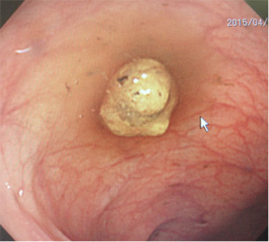
Figure 1a: Soybean-like small and round stool related to diverticula.
In addition, an endoscopist and a nurse assisting in the procedure objectively determined whether stools characteristic of diverticulosis were present or not. In this research, the information which had been obtained in a general clinical practice such as CS was accumulated and analyzed. For statistical analysis, the Mann-Whitney's U test and Fisher's exact test were used. Data were presented as a mean ± standard deviation, and differences with p-values of less than 0.05 were considered significant.
RESULTS
Diverticula were present in 30 of the 78 patients (38.5%). The male:female ratio for patients with diverticula was 24:6 as compared to 28:20 for patients without diverticula. The average age of the patients with diverticula was 66.7 ± 13.2 years as compared to 60.4 ±17.0 years in those without diverticula. Patients with diverticula had an average of 9.8 ± 3.7 bowel movements prior to CS while patients with no diverticula had an average of 9.1 ± 3.0 bowel movements. Of the 30 patients with diverticula, 12 patients had 1-5 diverticula, 11 had 6-10 diverticula, and 7 had 11 or more diverticula (Table 1).
Table 1: The relationship between colonic and diverticulosis and soybean –like small and round stool
| Diverticulosis (+) (n=30) | Diverticulosis(-) (n=48) | P-Value | |
|---|---|---|---|
| Sex (maleFemale) | 24:6 | 28:20 | P=0.048 |
| Age of operation (years) | 66.7± 13.2 | 60.4 ± 17.0 | P = 0.055 |
| Number of bowel movementsprior to CS | 9.8 ± 3.7 | 9.1± 3.0 | P=0.178 |
| SLSRS* | 21 (70.0%) | 1(2.08%) | P=8.882e-11 |
| Number of diverticula (1 ~ 5/ 6 ~10/ 11 or more | 12(5)**/11(9)**/7(7)** | - | - |
| Location of diverticular (R/L/R&L)** | 8(5)**8(5)**/14(11)** | - | - |
*soybean - like small and round stool ( ) **shows the number of the patients with SLSRS ***R: only in right colon, L: only in Left colon, R & L: both
Locations of diverticula are shown in Table 1. SLSRS (Figure1a) were noted in 21 of the 30 patients (70.0%) with diverticula. Moreover, the discovery rate of SLSRS was high so that there were many diverticula, but had nothing to do with the diverticular position. Diverticula were not noted in 48 patients, and only one of these patients had SLSRS (2.08%). Thus, SLSRS had a sensitivity of 70%, a specificity of 97.9%, and an accuracy of 87.2% in indicating the presence of diverticula. Accordingly, the presence of diverticula strongly and positively correlated with the detection of SLSRS. This suggests that SLSRS may have been produced by diverticula.
DISCUSSION
SLSRS is often encountered during CS. Such stool is often noted when diverticula are present. In light of their shape, such stool is presumably produced when feces is caught and forged in diverticula. However, no studies have reported data indicating a causal relationship between diverticula and such stool. Small and round stool is referred to as “stool in separate hard lumps,” so there is no consistent designation for these stools in practice [1]. The cathartic and prokinetic agents taken prior to CS do not cause SLSRS to lose their shape. Such stool remains in the colon during CS, where it is often encountered. SLSRS was noted irrespective of the location and number of diverticula. However, the discovery rate of SLSRS was high so that there were many diverticula, but had nothing to do with the diverticular position (Table1). Thus, soft stool accumulates in the diverticula. As stool continues to enter the diverticula, it is compacted and forged into a relatively dense form (Figure 1b). This process way produce stool that are larger than the size of the diverticula [2-4]. The reason why stool characteristic of diverticula is round is presumably because the diverticula and SLSRS have a relationship like that of potholes and whirling stones in nature [5]. Stool that remains in diverticula instead of being expelled calcifies and becomes fecaliths in diverticula. These fecaliths appear as calcifications in diverticula on a CT scan [6, 7].
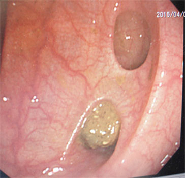
Figure 1b: Soybean-like small and round stool related to diverticula. Thus, soft stool accumulates in the diverticula. As stool continues to enter the diverticula, it is compacted and forged into a relatively dense form.
The large dose of the cathartic and prokinetic agents used during CS may expel SLSRS from diverticula. In addition, the cathartic and prokinetic agents result in frequent bowel movements. SLSRS remains in the colon even after bowel movements, and they are subsequently discovered during CS. This is because the form of SLSRS is presumably related to the semilunar folds of the colon. A rounded shape readily rotates. As stool travels through the colon, it rotates in the opposite direction when it reaches the location of the semilunar folds. SLSRS have a high specific gravity and readily accumulates in the location of the semilunar folds [8]. SLSRS did not indicate the presence of diverticula with a specificity of 100%. This is presumably because diverticula were overlooked during CS or because normal stools was deemed to be SLSRS due to their similar morphological features. SLSRS indicated the presence of diverticula with a specificity of 97.9%. If SLSRS are noted, small diverticula may be present but hidden in the semilunar folds of the colon even if endoscopic findings do not reveal diverticula. If the colon is believed to be responsible for bloody stool, anemia, or abdominal pain, CS is performed. If CS fails to reveal diverticula but it nonetheless reveals SLSRS, then diverticula need to be suspected as the cause of such symptoms. In summary, SLSRS is encountered during CS. Such stool is likely to have been produced in the diverticula, and therefore is a sign suggestive of the presence of diverticula. Therefore, we could name SLSRS as “Diverticular Stool”.
REFERENCES
- Muhammad A, Lamendola O, Daas A, Kumar A, et al. (2014). Association between colonic diverticulosis and prevalence of colorectal polyps. Int J Colorectal Dis. 29(8):947-951.
- Steenvoorde P, Vogelaar FJ, Oskam J and Tollenaar RA. (2004). Giant colonic diverticula review of diagnostic and therapeutic options. Dig Sug. 21(1): 1-6.
- Choong CK and Frizelle FA. (1998). Giant colonic diverticulum: report of four cases and review ofthe literature. DisColon Rectum. 41(9): 1178-1185.
- Nagata N, Niikura R and Aoki T. (2015). Association between colonic diverticulosis and bowel symptoms: a case-control study of 1629 Asian patients. J Gastroenterol Hepatol. 30(8): 1252-1259.
- Gilbert GK. (1906). “Moulin work under glaciers.” Geological Society of America Bulletin. 17(1): 317-320.
- Halligan S and Saunders B. (2002). Imaging diverticular disease. Best Pract Res Clin Gastroenterol 16(4): 595-610.
- Nigri G, Petrucciani N and Giannini G. (2015). Giant colonic diverticulum: clinical presentation, diagnosis and treatment: systematic review of 166 cases. World J Gastroenterol. 21(1): 360-368.
- Shangfu K. (1993). Formation mechanisms and prediction models of debris flow due to natural dam failures. J Sediment Res. 4: 42-57.