Information Links
Related Conferences
Current Issue Volume 11, Issue 1 - 2026
Bardet-Biedl Syndrome: A Case Report from Dalal Jamm Hospital in Dakar
Sow AS*, Niasse N, Diallo R, BA AO, Camara A, Mendy GKJI, Ndiaye JMM, Ndoye Roth PA
Bardet-Biedl Syndrome: A Case Report from Dalal Jamm Hospital in Dakar, Université Cheikh Anta Diop, Senegal
*Corresponding Author: Dr. Aboubacry Sadikh Sow, Bardet-Biedl Syndrome: A Case Report from Dalal Jamm Hospital in Dakar, Université Cheikh Anta Diop, Senegal, Phone: 00221775659760, Email: [email protected]
Received Date: May 30, 2026
Published Date: June 29, 2026
Citation: Sow AS, et al. (2026). Bardet-Biedl Syndrome: A Case Report from Dalal Jamm Hospital in Dakar. Mathews J Ophthalmol. 11(1):39.
Copyrights: Sow AS, et al. © (2026).
ABSTRACT
Introduction: Retinitis pigmentosa is an inherited retinal degeneration characterized by dysfunction of the retinal pigment epithelium and loss of photoreceptors, associated with pigment deposits visible on fundus examination. It may occur in isolation or in association with extraocular manifestations. Objective: After the informed and signed consent of the patient's parents, we report a Bardet Biedl Syndrom (BBS) case, recalling the epidemiological, clinical aspects, particularly ophthalmological and therapeutic aspects. Observation: The patient was a 15-year-old girl presented with bilateral, progressive myopia and night blindness. Her medical history included psychomotor developmental delay, polyuria, enuresis, and surgery for hexadactyly. The ophthalmological examination revealed: a corrected visual acuity of 3/10 in the right eye and 2/10 in the left eye, strabismus, rotatory nystagmus, and epicanthus. Funduscopy showed diffuse retinopathy. On general examination, the patient weighed 80 kg, had a BMI of 36 kg/m², exhibited slow movements and head jerks, and slowed thought processes. The visual field, the visual evoked potential, and the electroretinogram were significantly impaired. Abdominal and pelvic ultrasound was not performed and karyotyping was not available. The patient met 5 major criteria confirming the diagnosis of BBS. Conclusion: BBS is an inherited ciliopathy characterized by multiorgan involvement and intellectual disability. Its management is multidisciplinary, and the visual prognosis is very poor. It is important to emphasize the importance of ophthalmological examinations in newborns with a family history of ciliopathy and to conduct regular monitoring, genetic counseling for families of patients with BBS and a pediatric assessment is necessary for all cases of retinitis pigmentosa.
Keywords: Bardet-Biedl Syndrome, Hexadactyly, Pigmentary Retinopathy.
INTRODUCTION
Bardet-Biedl syndrome is a condition first described by Bardet in 1920 following his observation of an obese girl, and later by Biedl, who reported a family with adipose and genital dystrophy [1,2]. Bardet-Biedl syndrome (BBS) is a rare autosomal recessive ciliopathy characterized by retinal dystrophy, obesity, postaxial polydactyly, renal dysfunction, learning disabilities, and hypogonadism. The diagnosis is based on clinical findings and can be confirmed by sequencing the genes known to cause the disease in 80% of patients. The BBS genes encode proteins that are localized in the cilia and the basal body and are involved in cilia biogenesis and function. Mutations lead to defective cilia, which partly explains the pleiotropic effects observed in BBS [3].
Objective
After the informed and signed consent of the patient's parents, we report this BBS case, recalling the epidemiological, clinical aspects, particularly ophthalmological and therapeutic aspects.
CASE REPORT
The patient was a 15-year-old girl who presented for an outpatient consultation due to progressive bilateral visual acuity loss (VAL) associated with night blindness. Her medical history included hospitalization at birth due to refusal to breastfeed, psychomotor delay with academic difficulties, polyuria, and enuresis, as well as surgery for right and left hexadactyly. She is the second of five siblings, including one stillborn and three other living, healthy children, with no similar cases in the family. A second-degree parental consanguinity was identified. The ophthalmological examination revealed visual acuity of 3/10 in the right eye following optical correction for myopic astigmatism of -9.00 (+0.25; 20°), epicanthus, esotropia, and persistent rotatory nystagmus. The anterior segment and ocular pressure were normal. The Fundus showed a grayish retinal field with diffuse osteoblasts and narrowing of the retinal vessels (Figure 1). The macula was dull with loss of the foveolar reflex. In the left eye, visual acuity was 2/10 after correction for myopic astigmatism of -10.00 (+0.25; 90°); the rest of the examination was the same as for the right eye.
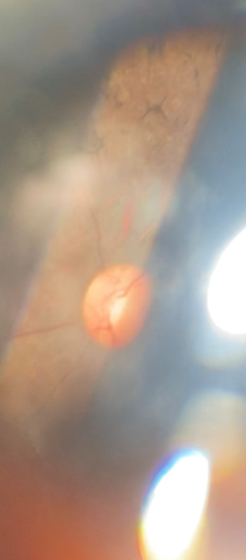
Figure 1. Osteoblasts, vascular narrowing in the fundus.
During the pediatric examination, vital signs and measurements revealed:
Normal blood pressure of 120/88 mm Hg; a heart rate of 80 beats per minute; a respiratory rate of 24 breaths per minute; a weight of 80 kg; a height of 150 cm, with a body mass index (BMI) of 36 kg/m² (Figure 2); and a capillary blood glucose level of 1.06 g/L on two separate occasions.
The general physical examination revealed good overall health and clear consciousness.
General examination revealed, scars from polydactyly surgery on the upper and lower extremities (Figure 3), mental retardation, slow thinking, and behavioral disturbances; severe obesity (Grade II); a normal ENT examination; and no abnormalities in the nervous system or other systems.
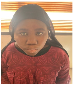
Figure 2. Head and trunk obesity.
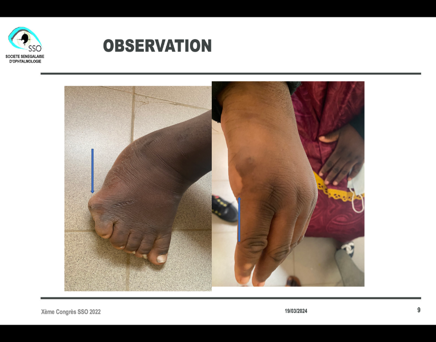
Figure 3. Scars from surgery for polydactyly of the limbs.
The paraclinical findings revealed bilateral visual pathway conduction abnormalities on the visual evoked potential (Figure 4), bilateral central retinal damage on the electroretinogram (Figure 5), and an agonistic visual evoked potential in both eyes. The brain CT scan and EEG were normal. Abdominal and pelvic ultrasound was not performed and karyotyping was not available.
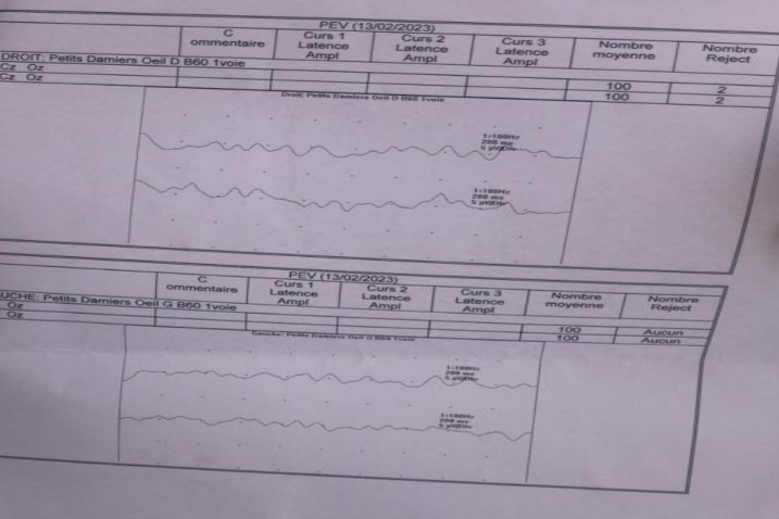
Figure 4. Impaired conduction in the visual pathways.
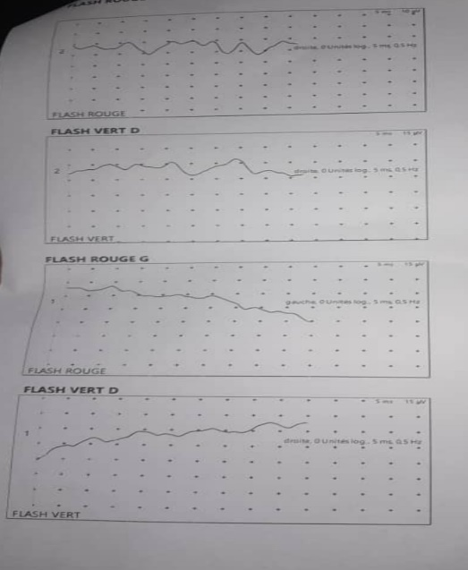
Figure 5. Central retinal damage.
Laboratory results showed a serum creatinine level of 10 mg/L with a glomerular filtration rate of 83 mL/min/1.72 m²; normal serum uric acid at 0.16 g/L; serum sodium at 135 mmol/L; serum potassium at 4.06 mmol/L; serum chloride at 104.4 mmol/L; normal serum calcium at 92 mg/L; and serum phosphorus at 39.6 mg/L. LH and FSH levels had been requested but not performed due to a shortage of specific reagents. The rest of the laboratory tests, including the complete blood count and lipid panel, were unremarkable. Medical imaging revealed a normal ECG and EEG while awake. An abdominal-pelvic ultrasound was not performed because the ultrasound was out of order at the same time. The karyotype was not available.
The recommended treatment included dietary advice, corrective eyewear, wearing protective and filtering sunglasses, wearing a hat, vitamin A and E therapy, and multidisciplinary clinical and laboratory monitoring by nephrologists, pediatricians, and ophthalmologists.
Table 1. Diagnostic criteria for BBS in our patient
|
Major Criteria |
Minor Criteria |
|
Obesity (BMI= 32 kg/m2) |
Psychomotor developmental delay |
|
Retinitis pigmentosa (RP) |
Refractive error |
|
Polydactyly |
Strabismus |
|
Learning disability |
Coordination disorder |
DISCUSSION
BBS is a relatively rare condition in Africa, and its exact prevalence remains unknown [4]. At Abass NDAO Hospital in Dakar, three cases were recorded over a 10-year period.
The prevalence has been estimated at 1 in 160.000 in European populations. However, it appears to be much more common in the Middle East, with a prevalence of 1 in 13.000 among Bedouins in Kuwait and 1 in 17.000 in Newfoundland. A preliminary epidemiological study of BBS in Tunisia reports 67 diagnosed cases between 1984 and 2009 [5].
A study conducted in a Congolese family identified children aged 16 and 9 [6]. The study by FERIANI [7] reported even older ages, exceeding 16 years. This could be explained by traditional beliefs observed in our regions, where people are reluctant to seek care at health facilities. Night blindness, photophobia, and decreased visual acuity are the most common symptoms and typically appear around age 10 [8]. By their thirties, all patients with BBS are virtually blind [8,9]. The study by Laurier et al. [10] shows that an abnormality in oculomotor function is present in 64% of cases following recording of optokinetic nystagmus. Our patient presented with myopic astigmatism, as confirmed by Grenn et al. [11] in their study on the cardinal manifestations of BBS. This high myopia explains the severe amblyopia noted in our patient, especially since she was seen at an advanced age. Fundus examination reveals the initial sign of macula pigmentation with constricted retinal vessels and optic disc pallor; mid-peripheral pigmentation appears later and consists of osteoblasts, or rounded clusters of pigment [12]. The ERG is the key test for diagnosis in the early stages [8]. The VEP and visual field tests are performed only if the ERG is abnormal. Renal involvement is the leading cause of mortality in this syndrome [10]; the prevalence of renal failure in BBS varies across studies from 42% to 100% of cases [9]. According to other authors, renal failure is diagnosed in 30% to 60% of cases [11,13]. Obesity affects 72% of patients according to the review by Tobin and Beales [14]. Glycemic imbalance may be linked to glucose intolerance due to insulin resistance [8]. In the study by FISLI et al. [15], only one in seven patients had diabetes. In contrast, in the study by MOORE et al. [16], type 2 diabetes occurred in 48% of patients, and impaired glucose tolerance was diagnosed in 4 out of 46 patients. The frequency of polydactyly in SBB is estimated differently by various authors and ranges from 58% to 100% of cases [13,17]. Genitourinary disorders are among the major criteria for SBB and are considered constant in this condition [8]. Hypogonadism is present in 98% of boys [16]. Girls present with menstrual cycle disorders or, more rarely, vaginal atresia with hydrometrocervix at birth, or fallopian tube hypoplasia [9]. Cognitive impairment is almost always present. The major sign of these manifestations is intellectual disability [16]. Our patient was observed to have intellectual disability, which was marked by learning difficulties at school and behavioral problems.
The treatment is multidisciplinary. It essentially includes wearing glasses, vitamin A and E supplementation, monitoring of renal function and dietary measures. Hexadactyly and certain genital or cardiac malformations can be surgically corrected. Psychological treatment is useful in order to optimize socio-professional integration. Genetic counseling is recommended for parents [9].
CONCLUSION
SBB is a rare and polymorphic genetic disorder that belongs to a large group of syndromes known as ciliopathies, which are characterized by abnormalities of the primary cilium. It is an autosomal recessive disorder that presents with facial dysmorphism, obesity, polydactyly, multiorgan involvement, and intellectual disability [6].
ACKNOWLEDGEMENTS
None.
CONFLICT OF INTEREST
The authors declare no conflict of interest.
REFERENCES
- Bardet G. (1995). On congenital obesity syndrome with polydactyly and retinitis pigmentosa (a contribution to the study of clinical forms of hypophyseal obesity). 1920. Obes Res. 3(4):387-399.
- Biedl A. (1995). A pair of siblings with adiposo-genital dystrophy. 1922. Obes Res. 3(4):404.
- Forsythe E, Beales PL. (2013). Bardet-Biedl syndrome. Eur J Hum Genet. 21(1):8-13.
- Wolf B. (1991). Bardet-Biedl syndrome in a Zimbabwean child. Cent Afr J Med. 37(10):341-342.
- M’hamdi O, Ouertani I, Maazoul F, Chaabouni-Bouhamed H. (2011). Prevalence of Bardet–Biedl syndrome in Tunisia. J Community Genet. 2(2):97‑99.
- Elenga-Bongo CL, Manvouri L, Monabeka HG. (2014). Le syndrome de Bardet-Biedl dans une famille congolaise : à propos de 2 cas, diagnostiqués au décours d’un coma diabétique. Ann Endocrinol. 75(5):391.
- Feriani H, Elloumi Z, Amri N, Hedri H, Kheder A. (2014). Syndrome de Bardet-Biedl et insuffisance rénale – À propos de 4 cas. Néphrologie Thérapeutique. 10(5):359.
- Ingster-Moati I, Rigaudiere F, Choltus-De Petigny MC, Bremond-Gignac D, Lestrade C, Grall Y. (2000). [Functional visual explorations of Bardet-Biedl syndrome. A study of three cases]. J Fr Ophtalmol. 23(8):802‑808.
- Rooryck C, Lacombe D. (2008). [Bardet-Biedl syndrome]. Ann Endocrinol. 69(6):463‑471.
- Laurier V, Stoetzel C, Muller J, Thibault C, Corbani S, Jalkh N, et al. (2006). Pitfalls of homozygosity mapping: an extended consanguineous Bardet-Biedl syndrome family with two mutant genes (BBS2, BBS10), three mutations, but no triallelism. Eur J Hum Genet EJHG. 14(11):1195‑1203.
- Green JS, Parfrey PS, Harnett JD, Farid NR, Cramer BC, Johnson G, et al. (1989). The cardinal manifestations of Bardet-Biedl syndrome, a form of Laurence-Moon-Biedl syndrome. N Engl J Med. 321(15):1002-1009.
- Riise R, Andréasson S, Wright AF, Tornqvist K. (1996). Ocular findings in the Laurence-Moon-Bardet-Biedl syndrome. Acta Ophthalmol Scand. 74(6):612-617.
- David A, Bitoun P, Lacombe D, Lambert JC, Nivelon A, Vigneron J, et al. (1999). Hydrometrocolpos and polydactyly: a common neonatal presentation of Bardet-Biedl and McKusick-Kaufman syndromes. J Med Genet. 36(8):599-603.
- Tobin JL, Beales PL. (2007). Bardet-Biedl syndrome: beyond the cilium. Pediatr Nephrol. 22(7):926-936.
- Fisli N, Khodja A, Nouri N, Khensal S, et al. (2017). Phénotype du syndrome de Bardet–Biedl : à propos de 7 cas. Ann Endocrinol. 78(4):302.
- Moore SJ, Green JS, Fan Y, Bhogal AK, Dicks E, Fernandez BA, et al. (2005). Clinical and genetic epidemiology of Bardet-Biedl syndrome in Newfoundland: a 22-year prospective, population-based, cohort study. Am J Med Genet A. 132A(4):352-360.
- Jeddi A, M'Rabet A, Ruhaumoly Y, Soussi H, Ben Brahim W, Gharbi M, et al. (1997). Le syndrome de Laurence Moon Bardet Biedl: à propos de 2 cas familiaux [Laurence Moon Bardet Biedl syndrome: review of 2 familial cases]. Tunis Med. 75(11):874-879.
