Previous Issues Volume 1, Issue 1 - 2016
Distinguish Between Benign and Malignant Prostate Using the Trace Element Content Ratios in Prostatic Tissue as Tumor Markers
Vladimir Zaichick1,Sofia Zaichick2
Department of Radionuclide Diagnostics, Medical Radiological Research Centre, Russia.
2Department of Medicine, University of Illinois College of Medicine, USA.
Corresponding Author:Vladimir Zaichick, Department of Radionuclide Diagnostics, Medical Radiological Research Centre, Koroleva Str- 4, Obninsk 249036, Kaluga Region, Russia, Tel: +7 (48439) 60289; E-Mail: [email protected]
Received Date: 28 Jul 2016 Accepted Date: 18 Aug 2016 Published Date: 24 Aug 2016
Copyright © 2016 Zaichick V
Citation:Zaichick V and Zaichick S. (2016). Distinguish Between Benign and Malignant Prostate Using the Trace Element Content Ratios in Prostatic Tissue as Tumor Markers. Mathews J Cancer Sci. 1(1): 006.
ABSTRACT
The aim of the study was the development of new highly precise testing methods for early diagnosis of prostate cancer. For this purpose the values of the zinc/silver (Zn/Ag), zinc/cobalt (Zn/Co), zinc/chromium (Zn/Cr), zinc/iron (Zn/Fe), zinc/ mercury (Zn/Hg), zinc/rubidium (Zn/Rb), zinc/antimony (Zn/Sb), zinc/scandium (Zn/Sc), and zinc/selenium (Zn/Se) mass fraction ratios in normal (n = 37), benign hypertrophic (n = 43) and cancerous tissues (n = 60) of the human prostate gland were calculated using data obtained by instrumental neutron activation analysis. Mean values ± standard error of mean (M ± SΕΜ) for trace element mass fraction ratios in normal tissue were: Zn/Ag – 32270 ± 5360, Zn/Co – 27010 ± 3720, Zn/Cr – 2650 ± 360, Zn/Fe – 10.9 ± 1.4, Zn/Hg – 27010 ± 3720, Zn/Rb – 73.6 ± 6.6, Zn/Sb – 34330 ± 6160, Zn/Sc – 46790 ± 7870, and Zn/Se – 1550 ± 170, respectively. It was observed that in benign hypertrophic tissues the Zn/Cr, Zn/ Hg, Zn/Sb, and Zn/Se mass fraction ratios were lower than in normal tissues. In cancerous tissue all investigated mass fraction ratios were significantly (p < 0.001) lower than in normal and benign hypertrophic tissues of the prostate. Finally, we propose to use the estimation of Zn/Ag, Zn/Cr, Zn/Fe, Zn/Hg, and Zn/Sb mass fraction ratios in a needle-biopsy core as an accurate tool to diagnose prostate cancer. Sensitivity, specificity, and accuracy of these tests were in ranges 96-100%, 89-100%, and 94-100%, respectively. Application of the Zn/trace element mass fraction ratios instead the Zn mass fraction is more suitable for PCa diagnosis, because accuracy of Zn/trace element mass fraction ratios does not depend on sample mass and changes in moisture content.
KEYWORDS
Trace Element; Trace Element Mass Fraction Ratios; Prostate; Benign Prostatic Hypertrophy; Prostatic Carcinoma; Neutron Activation Analysis.
INTRODUCTION
The prostate gland may be a source of many health problems in men past middle age, the most common being benign prostatic hyperplasia (BPH), and prostatic carcinoma (PCa). BPH is a noncancerous enlargement of the prostate gland leading to obstruction of the urethra and can significantly impair quality of life [1]. The prevalence of histological BPH is found in approximately 50-60% of males age 40-50, in over 70% at 60 years old and in greater than 90% of men over 70 [2, 3]. In many Western industrialized countries, including North America, PCa is the most frequently diagnosed form of noncutaneous malignancy in males and, except for lung cancer, is the leading cause of death from cancer [4-9]. Although the etiology of BPH and PCa is unknown, many trace elements have been highlighted in the literature in relation to the development of these prostate diseases [10-29]. Trace elements have essential physiological functions such as maintenance and regulation of cell function and signalling, gene regulation, activation or inhibition of enzymatic reactions, neurotransmission, and regulation of membrane function. Essential or toxic (mutagenic, carcinogenic) properties of trace elements depend on tissue-specific need or tolerance, respectively [30]. Excessive accumulation, deficiency or an imbalance of the trace elements may disturb the cell functions and may result in cellular degeneration, death and malignant transformation [31]. In previous studies significant changes of trace element contents in hyperplastic and cancerous prostate in comparison with those in the normal prostatic tissue were observed [32- 53]. Moreover, a significant informative value of Zn content as a tumor marker for PCa diagnostics was shown [54]. Hence it is possible that besides Zn, some other trace elements also can be used as tumor markers for distinguish between benign and malignant prostate. Current methods applied for measurement of trace element contents in samples of human tissue include a number of methods. Among these methods the instrumental neutron activation analysis with high resolution spectrometry of longlived radionuclides (INAA-LLR) is a non-destructive and one of the most sensitive techniques. It allows measure the trace element contents in a few milligrams tissue without any treatment of sample. Analytical studies of the Ag, Co, Cr, Fe, Hg, Rb, Sb, Sc, Se, and Zn contents in normal, BPH and PCa tissue were done by us using INAA-LLR [16, 21, 29, 48, 51, 55]. Nondestructive method of analysis avoids the possibility of changing the content of chemical elements in the studied samples, which allowed for the first time to obtain reliable results [56- 59]. In particular, it was shown that the average mass fraction of Zn in PCa tissues is 7 times lower than in healthy or BPH tissue [55]. Obtained results formed the basis for a new method for differential diagnosis of BPH and PCa, the essence of which was to determine the content of zinc in the material of trans-rectal needle biopsy of prostate indurated site. For the first time it was proposed to use INAA-LLR to determine zinc and other trace element contents in needle-biopsy cores [55]. Moreover, it was shown in the study that in a normal prostate tissue mass fraction of some trace elements tend to be correlated with Zn, while in BPH and PCa tissues these relationships are partially broken or changed [55]. These findings open the additional possibilities for developing new methods of PCa diagnostics using Zn content/trace element content ratio. Thus, this work had three aims. The first was to calculate individual Zn/Ag, Zn/Co, Zn/Cr, Zn/Fe, Zn/Hg, Zn/Rb, Zn/Sb, Zn/ Sc, and Zn/Se mass fraction ratios using data about the Ag, Co, Cr, Fe, Hg, Rb, Sb, Sc, Se, Zn contents in intact prostate of healthy men aged over 40 years obtained by INAA. The second aim was to compare the levels of mass fraction ratios in the prostate gland of age-matched patients, who had either BPH or PCa, and the third was to evaluate mass fraction ratios to aid diagnosis of prostate cancer. All studies were approved by the Ethical Committees of the Medical Radiological Research Centre, Obninsk.
MATERIAL AND METHODS
Samples
All patients studied (n = 103) were hospitalized in the Urological Department of the Medical Radiological Research Centre. Trans-rectal puncture biopsy of suspicious indurated regions of the prostate was performed for every patient, to permit morphological study of prostatic tissue at these sites and to estimate their chemical element contents. In all cases the diagnosis BPH (n = 43) and PCa (n = 60) has been confirmed by clinical and morphological results obtained during studies of biopsy and resected materials. Intact (normal) prostates were removed at necropsy from 37 men who had died suddenly. The majority of deaths were due to trauma. Tissue samples were collected from the peripheral zone of dorsal and lateral lobes of their prostates, within 2 days of death and then the samples were divided into two portions. One was used for morphological study while the other was intended for chemical element analysis. A histological examination was used to control the age norm conformity, as well as to confirm the absence of microadenomatosis and latent cancer [16, 21, 29].
Sample preparation, instrumentation, methods and certified reference materials
Details of sample preparation, the relevant nuclear reactions, radionuclides, gamma energies, methods of analysis and the results of quality control were presented in our earlier publications concerning the chemical elements of human prostate tissue investigated by INAA-LLR [16, 21, 29, 48, 51, 60].
Computer programs and statistic A dedicated computer program for INAA mode optimization was used [61]. All prostate samples for INAA-LLR were prepared in duplicate and mean values of trace element contents were used in final calculation. Using the Microsoft Office Excel software, the summary of statistics, arithmetic mean, standard deviation, standard error of mean, minimum and maximum values, median, percentiles with 0.025 and 0.975 levels was calculated for trace element mass fraction ratios in normal, benign hyperplastic and cancerous prostate tissue. The difference in the results between BPH and Norm, PCa and Norm, and PCA and BPH was evaluated by Student's t-test and nonparametric Wilcoxon-Mann-Whitney U-test. We applied a significance level of 0.05. For the construction of "individual data sets for Zn/Ag, Zn/Co, Zn/Cr, Zn/Fe, Zn/Hg, Zn/Rb, Zn/Sb, Zn/Sc, and Zn/Se mass fraction ratios in normal, benign hypertrophic and cancerous prostate tissue"diagrams the Microsoft Office Excel software was also used.
RESULTS
The age of 43 patients with BPH ranged from 38 to 83 years, the mean being 66 ± 8 years (M ± SD). The 60 patients aged 40-79 suffered from PCa. Their mean age was 65 ± 10 years. Mean age of men with intact prostates (Normal group) was 55 ± 11 years (range 41-79). Table 1 presents certain statistical parameters (arithmetic mean, standard deviation, standard error of mean, minimal and maximal values, median, percentiles with 0.025 and 0.975 levels) of the Zn/Ag, Zn/Co, Zn/Cr, Zn/Fe, Zn/Hg, Zn/Rb, Zn/ Sb, Zn/Sc, and Zn/Se mass fraction ratios in normal, benign hypertrophic and cancerous prostate tissue.
Table 1: Some statistical parameters of Zn/Ag, Zn/Co, Zn/Cr, Zn/Fe, Zn/Hg, Zn/Rb, Zn/Sb, Zn/Sc, and Zn/Se mass fraction ratios in normal, benign hypertrophic and cancerous prostate.
| Tissue Mass fraction ratio | Mean | SD | SEM | Min | Max | Median | Per. | Per. |
|---|---|---|---|---|---|---|---|---|
| 0.025 | 0.975 | |||||||
| Normal (n = 37) | ||||||||
| Zn/Ag | 32271 | 27330 | 5360 | 3036 | 113139 | 23563 | 4263 | 90351 |
| Zn/Co | 27011 | 20354 | 3716 | 4236 | 82272 | 21419 | 4798 | 69552 |
| Zn/Cr | 2654 | 1780 | 356 | 248 | 7073 | 2404 | 330 | 6369 |
| Zn/Fe | 10.9 | 8.0 | 1.4 | 1.68 | 30.5 | 8.66 | 2.06 | 29.9 |
| Zn/Hg | 27011 | 18957 | 3717 | 2533 | 69231 | 24331 | 4110 | 64394 |
| Zn/Rb | 73.6 | 37.1 | 6.6 | 18.4 | 181 | 72.1 | 72.1 | 149 |
| Zn/Sb | 34333 | 33149 | 6156 | 2935 | 122891 | 18500 | 4970 | 118907 |
| Zn/Sc | 46794 | 34288 | 7866 | 14274 | 121364 | 35309 | 15874 | 116182 |
| Zn/Se | 1548 | 908 | 166 | 421 | 3652 | 1335 | 442 | 3439 |
| BPH (n=43) | ||||||||
| Zn/Ag | 39748 | 19357 | 4328 | 13499 | 83073 | 39623 | 14448 | 76749 |
| Zn/Co | 20798 | 14641 | 3359 | 6285 | 58902 | 15637 | 7496 | 54869 |
| Zn/Cr | 1161 | 678 | 156 | 282 | 2720 | 902 | 359 | 2619 |
| Zn/Fe | 10.4 | 6.82 | 1.15 | 1.88 | 30.2 | 9.30 | 2.22 | 28.8 |
| Zn/Hg | 6490 | 3302 | 688 | 1575 | 14404 | 6588 | 1829 | 13492 |
| Zn/Rb | 79.4 | 40.2 | 7.0 | 24.8 | 182 | 73.6 | 26.1 | 175 |
| Zn/Sb | 10115 | 9947 | 2344 | 1765 | 39016 | 7255 | 1903 | 36005 |
| Zn/Sc | 39678 | 12156 | 3372 | 18545 | 60576 | 40462 | 21415 | 58160 |
| Zn/Se | 886 | 413 | 90 | 236 | 1562 | 825 | 283 | 1540 |
| PCa (n=60) | ||||||||
| Zn/Ag | 723 | 680 | 133 | 64.5 | 2968 | 492 | 72.3 | 2474 |
| Zn/Co | 4293 | 2933 | 554 | 926 | 12136 | 3186 | 1021 | 11264 |
| Zn/Cr | 78.1 | 68.2 | 13.4 | 11.4 | 256 | 58.2 | 12.4 | 247 |
| Zn/Fe | 1.08 | 0.79 | 0.12 | 0.054 | 2.97 | 0.84 | 0.114 | 2.83 |
| Zn/Hg | 1216 | 618 | 115 | 184 | 2301 | 1106 | 207 | 2172 |
| Zn/Rb | 15.7 | 8.0 | 1.2 | 1.30 | 32.4 | 15.4 | 1.69 | 31.1 |
| Zn/Sb | 334 | 253 | 44 | 37.1 | 857 | 256 | 40.8 | 843 |
| Zn/Sc | 13157 | 8593 | 1624 | 2222 | 33750 | 10693 | 2703 | 30178 |
| Zn/Se | 270 | 155 | 28 | 69.2 | 706 | 234 | 90.0 | 644 |
M - arithmetic mean; SD – standard deviation; SEM – standard error of mean; Min – minimum value; Max – maximum value; Per. 0.025 – percentile with 0.025 level; Per. 0.975 – percentile with 0.975 level, n - number of samples.
The ratios of means and the difference between mean values of Zn/Ag, Zn/Co, Zn/Cr, Zn/Fe, Zn/Hg, Zn/Rb, Zn/Sb, Zn/Sc, and Zn/Se mass fraction ratios in BPH and Norm, PCa and Norm, and PCA and BPH evaluated by Student's t-test and nonparametric Wilcoxon-Mann-Whitney U-test are presented in Table 2.
Table 2: Ratio of means and the difference between mean values of Zn/Ag, Zn/Co, Zn/Cr, Zn/Fe, Zn/Hg, Zn/Rb, Zn/Sb, Zn/Sc, and Zn/Se mass fraction ratios in normal, benign hypertrophic and cancerous prostate.
| Mass fraction ratio | BPH and Normal (N) | PCa and Normal (N) | PCa and BPH | ||||||
|---|---|---|---|---|---|---|---|---|---|
| Ratio | p | p | Ratio | p | p | Ratio | p | p | |
| BPH/N | t-test | U-test | PCa/N | t-test | U-test | PCa/BPH | t-test | U-test | |
| Zn/Ag | 1.23 | =0.28. | >0.05 | 0.022 | < 0.001 | =0.01 | 0.018 | < 0.001 | =0.01 |
| Zn/Co | 0.77 | =0.22 | >0.05 | 0.16 | < 0.001 | =0.01 | 0.21 | < 0.001 | =0.01 |
| Zn/Cr | 0.44 | < 0.001 | =0.01 | 0.029 | < 0.001 | =0.01 | 0.067 | < 0.001 | =0.01 |
| Zn/Fe | 0.95 | =0.78 | >0.05 | 0.099 | < 0.001 | =0.01 | 0.104 | < 0.001 | =0.01 |
| Zn/Hg | 0.24 | < 0.001 | =0.01 | 0.045 | < 0.001 | =0.01 | 0.19 | < 0.001 | =0.01 |
| Zn/Rb | 1.08 | =0.55 | >0.05 | 0.21 | < 0.001 | =0.01 | 0.20 | < 0.001 | =0.01 |
| Zn/Sb | 0.29 | < 0.001 | =0.01 | 0.0097 | < 0.001 | =0.01 | 0.033 | < 0.001 | =0.01 |
| Zn/Sc | 0.85 | =0.41 | >0.05 | 0.28 | < 0.001 | =0.01 | 0.33 | < 0.001 | =0.01 |
| Zn/Se | 0.57 | =0.001 | =0.01 | 0.18 | < 0.001 | < 0.001 | 0.30 | < 0.001 | =0.01 |
t-test - Student's t-test, U-test - Wilcoxon-Mann-Whitney U-test, Bold significant differences.
Table 3 contains parameters of the importance (sensitivity, specificity and accuracy) of some trace element mass fraction ratios for the diagnosis of PCa calculated in this work.
Table 3: Parameters of the importance (sensitivity, specificity and accuracy) of some trace element mass fraction ratios for the diagnosis of PCa (an estimation is made for "PCa or intact and BPH prostate").
| Mass fraction ratio | Upper limit for PCa | Sensitivity | Specificity | Accuracy |
|---|---|---|---|---|
| (M+2.5SD) | % | % | % | |
| Zn/Ag | 2420 | 96±4 | 100-2 | 99±1 |
| Zn/Cr | 250 | 96±4 | 98±2 | 97±2 |
| Zn/Fe | 3.0 | 100-2 | 89±4 | 94±2 |
| Zn/Hg | 2760 | 100-3 | 94±3 | 96±2 |
| Zn/Sb | 970 | 100-3 | 100-2 | 100-1 |
M - arithmetic mean, SD – standard deviation.
Figure 1 depicts individual data sets for Zn/Ag, Zn/Co, Zn/Cr, Zn/Fe, Zn/Hg, Zn/Rb, Zn/Sb, Zn/Sc, and Zn/Se mass fraction ratios in all investigated samples of normal, benign hypertrophic and cancerous prostate tissue.
DISCUSSION
As was shown by us the use of CRM IAEA H-4 as a CRM for the analysis of samples of prostate tissue can be seen as quite acceptable [16, 21, 29]. Good agreement of the trace element mass fractions analyzed by INAA-LLR with the certified data of CRM IAEA H-4 indicates an acceptable accuracy of the results obtained in the study of trace elements of the prostate presented in Tables 1-3 and Figure 1. The mean values and all selected statistical parameters were calculated for 9 mass fraction ratios (Zn/Ag, Zn/Co, Zn/Cr, Zn/ Fe, Zn/Hg, Zn/Rb, Zn/Sb, Zn/Sc, and Zn/Se) (Table 1). The mass fraction ratio of these elements were calculated for all, or a major portion of normal, BPH and PCa samples. No published data referring to mass fraction ratios of trace elements in the human prostate was found. From Tables 1 and 2, it is observed that in benign hypertrophic tissues the mass fraction ratios of Zn/Cr (p < 0.001), Zn/Hg (p < 0.001), Zn/Sb (p < 0.001), and Zn/Se (p < 0.01) are lower than in normal tissues. In cancerous tissue all investigated mass fraction ratios are lower (p < 0.001) than in normal and benign hypertrophic tissues of the prostate. Analysis of trace element mass fraction ratios in prostate tissue could become a powerful diagnostic tool. To a large extent, the resumption of the search for new methods for early diagnosis of PCa was due to experience gained in a critical assessment of the limited capacity of the prostate specific antigen (PSA) serum test [62]. In addition to the PSA serum test and morphological study of needle-biopsy cores of the prostate, the development of other highly precise testing methods seems to be very useful. Experimental conditions of the present study were approximated to the hospital conditions as closely as possible. In all cases we analyzed a part of the material obtained from a puncture transrectal biopsy of the indurated site in the prostate. Therefore, our data allow us to evaluate adequately the importance of trace element ratios information for the diagnosis of PCa. As is evident from individual data sets (Figure 1), the Zn/Ag, Zn/Cr, Zn/Fe, Zn/Hg, and Zn/Sb mass fraction ratios are the most informative for a differential diagnosis. For example, if 2420 (M ± 2.5SD) is the value of Zn/Ag mass fraction ratio assumed to be the upper limit for PCa (Figure 1) and an estimation is made for “PCa or intact and BPH tissue”, the following values are obtained: Sensitivity = {True Positives (TP)/[TP + False Negatives (FN)]} •100% = 96 ± 4%; Specificity = {True Negatives (TN)/[TN + False Positives (FP)]} •100% = 100-2%; Accuracy = [(TP+TN)/(TP+FP+TN+FN)] •100% = 99 ± 1%. The number of people (samples) examined was taken into account for calculation of confidence intervals [63]. In other words, if Zn/Ag mass fraction ratio in a prostate biopsy sample do not exceed 2420, one could diagnose a malignant tumor with an accuracy of 99 ± 1%. Thus, using the (Zn/Ag)-test makes it possible to diagnose cancer in 96 ± 4% cases (sensitivity). The same way parameters of the importance (sensitivity, specificity and accuracy) of Zn/Cr, Zn/Fe, Zn/Hg, and Zn/Sb mass fraction ratios for the diagnosis of PCa were calculated (Table 3).
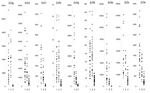
Figure 1:Individual data sets for Zn/Ag, Zn/Co, Zn/Cr, Zn/Fe, Zn/Hg, Zn/Rb, Zn/Sb, Zn/Sc, and Zn/Se mass fraction ratios in samples of normal (1), benign hypertrophic (2) and cancerous (3) prostate.
Mass fraction ratios of Zn/Ag, Zn/Cr, Zn/Fe, Zn/Hg, and Zn/Sb in the needle-biopsy cores could be used as a tool to diagnose PCa and are comparable with characteristics of the Zn mass fraction-test [55]. However, it is our opinion that application of the trace element mass fraction ratios is more suitable for PCa diagnosis. Trace element mass fraction depends on the sample mass, which decreases with loss of its moisture. The needle-biopsy core is a small piece of tissue with a relatively high "surface/volume" ratio. After sampling, it begins to lose mass very fast. Weight loss of samples depends on the humidity of operating and store rooms [56]. Thus, it is very difficult to determine the fresh mass of needle-biopsy cores and to calculate the precise mass fraction of trace elements. Sample freeze-dry, storage in air-tight vials until weighing, and then calculating mass fraction on dry weight basis is the only possible method that eliminates the variation in sample weight. Conversely, accuracy of trace element mass fraction ratios does not depend on sample mass and changes in moisture content. Therefore, this method does not require dry samples. Moreover, the use of the relations between mass fractions of chemical elements is particularly promising for the development of in vivo diagnostic methods, including the diagnosis of PCa.
CONCLUSION
In this work, mean values of Zn/Ag, Zn/Co, Zn/Cr, Zn/Fe, Zn/Hg, Zn/Rb, Zn/Sb, Zn/Sc, and Zn/Se mass fraction ratios in normal, benign hypertrophic and cancerous prostate tissue were calculated using data of INAA-LLR. It was observed that in benign hypertrophic tissues the Zn/Cr, Zn/Hg, Zn/Sb, and Zn/Se mass fraction ratios are lower than in normal tissues. In cancerous tissue all investigated mass fraction ratios are lower than in normal and benign hypertrophic tissues of the prostate. It was shown that the Zn/Ag, Zn/Cr, Zn/Fe, Zn/Hg, and Zn/Sb mass fraction ratios are the most informative for a differential diagnosis among all investigated mass fraction ratios. Finally, we propose to use the estimation of Zn/Ag, Zn/Cr, Zn/ Fe, Zn/Hg, and Zn/Sb mass fraction ratios in a needle-biopsy core as an accurate tool to diagnose prostate cancer.
REFERENCES
- Kirby RS. (2000). The natural history of benign prostatic hyperplasia: what have we learned in the last decade? Urology. 56(5 Suppl 1), 3-6.
- Roehrborn C and McConnell J. (2002). Etiology, pathophysiology, epidemiology and natural history of benign prostatic hyperplasia. In: Walsh P, Retik A, Vaughan E, Wein A, editors. Campbell's Urology. 8th ed. Philadelphia, Saunders. 1297- 1336.
- Lepor H. (2005). Pathophysiology of benign prostatic hyperplasia in the aging male population. Rev Urol. 7(Suppl 4), S3-S12.
- Oliver SE, Gunnell D and Donovan JL. (2000). Comparison of trends in prostate-cancer mortality in England and Wales and the USA. Lancet. 355, 1788-1789.
- Kumar RJ, Barqawi AB and Crawford ED. (2004). Epidemiology of prostate cancer. Business Briefing: US Oncology Review. 1-6.
- Maddams J, Brewster D, Gavin A, Steward J, et al. (2009). Cancer prevalence in the United Kingdom: estimates for 2008. Br J Cancer. 101(3), 541-547.
- Lutz JM, Francisci S, Mugno E, Usel M, et al. (2003). Cancer prevalence in Central Europe: the EUROPREVAL Study. Ann Oncol. 14(2), 313-322.
- Moller T, Anderson H, Aareleid T, Hakulinen T, et al. (2003). Cancer prevalence in Northern Europe: the EUROPREVAL study. Ann Oncol. 14(6), 946-957.
- De Angelis R, Grande E, Inghelmann R, Francisci S, et al. (2007). Cancer prevalence estimates in Italy from 1970 to 2010. Tumori. 93(4), 392-397.
- Waalkes MP and Rehm S. (1994). Cadmium and prostate cancer. J Toxicol Environ Health. 43, 251-269.
- Zaichick V and Zaichick S. (1999). Role of zinc in prostate cancerogenesis. In: Anke M, et al., editors. Mengen und Spurenelemente. 19. Arbeitstagung. Jena, Friedrich-Schiller- Universitat. 104-115.
- Platz EA and Helzlsouer KJ. (2001). Zinc, Selenium, and prostate cancer. Epidemiol Rev. 23, 93-101.
- Zaichick V. (2004). INAA and EDXRF applications in the age dynamics assessment of Zn content and distribution in the normal human prostate. J Radioanal Nucl Chem. 262(1), 229- 234.
- Gray MA, Centeno JA, Slaney DP, Ejnik JW, et al. (2005). Environmental exposure to trace elements and prostate cancer in three New Zealand ethnic groups. Int J Environ Res Public Health. 2, 374-384.
- Zaichick S and Zaichick V. (2011). INAA application in the age dynamics assessment of Br, Ca, Cl, K, Mg, Mn, and Na content in the normal human prostate. J Radioanal Nucl Chem. 288(1), 197-202.
- Zaichick S and Zaichick V. (2011). The effect of age on Ag, Co, Cr, Fe, Hg, Sb, Sc, Se, and Zn contents in intact human prostate investigated by neutron activation analysis. Appl Radiat Isot. 69(6), 827-833.
- Zaichick S and Zaichick V. (2011). The Br, Fe, Rb, Sr, and Zn content and interrelation in intact and morphologic normal prostate tissue of adult men investigated by energy dispersive X-ray fluorescent analysis. X-Ray Spectrom. 40(6), 464-469.
- Zaichick V, Nosenko S and Moskvina I. (2012). The effect of age on 12 chemical element contents in intact prostate of adult men investigated by inductively coupled plasma atomic emission spectrometry. Biol Trace Elem Res. 147(1-3), 49-58.
- Zaichick S, Zaichick V, Nosenko S and Moskvina I. (2012). Mass Fractions of 52 Trace Elements and Zinc Trace Element Content Ratios in Intact Human Prostates Investigated by Inductively Coupled Plasma Mass Spectrometry. Biol Trace Elem Res. 149(2), 171-183.
- Zaichick V and Zaichick S. (2014). Age-related histological and zinc content changes in adult nonhyperplastic prostate glands. Age. 36(1), 167-181.
- Zaichick V and Zaichick S. (2014). INAA application in the assessment of chemical element mass fractions in adult and geriatric prostate glands. Appl Radiat Isot. 90, 62-73.
- Zaichick V and Zaichick S. (2014). Determination of trace elements in adults and geriatric prostate combining neutron activation with inductively coupled plasma atomic emission spectrometry. Open Journal of Biochemistry. 1(2), 16-33.
- Zaichick V and Zaichick S. (2014). Use of INAA and ICP-MS for the assessment of trace element mass fractions in adult and geriatric prostate. J Radioanal Nucl Chem. 301(2), 383- 397.
- Zaichick V. (2015). The variation with age of 67 macro- and microelement contents in nonhyperplastic prostate glands of adult and elderly males investigated by nuclear analytical and related methods. Biol Trace Elem Res. 168(1), 44-60.
- Zaichick V and Zaichick S. (2015). Dietary intake of minerals and prostate cancer: insights into problem based on the chemical element contents in the prostate gland. J Aging Res Clin Practice. 4(3), 164-171.
- Zaichick V and Zaichick S. (2015). Global contamination from uranium: insights into problem based on the uranium content in the human prostate gland. J Environ Health Sci. 1(4), 1-5.
- Zaichick V and Zaichick S. (2016). Variations in concentration and distribution of several androgen-dependent and -independent trace elements in nonhyperplastic prostate gland tissue throughout adulthood. J Androl Gynaecol. 4(1), 1-10.
- Zaichick V and Zaichick S. (2016). Age-related Changes in Concentration and Histological Distribution of Br, Ca, Cl, K, Mg, Mn, and Na in Nonhyperplastic Prostate of Adults. European Journal of Biology and Medical Science Research. 4(2), 31-48.
- Zaichick V and Zaichick S. (2016). Variations in concentration and histological distribution of Ag, Co, Cr, Fe, Hg, Rb, Sb, Sc, Se, and Zn in nonhyperplastic prostate gland throughout adulthood. Jacobs Journal of Cell and Molecular Biology. 2(1), 1-16.
- Zaichick V. (2006). Medical elementology as a new scientific discipline. J Radioanal Nucl Chem. 269(2), 303-309.
- Schwartz MK. (1975). Role of trace elements in cancer. Cancer Res. 35(11 pt 2), 3481-3487.
- Kubo H, Hashimoto S, Ishibashi A, Chiba R, et al. (1976). Simultaneous determinations of Fe, Cu, Zn, and Br concentrations in human tissue sections. Medical Physics. 3, 204-209.
- Forssen A. (1972). Inorganic elements in the human body. I. Occurrence of Ba, Br, Ca, Cd, Cs, Cu, K, Mn, Ni, Sn, Sr, Y and Zn in the human body. Ann Med Exp Biol (Finland). 50, 99-162.
- Sangen H. (1967). The influence of the trace metals upon the aconitase activity in human prostate glands. Jap J Urol. 58(11), 1146-1159.
- Jafa A, Mahendra NM, Chowdhury AR and Kamboj VP. (1980). Trace elements in prostatic tissue and plasma in prostatic diseases of man. Indian J Cancer. 17, 34-37.
- Stitch SR. (1957). Trace elements in human tissue. I. A semi-quantitative spectrographic survey. Biochem J. 67, 97- 103.
- Soman SD, Joseph KT, Raut SJ, Mulay GD, et al. (1970). Studies of major and trace element content in human tissues. Health Phys. 19(5), 641-656.
- Tipton IH and Cook MJ. (1963). Trace elements in human tissue. Part II. Adult subjects from the United States. Health Phys. 9(2), 103-145.
- Brys M, Nawrocka AD, Miekos E, Zydek C, et al. (1997). Zinc and cadmium analysis in human prostate neoplasmas. Biol Trace Element Res. 59(1-3), 145-152.
- Lindholmer C and Glaumann H. (1972). Zinc and magnesium in human male reproductive tract. Andrologie (Berlin). 4(3), 231-237.
- Raju GJN, Padala S, Gummuluri AVRM, Muktineni RK, et al. (2007). Trace elemental analysis of normal, benign, hypertrophic and cancerous tissues of the prostate gland using the particle-induced X-ray emission technique. Eur J Cancer Prev. 16(2), 108-115.
- Kiziler AR, Aydemir B, Guzel S, Alici B, et al. (2010). May the level and ratio changes of trace elements be utilized in identification of disease progression and grade in prostatic cancer? Trace Elements and Electrolytes. 27(4), 65-72.
- Gyorkey F, Min K-W, Huff JA and Gyorkey P. (1967). Zinc and magnesium in human prostate gland: Normal, hyperplastic, and neoplastic. Cancer Res. 27(8), 1349-1353.
- Kwiatek WM, Hanson AL, Paluszkiewicz C, Galka M, et al. (2004). Application of SRIXE and XANES to the determination of the oxidation state of iron in prostate tissue sections. Journal of Alloys and Compounds, 362, 83-87.
- Zaichick V and Zaichick S. (2015). Differences and relationships between morphometric parameters and zinc content in nonhyperplastic and hyperplastic prostate glands. British Journal of Medicine and Medical Research. 8(8), 692-706.
- Zaichick V and Zaichick S. (2016). Trace element contents in adenocarcinoma of human prostate investigated by energy dispersive X-ray fluorescent analysis. Journal of Adenocarcinoma. 1(1), 1-7.
- Zaichick V and Zaichick S. (2016). The Bromine, Calcium, Potassium, Magnesium, Manganese, and Sodium Contents in Adenocarcinoma of Human Prostate Gland. J Hematology and Oncology Research. 2(2), 1-12.
- Zaichick V and Zaichick S. (2016). Trace element contents in adenocarcinoma of the human prostate gland investigated by neutron activation analysis. Cancer Research and Oncology. 1(1), 1-10.
- Zaichick V and Zaichick S. (2016). Prostatic tissue levels of 43 trace elements in patients with prostate adenocarcinoma. Cancer and Clinical Oncology. 5(1), 79-94.
- Zaichick V and Zaichick S. (2016). Chemical elemental content / Calcium ratios in tissues of human hyperplastic prostate gland. Journal of Applied Life Sciences International. 4(4), 1-11.
- Zaichick V and Zaichick S. (2016). Neutron activation analysis of Ag, Co, Cr, Fe, Hg, Rb, Sb, Sc, Se, and Zn contents in benign prostatic hypertrophic tissue. In: Neutron Spectroscopy, Nuclear Structure, Related Topics (Proceedings of ISINN-23). Dubna, Moscow Region, Russia, Joint Institute for Nuclear Research, 437-442.
- Zaichick V and Zaichick S. (2016). Prostatic tissue level of some major and trace elements in patients with BPH. Jacobs Journal of Nephrology and Urology. 3(1), 1-10.
- Zaichick V and Zaichick S. (2016). Levels of 43 Trace Elements in Hyperplastic Prostate Tissues. British Journal of Medicine and Medical Research. 15(2), 1-12.
- Zaichick V, Sviridova T and Zaichick S. (1997). Zinc in human prostate gland: normal, hyperplastic and cancerous. Int Urol Nephrol. 29(5), 565-574.
- Zaichick S and Zaichick V. (2012). Trace elements of normal, benign hypertrophic and cancerous tissues of the human prostate gland investigated by neutron activation analysis. Appl Radiat Isot. 70(1), 81-87.
- Zaichick V. (1997). Sampling, sample storage and preparation of biomaterials for INAA in clinical medicine, occupational and environmental health. In: Harmonization of Health-Related Environmental Measurements Using Nuclear and Isotopic Techniques. Vienna, IAEA. 123-133.
- Zaichick V and Zaichick S. (1996). Instrumental effect on the contamination of biomedical samples in the course of sampling. The Journal of Analytical Chemistry. 51(12), 1200- 1205.
- Zaichick V and Zaichick S. (1997). A search for losses of chemical elements during freeze-drying of biological materials. J Radioanal Nucl Chem. 218(2), 249-253.
- Zaichick V. (2004). Losses of chemical elements in biological samples under the dry aching process. Trace Elements in Medicine. 5(3), 17-22.
- Zaichick V. (1995). Applications of synthetic reference materials in the medical Radiological Research Centre. Fresenius J Anal Chem. 352, 219-223.
- Korelo AM and Zaichick V. (1993). Software to optimize the multielement INAA of medical and environmental samples. In: Activation Analysis in Environment Protection. Dubna, Moscow Region, Russia, Joint Institute for Nuclear Research. 326- 332.
- Catalona WJ. (1996). Clinical utility of measurements of free and total prostate-specific antigen (PSA): A review. Prostate. 7, 64-69.
- Genes VS. (1967). Simple methods for cybernetic data treatment of diagnostic and physiological studies. Moscow, Nauka.