Information Links
Related Conferences
Previous Issues Volume 7, Issue 2 - 2023
Use of Mesenchymal Stem Cells in Corneal Ulcers in Dogs: A Case Report
Perla Palafox-Herrera1, Yomira N Ortiz-Avilez1, Ramón Ruelas-Aviles2, Daniela Ruelas-Vogel2, Diana Mosco-Fierro2, Darien Mesa-Diaz2, Selem Torres1,3, Miguel Ramirez-Amezcua1,3, Diana Esquivel1,3,4,*
1PetCell, Mexico
2Instituto de Terapia Celular (ITC), Mexico
3Central Medica Veterinaria, Mexico
4Global Institute of Stem Cell and Research, Mexico
*Corresponding author: M.Sc. Diana Esquivel, Head of the laboratories of GIOSTAR, Mexico; E-mail: [email protected].
Received Date: April 14, 2023
Published Date: May 02, 2023
Citation: Esquivel D, et al. (2023). Use of Mesenchymal Stem Cells in Corneal Ulcers in Dogs: A Case Report. Mathews J Vet Sci. 7(1):20.
Copyrights: Esquivel D, et al. © (2023).
ABSTRACT
The development and complication of corneal ulcers (CU) in dogs has become a serious burden in pet health. Dogs with exaggerated juvenile-like craniofacial conformations, such as pug breed, are twenty times more prone to develop this condition. Although most of the cases are treated without surgical intervention, complicated and infected cases could lead to permanent blindness and other related complications. New approaches in treating this condition with mesenchymal stem cells (MSC) have been proposed with great results. Given their immunomodulation and regenerative properties, local administration of MSCs has proven to be safe and efficient in treating ocular related health conditions. This manuscript presents a complicated case of a canine CU treated with local infiltration of 1x10⁶ canine dental pulp MSCs (cDMSCs). Stem cell therapy was performed at the Central Medica Veterinaria in Colima, Mexico with the consent of the owner, as it was hypothesized that the regenerative properties of MSCs could potentially alleviate the patient´s condition. Notorious improvement was reported after 12 hours of infiltration, and neovascularization process was observed within the next few days. The patient was discharged on the 29th day of treatment, with minimal scar and fully regain normal vision. Therefore, using MSCs is a viable option for treating corneal ulcers in dogs.
Keywords: Corneal Ulcer, Dogs, Mesenchymal Stem Cells, Regenerative Medicine.
ABBREVIATIONS
CU: Corneal Ulcers; cDMSCs: Canine Dental Pulp MSCs; MSCs: Mesenchymal Stem Cells; cATMSCs: Canine Adipose Tissue-Derived Mesenchymal Stem Cells.
INTRODUCTION
One of the most common conditions regarding canine ocular problems is the development of corneal ulcers. Along with corneal melanosis and the growth of cataracts, CU has become a serious burden in pet health [1]. These lesions are often accompanied by pain, redness sore in the ocular area, some degree of squinting and excessive tear production, affecting the overall wealth of the dog and even their domestic behavior [2]. Often called ulcerative keratitis, CU is keratopathy-associated lesions, in which loss of the epithelium and hyperactive inflammation processes are commonly related to corneal conditions [3]. Among the variety of causes leading to the development of CU, direct trauma in the eye during playtime is one of the most reported at veterinary consultation. In a similar way, entropion, distichiasis conditions, as well as viral and bacterial infections are frequently associated to this ocular condition [2].
Moreover, exaggerated juvenile-like craniofacial conformations, including small wide-open eyes, heightens the risk of presenting complicated cases of CU. Of special interest pugs are one of the most afflicted breed; given their craniofacial conformation pugs are twenty times more prone to present complicated cases of CU [4]. Although simple CU lesions are expected to heal with antibiotic prescriptions, antiinflammatory drugs or even without any veterinary intervention, severe complex cases often need surgical intervention. If not treated properly, complicated cases of CU could lead to permanent blindness as well as other health complications [1,5]. Therefore, currently innovative proposals in treating complicated and infected CU have been research. Tissue adhesives, conjunctival grafts, corneal transplants and the development of compatible biomaterials have been performed as an alternative with moderate successful results. [6]. In the need for safe and efficient alternatives, the use of mesenchymal stem cells (MSCs) both in human and animal related health problems has gained interest in the last decade. Previous studies have demonstrated the remarkable properties of MSCs to modulate the inflammatory response of the patient, along with their differentiation capacity. These mechanisms of action could accelerate and promote corneal recovery, due to their proven ability to differentiate into keratocytes in vitro and in vivo, while producing new collagen within the host stroma [7, 8]. Given the MSCs properties within the scope of regenerative medicine, this manuscript aims to present the promising results obtained in a complicated case of canine CU.
CASE REPORT
A female pug with 5 years and 9 months old weighting 11kg was diagnosed with a severe case of CU in the right eye on September 2022. The patient has already been prescribed with combined ulcer kit (midriavet, proxiflox, cloranfivet), along with an ophthalmologic nutraceutical 1 tab a day for 90 days, doxicicline half a tablet a day for 8 days, and ceftriaxone 1g/3mL every 24 hours trough intramuscular injection for 4 days as pharmacological treatment, which controlled the infection but failed to alleviate the CU. When the patient came to Central Medica Veterinaria in Colima, Mexico, it presented stitches in the area due to two failed tarsorrhaphy surgeries and stenosis. A revision fluorescein test was performed and notorious damage to the cornea was detected. The internal diagnosis confirmed the CU lesion, resulting in keratoconjunctivitis sicca previously diagnosed with entropion.
In order to promote fully regeneration of the ocular tissue, stitches of previous surgeries were removed and local administration of 1x10⁶ canine dental pulp mesenchymal stem cells (cDMSCs) was carried out. The procedure was performed under general anesthesia, infiltrating at the conjunctival tissues in four different points as observed in Figure 1. Significant improvements in the inflamed region as well as increased vascularization in the corneal area were observed after 12 hours. Follow-up consultations were performed with good evolution signs; and saline solution cleanings along with the combined ulcer kit was prescribed for the following 7 days to prevent reinfection. 18 days after the cDMSCs infiltration, a fluorescein test showed no signs of lesion, outstanding close of the CU, improvement in the photosensitivity along with adequate ocular rotation. Complete healing of the CU was observed after 25 days of MSCs intervention, with minimum corneal scar. Combined ulcer kit dry eye treatment was prescribed for the following 4 days to prevent local dehydration, and on the 29th day medical discharge was made (Figure 1).
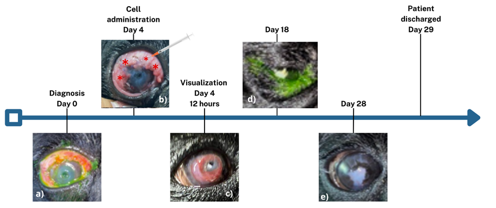
Figure 1. Clinical course and improvement after MSCs application. a) Positive fluorescence test for corneal injury, b) Corneal ulcer prior to application c) Vascularity increased after 12 hours of MSCs application, d) Negative fluorescence test for corneal injury, e) Corneal ulcer healed.
DISCUSSION AND CONCLUSION
CU is one of the most common ocular health concerns in dogs; which primary causes include trauma, foreign bodies, infection, inadequate lacrimal secretion, corneal endothelial dysfunction, spontaneous, toxic, and anatomical functional abnormalities in adnexa [9]. It has been hypothesized that the anatomical facial features of some dogs play a role in the development of sever cases of CU. Recently, a large population study reported that brachycephalic dogs with prominent eyes, pug breed included, have a higher overall susceptibility to CU [10]. Conventional CU medical management approaches different symptomatology; including proper antimicrobials, aqueous suppressants, anti-collagenases, anti-inflammatory therapy and promoting epithelial healing [11]. Nevertheless, sever and an infected case of this condition usually leads to the loss of more than half of the corneal thickness, as well as corneal perforation. These complications can have devastating visual consequences if not treated properly. Anatomical integrity of the cornea is desired but most of the time, surgical intervention failed to preserve corneal functionality [9].
Therefore, an urgent need to find new therapeutic approaches for CU, has gained the attention of veterinary clinics. Alternative treatments such as platelet-rich plasma [12], amniotic membrane graft [13], matrix regenerating agents [14,15] have been reported. Recently MSCs has been used as an alternative for CU in humans, with outstanding outcomes [16]. Likewise, other animals such as cats [17], horses [18] and dogs [19, 20] have been documented with promising results. On a recent case report a Poodle diagnose with CU was administered two doses of allogeneic stem cell through topical eye application; each dose containing 3x10⁶ canine adipose tissue-derived mesenchymal stem cells (cATMSCs). Clinical evaluation, along with Fluorescein test, Schirmer test and ocular fundus exam showed remarkable improvement with lesion resolution [19]. In addition, on a very interesting study 26 breed-not-specified dogs were diagnosed with deep corneal ulcers and have been unsuccessfully treated for 15 days with allopathic drops. Dogs received an application of 3x10⁶ allogeneic cATMSCs via subconjunctival in 12 instillations (one per hour) of 0.25 mL of transport medium containing 250,000 cells. After clinical evaluation, considering general discomfort and deepen of the CU, 84.6% of the cases reversed without the need of surgical intervention [20].
MSCs enhances an immunomodulation process via paracrine mechanisms, leading to an observable therapeutic action in corneal wound re-epitheliazation [21]. Local administration of MSCs attenuates inflammation, stimulates neovascularization, accelerates corneal wound healing [22], and promotes endogenous regenerations [23]. These mechanisms of action places MSCs therapy as a safe and efficient approach in treating corneal conditions in the veterinary area. In this manuscript, we have presented an outstanding healing of a CU in a canine patient using stem cell therapy. After the infection was controlled and MSCs infiltration was performed, the patient shown significant improvement in the first 12 hours. A notorious process of ulcer closure along with neovascularization process was observed within the first weeks of treatment. The patient was discharged on day 29th with no signs of infection. Our results are in accordance with has been previously reported [19,20], posing MSCs therapy as a therapeutic alternative in treating this condition.
ACKNOWLEDGEMENTS
The authors would like to acknowledge the valuable contribution and data collection of Central Medica Veterinaria, Hospital Veterinario de Especialidades.
CONFLICT OF INTEREST
The authors declare no conflict of interest.
AUTHOR CONTRIBUTIONS
PP-H, YO-A have participated in the analyses and draft of the manuscript. RR-A, DR-V, DM-F, DM-D and MR-A has made significant contribution with the data and presentation of the case report. ST, DE has participated in the design and revision of the manuscript. All authors have read and approved the final manuscript.
REFERENCES
- Patel KP, Parikh PV, Mahla JK, Ashwath SN, Kelawala, DN. (2020). Incidence of Corneal Ulcer in Dogs–A Retrospective Study. Int J Curr Microbiol App Sci. 9(8):3174-3179.
- Germensky-Metzlet AJ. Section I Diseases and Disorders By Discipline–Corneal Ulceration. In: Côté E, editors. Clinical veterinary advisor: Dogs and cats. 3rd edn. USA, Missouri: Elsevier Mosby. 2015. p. 229-231.
- Maggs DJ. Cornea and Sclera. In: Maggs DJ, Miller PE, Ofri R Slatter DH, editors. Slatter's fundamentals of veterinary ophthalmology. 4th edn. Sounders Elsevier. 2008. p. 175-204.
- Packer RM, Hendricks A, Burn CC. (2015). Impact of facial conformation on canine health: corneal ulceration. PLoS One. 10(5):e0123827.
- Ledbetter EC, Gilger BC. Disease and surgery of the canine cornea and Sclera. In: Gelatt KN, Gilger BC, Kern T J, Gilger BC, Ledbetter EC, editors. Veterinary ophthalmology Volume 2. 5th edn. Germany: Wiley-Blackwell. 2013 p. 980-981.
- Jaksz M, Fischer MC, Fenollosa-Romero E, Busse C. (2021). Autologous corneal graft for the treatment of deep corneal defects in dogs: 15 cases (2014-2017). J Small Anim Pract. 62(2):123-130.
- Arnalich-Montiel F, Pastor S, Blazquez-Martinez A, Fernandez-Delgado J, Nistal M, Alio JL, et al. (2008). Adipose-derived stem cells are a source for cell therapy of the corneal stroma. Stem Cells. 26(2):570-579.
- Alio del Barrio JL, Chiesa M, Garagorri N, Garcia-Urquia N, Fernandez-Delgado J, Bataille L, et al. (2015). Acellular human corneal matrix sheets seeded with human adipose-derived mesenchymal stem cells integrate functionally in an experimental animal model. Exp Eye Res. 132:91-100.
- Gogova S, Leiva M, Ortillés Á, Lacerda RP, Seruca C, Laguna F, et al. (2020). Corneoconjunctival transposition for the treatment of deep stromal to full-thickness corneal defects in dogs: A multicentric retrospective study of 100 cases (2012-2018). Vet Ophthalmol. 23(3):450-459.
- Cebrian P, Escanilla N, Lowe RC, Dawson C, Sanchez RF. (2021). Corneo-limbo-conjunctival transposition to treat deep and perforating corneal ulcers in dogs: A review of 418 eyes and corneal clarity scoring in 111 eyes. Vet Ophthalmol. 24(1):48-58.
- Stamate AC, Tătaru CP, Zemba M. (2019). Update on surgical management of corneal ulceration and perforation. Rom J Ophthalmol. 63(2):166-173.
- Alio JL, Rodriguez AE, De Arriba P, Gisbert S, Abdelghany AA. (2018). Treatment with platelet-rich plasma of surgically related dormant corneal ulcers. Eur J Ophthalmol. 28(5):515-520.
- Costa D, Leiva M, Sanz F, Espejo V, Esteban J, Vergara J, et al. (2019). A multicenter retrospective study on cryopreserved amniotic membrane transplantation for the treatment of complicated corneal ulcers in the dog. Vet Ophthalmol. 22(5):695-702.
- Cochener B, Zagnoli C, Hugny-Larroque C, Derrien S. (2019). Healing of resistant corneal neurotrophic ulcers using a matrix regenerating agent. J Fr Ophtalmol. 42(2):159-165.
- Martinez JA, Chiappini F, Barritault D. (2019). Case Reports for Topical Treatment of Corneal Ulcers with a New Matrix Therapy Agent or RGTA® in Dogs. Vet Sci. 6(4):103.
- Agorogiannis GI, Alexaki VI, Castana O, Kymionis GD. (2012). Topical application of autologous adipose-derived mesenchymal stem cells (MSCs) for persistent sterile corneal epithelial defect. Graefes Arch Clin Exp Ophthalmol. 250(3):455-457.
- Zakirova EY, Valeeva AN, Faizullina RR, Akhmetshin RF, Nefedovskaya LV, Rizvanov AA. (2015). Transplantation of allogeneic mesenchymal stromal cell for treating corneal ulcers in cats. Genes & Cells. 10(3):49-55.
- Marfe G, Massaro-Giordano M, Ranalli M, Cozzoli E, Di Stefano C, Malafoglia V, et al. (2012). Blood derived stem cells: an ameliorative therapy in veterinary ophthalmology. J Cell Physiol. 227(3):1250-1256.
- Arantes-Tsuzuki PdM, Mazzonetto PC, Lo Turco EG. (2019). Treatment for Canine Corneal Ulcer using Adipose Tissue-derived Mesenchymal Stem Cell Therapy-Case Report. Preprints.org. DOI: 10.20944/preprints201912.0185.v2.
- Falcão MSA, Brunel HDSS, Peixer MAS, Dallago BSL, Costa FF, Queiroz LM, et al. (2019). Effect of allogeneic mesenchymal stem cells (MSCs) on corneal wound healing in dogs. J Tradit Complement Med. 10(5):440-445.
- Yang YH, Hsieh TL, Ji AT, Hsu WT, Liu CY, Lee OK, et al. (2016). Stromal Tissue Rigidity Promotes Mesenchymal Stem Cell-Mediated Corneal Wound Healing Through the Transforming Growth Factor β Signaling Pathway. Stem Cells. 34(10):2525-2535.
- Li F, Zhao SZ. (2014). Mesenchymal stem cells: Potential role in corneal wound repair and transplantation. World J Stem Cells. 6(3):296-304.
- Harrell CR, Jankovic MG, Fellabaum C, Volarevic A, Djonov V, Arsenijevic A, et al. (2019). Molecular Mechanisms Responsible for Anti-inflammatory and Immunosuppressive Effects of Mesenchymal Stem Cell-Derived Factors. Adv Exp Med Biol. 1084:187-206.