Information Links
Related Conferences
Previous Issues Volume 1, Issue 1 - 2016
Surgical Management of Abdominal Hernia in a Duck (Anas Platyrhynchos)
J.D Parrah1, Khadim Hussain Dar2, Hakim Athar3, Beenish Qureshi4
1Associate Professor Faculty of Veterinary Sciences and Animal Husbandry, Ganderba, Sher-e-Kashmir University of Agricultural Sciences and Technology, Kashmir-190006.
2Ph.D Research Scholar Division of Veterinary Surgery and Radiology Ganderba, Sher-e-Kashmir University of Agricultural Sciences and Technology, Kashmir-190006.
3Assistant Professor Faculty of Veterinary Sciences and Animal Husbandry, Ganderba, Sher-e-Kashmir University of Agricultural Sciences and Technology, Kashmir-190006.
4M.V.Sc Scholar Division of Veterinary Surgery and Radiology Ganderba, Sher-e-Kashmir University of Agricultural Sciences and Technology, Kashmir-190006.
Corresponding Author: Khadim Hussain Dar, Ph.D Research Scholar, Division of Veterinary Surgery and Radiology Ganderba, Sher-e-Kashmir University of Agricultural Sciences and Technology, Kashmir-190006.Tel: 0194 246 2758;Email: [email protected]
Received Date: 22 April 2016 Accepted Date: 03 May 2016 Published Date: 31 May 2016
Copyright © 2016 Dar KH
Citation: Dar KH. (2016). Surgical Management of Abdominal Hernia in a Duck (Anas Platyrhynchos).Mathews J Vet Sci. 1(1): 002.
ABSTRACT
A 2-year old female duck weighing 3.75kg was presented to the Division of Veterinary Surgery & Radiology, SKUAST-K with a history of painless reducible abdominal swelling from 15 days present in the ventral abdominal region close to the cloaca. After clinical examination the ventral abdominal hernia was diagnosed. The bird was anaesthetized with diazepam followed 5 minutes later by Ketamine. A ventral midline celiotomy was performed. For repair of the abdominal hernia the abdominal muscles and skin were sutured in a standard two-layer closure using a simple continuous suture pattern. The bird was confined in a cage to restrict movement for a period of two weeks. Postoperatively, Meloxicam and Enrofloxacin were administered along with antiseptic wound dressing for 10 days. On removal of skin sutures on 10th day, complete healing of the area was observed and the bird recovered completely without any complications. The follow up study for 6 months revealed sound recovery.
KEYWORDS
Duck; Anaesthesia; Hernia; Repair.
INTRODUCTION
The etiology of abdominal hernias in birds is unknown. Abdominal hernia in birds can be either congenital or acquired [1]. However, abdominal hernia is characterized by separation of aponeurosis of abdominal muscles at ventral midline [2]. This gives the bird a pot-bellied appearance with protruding mass visible directly beneath the skin [3]. It may be related to breeding, hormonal influences or other space occupying coelomic masses [1]. Female ducks are more prone to abdominal hernia due to hormonal influences causing weakness of ab dominal muscles [4]. Altered calcium metabolism in chronic egg laying ducks may also lead to muscular atony and overdistention in the ventral abdomen [5]. Herniation can also occur due to trauma, an abdominal mass, egg binding, straining, and weight of viscera leads to marked enlargement so that the swelling becomes pendulant [6]. Occasionally herniation can occur secondary to abdominal lipoma, cystic masses, neoplasia and other space occupying masses [7]. Surgical repair by hernioraphy is indicated when the bird is stable clinically [2].
HISTORY AND CLINICAL OBSERVATIONS
A two-year old female duck (Anas platyrhynchos) weighing 3.75 kg was referred with a 15-day history of abdominal swelling. Physical examination revealed the bird to be dull and depressed with no indication of diarrhea observed in the cloacal region. Its diet consisted of a commercially available food. A spherical painless, reducible swelling about 7.5 cm extending from the keel to the pubic bones located in the ventral abdomen region close to the cloaca. The presented data enabled us to make a diagnosis of abdominal hernia (Figure 1). The bird was prepared for the aseptic surgery.
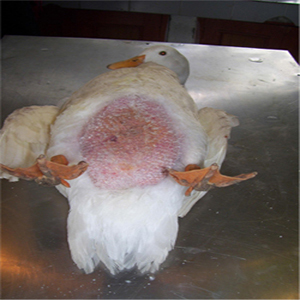
Figure 1: View of the abdominal swelling.
TREATMENT
After the pre-surgical starvation of 30 minutes, the duck was placed under general anaesthesia using the anesthetic regime of Diazepam/ Ketamine as described by Dar et al, [8]. Diazepam (Lori®, Neon laboratories Limited, Mumbai, India) @ 1mg/kg body weight was administered intramuscularly (IM) into the pectoral muscle then five minutes later when the sedation was achieved Ketamine (Aneket®, Neon laboratories Limited, Mumbai, India) @ 15 mg/kg was administered IM into the pectoral muscle. The bird was restrained in dorsal recumbence and the head raised about 30°. The wings were reflected dorsally while the legs were restrained and abducted in caudal direction. After preparation of the operation field, a ventral midline celiotomy were performed. Corrective surgery involved a procedure in which an elliptical transabdominal incision through the skin and abdominal muscles was performed to reduce the size of the distended hernial sac by removing part of the abdominal wall (Figure 2).
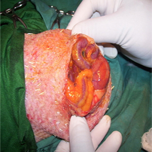
Figure 2: Contents seen after the initial incision.
The incision was then extended with fine scissors. The skin was the only structure of hernia sac holding the content of abdominal organs as the hernial sac. The hernia consisted of ileum and cecum loops glued with adhesions in the form of a fibrin mesh which formed one ball. All contents were returned back into the abdominal cavity. The linea-alba was sutured by a simple interrupted suture pattern using No. 2/0 catgut (Figure 3).
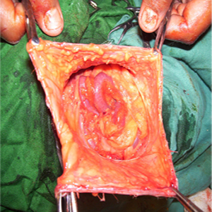
Figure 3: Content of hernial sac (intestinal loops covered with fibrin mesh).
Then the skin was sutured in a simple interrupted pattern using No. 1 silk as suture material (Figure 4).
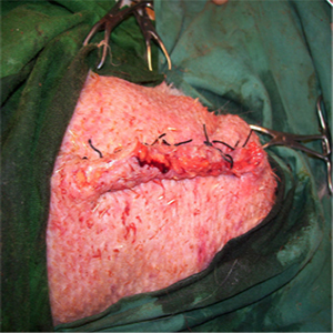
Figure 4: Suturing of the skin with silk.
The bird was confined in a cage to restrict movement for a period of two weeks. Post-operative Management Postoperative management consisted of diet modification and decreased exercise and activity for 2 weeks after surgery. Meloxicam (Melonex®, Intas pharmaceautical, India 0.25 mg/ kg IM, once daily for 5 days) and enrofloxacin (Bayrocin®, Pfizer- Bayer; 20 mg/kg IM, once daily for 7 days) were also administered IM along with antiseptic wound dressing for 10 consecutive days post operation with 5% povidone iodine solution. The skin sutures were removed on 10th post-operative day. The follow up study for 6 months revealed sound recovery with no complications.
DISCUSSION
There are only few published articles about abdominal hernias in birds particularly of ducks [7]. Abdominal hernia in birds is characterized by the separation in the aponeurosis of the abdominal musculature on the ventral midline [2]. The muscle is weakened by egg laying and infiltration of fat so muscle fibres separate [6]. Affected birds are most commonly middle-aged to older hens with variable degrees of abdominal swelling [7]. Some of the less serious cases may not be true hernias, but just an extended body wall and may be due to malnutrition, lack of exercise, chronic masturbation. All cases need thorough investigation before undertaking surgery [1]. However, the present case report describes the true abdominal hernia in duck (Anas platyrhynchos). Previously ventral abdominal hernias in turkey poults and pigeon (Columba livia) have been reported [9]. The abdominal hernia is frequently seen in female birds which may be related to hormonal imbalance causing weakness of abdominal muscles [4]. Prompt surgical repair of the hernia is important if the bird traumatizes its abdomen by rubbing on surfaces, experiences respiratory distress, has difficulty passing urates and feces from its cloaca, or has the entire abdominal viscera within the hernial sac [7]. In the present case surgical repair was performed because the bird was stable clinically and a hernioraphy was indicated. Ketamine-diazepam combination was found suitable for short term surgery in birds [8, 10]. The surgery lasted for 15 minutes during that time additional anaesthetic dose was not required and duck recovered uneventfully.
CONCLUSION
Hernioraphy is a safe and effective for the repair of abdominal hernia when presented at earlier stage procedure in birds under Diazepam-Ketamine anaesthesia.
ACKNOWLEDGEMENT
The authors are highly thankful to Dr. B. A. Moulvi, professor and Head, Division of Veterinary Surgery and Radiology F.V.Sc & A.H, SKUAST-K, for his valuable guidance and close supervision.
REFERENCES
- Bennet RA. (1994). Soft tissue surgery. In: Ritchie BW. Harrison, GJ. Harisson, LR. (eds.). Avian Medicine: Principles and application. Wingers Publishing, Lake Worth FL. 1097-1136.
- Smolec O, Kos J, Vnuk D, Babic D, et al. (2009). Abdominal ventral hernia in a pigeon (Columba livia): a case report. Veterinarni Medicina. 54(6), 291-294.
- Rosskopf WJ and Woerpel RW. (1987). Pet avian obstetrics. Proceeding of the First International Conference of Zoo and Avian medicine. 213-231.Ohau Hawaii, USA.
- Ranck FM. (1974). Umbilical hernias in turkeys from two flocks. Avian Diseases. 11(18), 477-483.
- Martin HD. (1986). Abdominal hernias with formation of urate concretions, Journal of American Veterinary Medical Association. 189(10), 1332-1333.
- Altman RB. (1997). Soft tissue surgical procedures. In: Avian Medicine and Surgery. WB Saunders, Philadelphia. 704- 731.
- MacWhirter P. (1994). A review of 60 cases of abdominal hernias in birds. Proceeding of the Annual Conference on Association Avian Veterinary. 27-37. Reno Nevada, USA.
- Khadim Hussain Dar, Mehraj-U-Din Dar, Adil S Baba, Mubashir Ahmad Baba, et al. (2015). Surgical management of compound metacarpal fracture in Black Kite (Milvus migrans): A Case Report. International Journal of Veterinary Sciences. 4 (2), 101-103.
- Carlson HC. (1962). Case report-abdominal hernia in turkey poults. The Canadian Veterinary Journal. 3, 263-264.
- Forbes NA. (2002). Avian gastrointestinal surgery. Seminars in Avian and Exotic Pet Medicine. 11(4), 196-207.