Information Links
Related Conferences
Previous Issues Volume 10, Issue 2 - 2025
Sturge-Kalischer-Weber Syndrome: A Historical Overview and Educational Imagery
Aamir Jalal Al-Mosawi*
Advisor Doctor and Expert Trainer, Baghdad Medical City and Iraqi Ministry of Health Baghdad, Iraq
*Corresponding Author: Aamir Jalal Al-Mosawi, Advisor Doctor and Expert Trainer, Baghdad Medical City and Iraqi Ministry of Health Baghdad, Iraq, Email: [email protected]
Received Date: July 21, 2025
Published Date: September 17, 2025
Citation: Al-Mosawi AJ. (2025). Sturge-Kalischer-Weber Syndrome: A Historical Overview and Educational Imagery. Mathews J Pediatr. 10(2):43.
Copyrights: Al-Mosawi AJ. © (2025).
ABSTRACT
Sturge-Kalischer-Weber syndrome is a rare, sporadic, non-hereditary congenital neurocutaneous condition characterized by a range of neurological and dermatological manifestations. The most common features include unilateral facial port-wine stains (nevi) and leptomeningeal angiomas, which may lead to seizures, typically during infancy in approximately 70% of cases. Associated features often include developmental delay, mental retardation, and glaucoma. This paper documents the first reported case of Sturge-Kalischer-Weber syndrome in Iraq, contributing to the limited body of literature on this condition in the region.
Keywords: Sturge-Kalischer-Weber Syndrome, Iraq.
INTRODUCTION
Sturge-Kalischer-Weber syndrome (also known as Sturge-Weber syndrome) is a rare, sporadic neurocutaneous disorder that manifests as a combination of skin and neurological abnormalities. The condition is most commonly associated with unilateral port-wine stains (nevus flammeus) on the face and leptomeningeal angiomas. Seizures occur in approximately 70% of cases, typically during infancy, and patients may also experience developmental delay, cognitive impairments, and glaucoma [1-6].
Neurocutaneous syndromes observed in Iraq include neurofibromatosis, tuberous sclerosis, and Sturge-Kalischer-Weber syndrome, though scientific documentation of the latter is scarce [7,8]. This paper aims to document the first clinical case of Sturge-Kalischer-Weber syndrome in Iraq.
PATIENTS AND METHODS
A six-year-old boy was referred for evaluation of developmental delay, including mental retardation and delayed speech development. He had no history of seizures or symptoms indicative of glaucoma. A well-defined unilateral hyperpigmented lesion, characteristic of a port-wine stain, was noted on his left cheek, extending from the periorbital region to the upper lip (Figure 1). The lesion had irregular, but distinct borders, and was present at birth. There were no signs of craniofacial asymmetry or other abnormalities.
.png)
Figure 1
To confirm the diagnosis of Sturge-Kalischer-Weber syndrome, brain magnetic resonance imaging (MRI) was recommended, but the patient has not returned for imaging as of this writing.
The absence of seizures and ocular symptoms at this stage does not exclude the diagnosis of Sturge-Kalischer-Weber syndrome, especially in the absence of any strong alternative diagnosis.
DISCUSSION
Sturge-Kalischer-Weber syndrome was first described in 1879 by the English physician William Allen Sturge (Figure 2A), who documented the condition in a six-and-a-half-year-old girl.
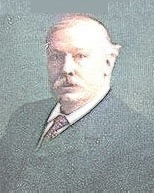
Figure 2A. William Allen Sturge (1850–1919), English Physician
The first pathological description of the condition was provided in 1901 by Siegfried Kalischer (Figure 2B), a German physician, who contributed crucial insights into the clinical presentation of the syndrome.
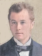
Figure 2B. Siegfried Kalischer (1862–1954), German Physician
In 1922, the English physician Frederick Parkes Weber (Figure 2C) further characterized the condition by describing its association with intracranial calcifications.
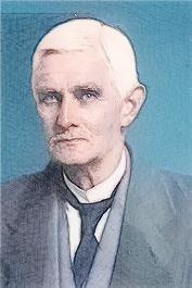
Figure 2C. Frederick Parkes Weber (1863–1962), English Physician
The historical contributions of these physicians helped establish the understanding of Sturge-Kalischer-Weber syndrome as a distinct clinical entity.
CONCLUSION
This report highlights the first known case of Sturge-Kalischer-Weber syndrome in Iraq, broadening the geographic documentation of this rare condition. Further diagnostic work-up, including brain MRI, is essential for definitive diagnosis and management.
ACKNOWLEDGEMENT
The author wishes to express sincere gratitude to the parents of the child, who have kindly permitted the publication of the patient’s photograph.The author holds the copyright to the sketches and images (Figures) included in this paper.
CONFLICT OF INTEREST
None.
REFERENCES
- Sturge WA. (1879). A case of partial epilepsy, apparently due to a lesion of one of the vasomotor centres of the brain. Trans Clin Soc London. 12:162.
- Kalischer S. (1901). Ein Fall von Telangiektasie (Angiom) des Gesichts und der weichen Hirnhäute. Arch Psychiatr Nervenkrankheiten Berl. 34:171-180.
- Parkes WF. (1922). Right-sided hemi-hypotrophy resulting from right-sided congenital spastic hemiplegia, with a morbid condition of the left side of the brain, revealed by radiograms. J Neurol Psychopathol. 3(10):134-139.
- Greenwald HM, Koota J. (1936). Associated facial and intracranial hemangiomas. Am J Dis Child. 51:868.
- Krabbe, Knud H. (1934). Facial and meningeal angiomatosis associated with calcifications of the brain cortex. Arch. Neurol & Psychiat. 32:737.
- Anderson HC. (1945). The Sturge-Weber syndrome; report of three cases. Yale J Biol Med. 18(2):103-106.
- Al-Mosawi AJ. (2019). Von Recklinghausen syndrome with right-sided basal ganglion abnormalities on brain magnetic resonance imaging. Journal of Clinical Research in Radiology. 2(1):1-3.
- Al-Mosawi AJ. (2019). Von Recklinghausen syndrome. 1st ed., Saarbrücken; LAP Lambert Academic Publishing. ISBN: 978-613-9-44487-8.