Information Links
Related Conferences
Previous Issues Volume 9, Issue 1 - 2025
Study of the Hepatoprotective Activity of the Hydroethanolic Extract of the Roots of Cochlospermum tinctorium Perr. A. Rich in Wistar Rats
Issa Oumarou BS*1,2, Hama Aghali Nouhou3, Hamani Issaka4, Hamadou Ibrahim4, Simon Azonbakin1, Yadji Guero Laila1,4, Sewadouno Faya Daniel1, Anatole Laleye1
1Histology, Biology of Reproduction, Cytogenetics and Medical Genetics Laboratory, Faculty of Health Sciences, University of Abomey-Calavi, Abomey-Calavi, Benin
2André Salifou University / Faculty of Health Sciences / Zinder National Hospital / Clinical Research and Health System Laboratory (LARECSS), Zinder, Niger
3Laboratory of Reference Hospital / Dan Dicko Dankoulodo University, Faculty of Health Sciences, Maradi, Niger
4Abdou Moumouni University / Faculty of Health Sciences / Laboratory of Histology-Embryology and Cellular Pathology “Pr Ag MOUMOUNI Hassane”, Niamey, Niger
*Corresponding author: Dr ISSA OUMAROU Boubacar Sidikou, André Salifou University, Faculty of Health Sciences, Zinder National Hospital, Clinical Research and Health System Laboratory (LARECSS), Zinder, Niger, Tel: (+227) 81013312, WhatsApp: (+227) 96728649; Email: [email protected]
Received Date: December 27, 2024
Published Date: January 21, 2025
Citation: Issa Oumarou BS, et al. (2025). Study of the Hepatoprotective Activity of the Hydroethanolic Extract of the Roots of Cochlospermum tinctorium Perr. A. Rich in Wistar Rats. Mathews J Cytol Histol. 9(1):30.
Copyrights: Issa Oumarou BS, et al. © (2025).
ABSTRACT
Title: Study of the Hepatoprotective Activity of the Hydroethanolic Extract of the Roots of Cochlospermum tinctorium Perr. A. Rich in Wistar Rats. Introduction: cochlospermum tinctorium Perr ex A Rich is a plant widely used by traditional African medicine. The main objective of our work is to study the hepatoprotective activity of the hydroethanolic extract of roots of cochlospermum tinctorium A rich. Material and Method: It was a prospective, descriptive and analytical study which took place over a period of 10 months from february to november 2021. We carried out a hydroethanolic extraction, then a phytochemical study of de the roots of cochlospermum tinctorium. To evaluate the hepatoprotective power in vivo of this extract, an animal model of toxicity induced by paracetamol in the wistar rat is used. The multiple comparisons are made by the ANOVA test. The differences are considered significant at p < 0, 05. Results: The extract showed a considerable hepatoprotective effect evidenced by a significant reduction in transaminases (ASAT, ALAT) and total bilirubin, an improvement in the tissue condition and hepatocyte morphology as well as reduction in signs of hepatic suffering resulting from intoxication with paracetamol. Conclusion: The results of study show that the hydroethanolic extracts of the roots of cochlospermum tinctorium protect the liver against attacks induced by paracetamol.
Keywords: Hepatoprotective, Cochlospermum tinctorium, Hydroethanolic Extract, Paracetamol.
ABBREVIATIONS
TM: Traditional Medicine; RCT: Roots of Cochlospermum tinctoriume A Rich; PCM: Paracetamol; CCl4: Carbon Tetrachloride.
INTRODUCTION
The use of medicinal plants by humans in general, and Africans in particular, dates back thousands of years. Medicinal plants represent a very important aspect in the history of medicine and have contributed enormously to the development of modern medicine. Many of the medicines in use today are derived solely from plants, some of which produce metabolites that can treat disorders of the human reproductive system [1].
Worldwide, the use of traditional medicine (TM), particularly herbal medicines, has increased over the last two decades. Many people now turn to them to treat a wide range of illnesses. For example, in Europe, the use of TM varies from 42% in Belgium to 90% in the UK; from 42% in the USA among adults to 70% in Canada. In Africa, TM use varies from 60% in Uganda and the United Republic of Tanzania, 70% in Ghana and Rwanda, to 80% in Benin and 90% in Burundi and Ethiopia [2]. In order to better promote the use of plants, numerous studies are needed to ensure their safety. To improve the use of plants, we were interested in cochlospermum tinctoriume A Rich, a plant widely used by the TM in West Africa. Apart from its very popular uses for the treatment of malaria, jaundice and bilious fevers mainly hematuric, the yellow rhizome is also used to treat oedema, urinary incontinence, dysmenorrhea, epilepsy, schistosomiasis, pneumonia, bronchial conditions, conjunctivitis, stomach problems, diarrhea, indigestion, stomach aches, hemorrhoids and skin conditions [2]. In Benin the roots of cochlospermum tinctoriume A Rich (RCT) are used for human food, the treatment of liver diseases, hemorrhoids, internal bleeding and malnutrition [3,4]. In Côte d'Ivoire, the pulp of the leaves is used as a moist dressing to ripen abscesses and boils while the decoction of the branches or rhizome is effective in the treatment of genitourinary disorders, kidney or intercostal pain [5,6]. In Mali, the plant is used to treat gastrointestinal diseases, jaundice, malaria and schistosomiasis [7]. In Nigeria, people drink a potion made from its fruit and tamarind to treat snake bites, in addition to its uses against jaundice and liver disease [8]. The richness of this plant in secondary metabolites gives it a wide range of therapeutic properties. This plant has interesting properties for protecting the liver. A review of the literature identified studies on the protective and curative effects of this plant against carbon tetrachloride (CCl4) poisoning, aflatoxin B and infectious hepatitis [9,10]. It would therefore be interesting to study the protective effects of RCT against other hepatotoxic xenobiotics to which exposure is frequent and repeated.
The liver, as a vital organ of the body, is mainly responsible for the metabolism of endogenous and exogenous agents. It plays an important role in the elimination, detoxification of drugs and repair of damage caused by xenobiotics, alcohol consumption, infections, malnutrition. Paracetamol (PCM) is widely used as an analgesic and antipyretic, but induces acute liver injury at very high doses [11,12]. Hepatic pathologies of drug origin are frequent and polymorphic in their PCM is one of the most widely used analgesics and antipyretics in the world. It is highly toxic to the liver, and sometimes fatal. The health risk is due to the fact that this product is sold in every country without a prescription, with easy access thanks to over-the-counter sales. It is practically present in every household. Overdose is responsible for cytolysis and dose-dependent hepatocyte necrosis, initiated by the formation of its reactive metabolite, N-acetyl-p-benzoquinone imine (NAPQI), produced by cytochrome P450-2E1. As the capacity of gluthation (GSH) to neutralise NAPQI is exceeded, oxidative stress is produced, leading to mitochondrial dysfunction, mediated by the activation of a cascade of cytosolic kinases and followed by DNA fragmentation. This is followed by an influx of inflammatory cells amplifying the production of cytokines and cytolytic enzymes [13,14]. The aim of the present study is to evaluate the hepatoprotective effect of hydroethanol extracts of RCT during PCM intoxication of Wistar rats.
METHOD AND MATERIALS
Framework
The study was carried out at the Institute of Applied Biomedical Sciences (ISBA) in Cotonou with:
- The team of the Laboratory of Histology, Reproductive Biology, Cytogenetics and Medical Genetics;
- The Biochemistry and Bioactive Natural Substances Team (EBSNB).
Materials
Plant material and drugs used
- Hepatic intoxication product: we used PCM (acetaminophen) for this experiment.
- Plant material: The plant material consisted of the roots of Cochlospermum tinctorium A Rich, which we acquired from a traditional therapist in the town of Kandi (Benin).
- Reference hepatoprotective molecule [12,15,16]: we used silymarin, a substance extracted from the fruit of milk thistle (silybum mariunum). Silymarin is actually made up of an isomeric mixture of active flavonolignans: silychristin, silydianine and two groups of diastereo isomeric flavonolignans: silybin A, silybin B, isosilybin A and isosilybin B. It is a highly hepatoprotective flavonoid complex.
Experimental animals
The experimental animals are female albino rats of the Wistar strain, 8 weeks old, weighing between 100 and 200 g and acclimatised to the rearing conditions in the animal house of the Human Biology Unit of the Faculty of Health Sciences (FSS). The animals are reared in a well-ventilated room of around 12 m2, with a day/night cycle of around 12 hours. The animals are kept in wire mesh cages (50×30×20) equipped with small feeders and drinkers. The bottom of the cages consists of a movable drawer system lined with wood shavings that collects faeces and urine through a lattice base. They have free access to drinking water and food. They are supplied with drinking water from the tap and their daily diet consists of a mixture of food in the form of kibbles marketed by Veto ServiceR. The breeding enclosure is regularly cleaned to ensure optimal development of the animals, free from any infection.
Methods
Type of study and duration of the study
It was a prospective, descriptive and analytical study which took place over a period of 10 months from February to November 2021.
Preparation of hydro-ethanolic extracts of
The hydroethanolic extracts are obtained by maceration of root powders in an ethanol-distilled water mixture (80% absolute ethanol and 20% distilled water) for seventy-two hours with stirring on an electromechanical device. The macerates are filtered through hydrophilic cotton then onto filter paper. The filters are evaporated under vacuum at 45ºC using a Rotavapor. The filtrates obtained were dried in a ventilated oven at 40ºC, then the extracts were collected in a glass bottle.
Phytochemical screening
The phytochemical study was carried out according to the methods described by Wagner and Bladt [17,18]. It is based on the detection of different families of secondary metabolites by thin layer chromatography. Thus, 100 μg of each extract (10 μl of 10 mg/ml) are placed on a plate. The latter is placed in a tank containing a migration system specific to the family of compounds sought. After migration, the plate is dried under the hood and observed under UV 254 min and/or 365 nm. The plate is then sprayed with specific chemical reagents to reveal the desired compounds. Conventional characterisation reagents were used to identify the following chemical groups: Alkaloids (Dragendorff and Mayer reagents), Coumarin (NH4OH 10%), tannins (ferric chloride), flavonoids (magnesium tournure and clhorydric acid), Naphthoquinones (99 volume of toluene + 1 volume of formic acid), saponosides (foam index), antrhracenic derivatives (ethyl acetate, methanol, water and ethanolic potassium hydroxide), sterols and triterpenes (Liebermann Buchard reaction).
Constitution of batches according to the test to be carried out
The hepatoprotective effect of the hydroethanolic extract of RCT was investigated by using the animal model of toxicity induced by paracetamol (acetaminophen). Wistar rats of both sexes, with body weight between 100 and 200 g, were divided into 4 groups of 6 rats as follows:
- Batch no. 1: control, each rat in this group had received 2 ml of a water-DMSO mixture (90/10) twice per day;
- Batch no. 2: positive control, these rats received 2 g/kg of paracetamol once in the morning per day for 14 days;
- Batch no. 3: The rats received 100 mg/kg of silymarin orally every day, then 4 hours later they were poisoned with 2 g/kg of paracetamol for 14 days;
- Batch no. 4: The rats received orally 300 mg/kg of the hydroethanolic extract of RCT every day, then 4 hours later they were poisoned with 2 g/kg of paracetamol for 14 days.
Methods of administration of extracts and medications
The administration of the products (paracetamol, silymarin and hydroethanolic extract of RCT) was carried out by gavage using a syringe fitted with a cannula for 14 days as follows:
- Intoxication: we used 500 mg paracetamol tablets which were reduced to powder then dissolved in distilled water. The rats are force-fed every morning at 10 a.m. and the dose of PCM (2 g/kg) to be received takes into account a fixed volume of 2 ml/gavage/rat and the average weight of the batch receiving the product. The administration was repeated daily to induce significant hepatic cytolysis in the animals. One hour after poisoning the animals had access to water, then to food;
- Reference hepatoprotective molecule: it was dissolved in a distilled water-DMSO mixture (90/10), gavage is done every day 4 hours before PCM intoxication. The dose of silymarin (100 mg/kg) to be given takes into account a fixed volume of 2 ml/gavage/rat and the average weight of the batch;
- Extract from RCT: it was dissolved in a DMSO-distilled water mixture (90/10), gavage is done every day 4 hours before PCM intoxication. The dose of the RCT extract takes into account a fixed volume of 2 ml/gavage/rat and the average weight of the batch.
Data from observations and examinations of rats
- Clinic:
During the experimental period, the rats were subjected to a daily clinical examination. Certain signs, such as loss of appetite, refusal to drink, coloring of the ocular mucosa, appearance of stools, weight loss, behavior of animals, have been recorded for a good assessment of possible toxic effects.
- Evaluation of the weights of rats and their organs
The animals and their organs were weighed using a precision balance. The weight of the animals is obtained on D0 (P0) before any administration, then on the 7th day (D7) and finally on the 15th day (D15) therefore giving the weight on Dn (Pn). For the organs, they were removed and weighed after the sacrifice of the animals. Thus, the weight of the livers of the treated animals was compared to that of the control animals.
The expression of gain or loss in weight of animals or organs is expressed as a percentage (P%), and is determined according to the following formula:
.png)
For each batch and each week of weighing, the percentage P (see formula above) is calculated where Po represents the weight on the day (D0) of treatment and Pn the weight n days later.
- Evaluation of the mortality rate:
For the different batches formed, we will evaluate the rate of dead animals per group according to the formula:
Mortality rate = .png)
with
- n: number of dead animals in the lot,
- N: the number of rats in the batch.
Sacrifice of animals and collection carried out
- Blood sampling:
Rats were fasted for 24 hours before being collected and sacrificed. The blood was collected in 2 different tubes on the 15th day by perforating the retro-orbital plexus under diethyl ether anesthesia:
- The dry tube was used to collect the blood in order to obtain the serum after centrifugation. The serum thus obtained was stored at room temperature for the determination of biochemical parameters.
- The EDTA tube made it possible to collect whole blood which was used within 24 hours to determine the different parameters of the blood count. These tubes, filled with blood, were transported in a cooler to the laboratory for the blood count.
- Organ collection: Just after blood collection, the livers of the rats were removed, washed with 0.9% NaCl physiological water, and immediately weighed. Then the removed organs were preserved in 10% buffered formaldehyde for histological sections.
Carrying out blood tests
Biochemical assays of γ-Glutamyl-Transferase, transaminases and total bilirubin were performed using ready-to-use commercial kits and absorbance readings were taken with a spectrophotometer (Spectrophotometer Selectra ProM). The hematological examination of the blood of the rats was carried out with an automated machine (Hematogical automate Mindray BC6200).
Histological studies
After the experimental period, the animals were sacrificed, the liver removed immediately, sliced and washed in 9% saline solution, weighed and then preserved in 10% buffered formalin. After 3 weeks of fixation in formalin, the organs underwent a succession of steps: Fixation, impregnation, inclusion, fine sectioning with a microtome. The ribbons thus obtained were spread on glass slides and stained with hematoxylin-eosin. The slides obtained were read and interpreted under an optical microscope (Leica DMLS trinocular microscope with 5 MP camera system). So we took the images of the histological sections obtained to compare them first with each other, then with the data in the literature.
Statistical analysis of data
Results are presented as mean ± Error of the Mean (ESM). The statistical analysis is carried out using the Excel software in its 2016 version. The multiple comparisons are carried out by the ANOVA test. Differences are considered significant at p < 0.05 (Tables 1-4).
RESULTS
Yield of hydroethanolic extraction of roots of cochlospermum tinctorium A Rich (RCT)
Table 1. Hydroethanolic extraction yield of RCT
We have a yield of 13.47% with the hydroethanolic extraction of RCT.
|
Plant material (g) |
Extraction Solvents |
Mass of extracts obtained (g) |
Yield (%) |
|
531,4 g |
Ethanol-water (80/20) |
71,6 g |
13,47 |
Table 2. Phytochemical analysis
|
N° |
Families |
Results |
|
1 |
Alkaloids |
(+) |
|
2 |
Coumarins |
(+) |
|
3 |
Tannins |
(+) |
|
4 |
Flavonoids |
(+) |
|
5 |
Naphthoquinones |
(-) |
|
6 |
Saponosides |
(+) |
|
7 |
Anthracenic Drifts |
(+) |
|
8 |
Triterpenes |
(+) |
|
9 |
Cardiac Glycosides |
(+) |
The hydroethanolic extract of RCT received for phytochemical analysis contains alkaloids, coumarins, tannins, flavonoids, triterpenes, anthracene derivatives, saponosides and cardiac glycosides. The extract does not contain naphthoquinones depending on the method used. The phytochemical study was carried out according to the methods described by Wagner and Bladt (2001). It is based on the detection of different families of secondary metabolites by thin layer chromatography.
Study of the hepatoprotective effect of the RCT extract
Signs presented by the animals during the experiment
Daily observation of the different experimental groups of rats allowed us to note the following symptoms in all animals poisoned with paracetamol: irritability, anorexia, nasal hemorrhage, apathy. In most cases, these signs appear from the 4th day. All these signs are related to paracetamol poisoning. The rats in the control group presented slight discomfort, torpor and motor impotence of the forelegs following force-feeding, but which disappeared a few minutes later.
Animal mortality
We did not record any deaths in the control batch and the batch treated with silymarin. On the other hand, the rats from the group treated with paracetamol alone and from the group treated with RCT extracts presented a respective mortality of 66.66% and 50%.
Effect of the extract on the average weight of rats
p<0.05 compared to the corresponding control group.
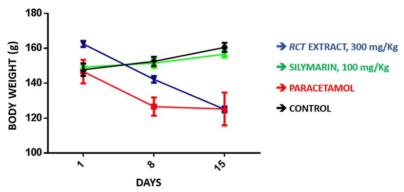
Figure 1. Curve of variation of the average weight of the animals.
During the experimental period, the weight loss of the rats of group n°4 treated with the RCT extracts is significant (p<0.05) compared to the control group and the group treated with silymarin.
Evaluation of the relative mean liver weight of rats
 p<0.05 compared to the corresponding control batch.
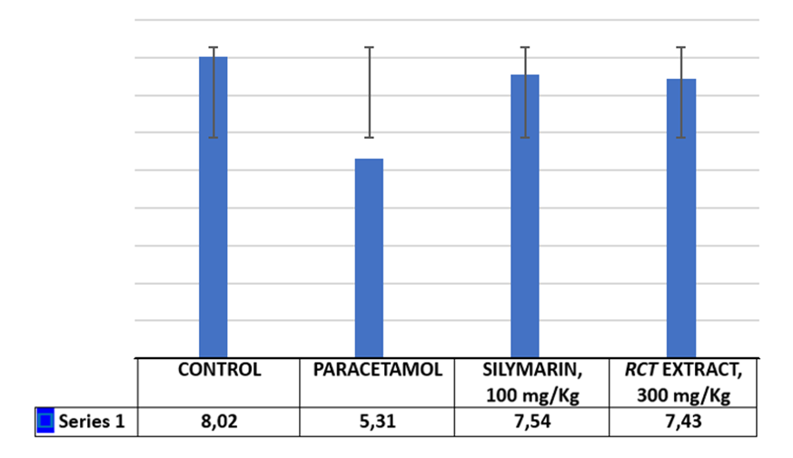
Figure 2. Effects of the hydro-ethanolic extract of RCT on the relative mean weight of the organs.
The statistical analysis showed us that there is:
- A significant decrease in the mean relative weight of the liver of all rats intoxicated with PCM (batch n°2, n°3 and n°4) compared to the control batch. On the other hand, the decrease in the mean relative weight of the liver of rats in the silymarin and extract batch is not significant compared to the PCM batch;
- A non-significant decrease in the mean relative weight of the liver of rats in the batch treated with RCT extracts compared to the silymarin batch;
- A significant decrease between the RCT extract batch compared to the PCM batch.
Effect of the extract on biochemical and hematological parameters
Biochemical parameters
Table 3. Effects of RCT extract on some blood biochemical markers of rat
|
Lots |
g-GT |
ASAT |
ALAT |
Total bilirubin |
|
2,05±0,47 |
92±24,25 |
52,33±6,64 |
1,71±0,27 |
|
18,67±0,88* |
179,33±16,18* |
104,67±9,39* |
86,67±6,12* |
|
7,27±0,23* |
119±4,18* |
75,67±4,18* |
6,67±1,45* |
|
12,13±1,21* |
97,67±4,33* |
97,67±4,33* |
11,13±1,95* |
*p<0.05 compared to the corresponding control group.
After two weeks of gavage, we observed an increase:
- Non-significant of γ-GT of all groups compared to the control group,
- Significant of transaminases of batches n°2 and n°4 compared to the control batch,
- Non-significant of transaminases of batch n°3 compared to batch n°2,
- Non-significant of total bilirubin of batches n°3 and n°4 compared to the control,
- Significant of total bilirubin of batch n°2 compared to the control batch.
Hematological parameters
Table 4. Effects of RCT extract on some hematological parameters of rats
|
Parameters evaluated |
Hematological parameters |
|||
|
Control |
Paracetamol |
Silymarin, 100 mg/Kg |
RCT Extract, 300 mg/Kg |
|
|
WBC (K/μl) |
7,77 ±0,06 |
2,56±0,67 |
5,44±0,49 |
4,33±0,40 |
|
RBC (M/μl) |
9,8±0,97* |
6,79±1,86* |
7,63±0,43* |
7,33±0,61* |
|
Hémoglobin (g/dl) |
16,1±0,70* |
8,13±0,69* |
13,17±0,84* |
13,37±1,51* |
|
MCV (fl) |
64±1,53* |
55,33±3,28* |
62,33±2,91* |
57,67±3,48* |
|
Platelets (M/μl) |
942,33±43,96* |
633,67±124,53* |
944±43,31* |
815,67±58,17* |
Mean values ± S.E.M. (n=6)
*p<0.05 compared to the corresponding control group
Paracetamol poisoning of rats resulted in a non-significant decrease compared to the control group in the levels of red blood cells, white blood cells and hemoglobin. The mean corpuscular volume did not vary in the different groups. Platelets significantly decreased in the group poisoned with paracetamol compared to the other groups.
Effects of the extract on the histological architecture of the liver under optical microscope
Histological images of rats in the control group
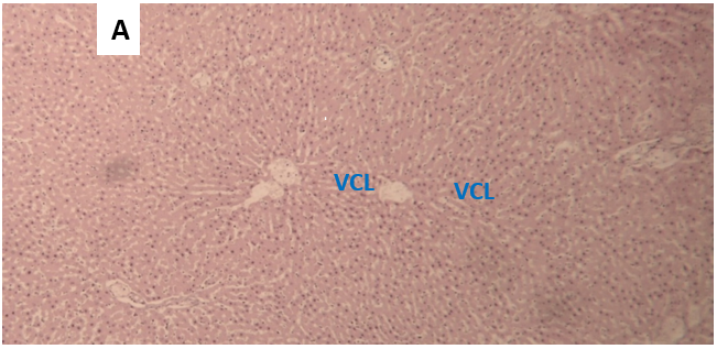
Figure 3. Photomicrograph of a histological section of the liver of a rat from the control group, transverse section, HE staining, magnification × 100.
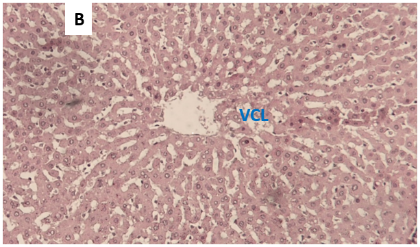
Figure 4. Photomicrograph of a histological section of the liver of a rat from the control group, transverse section, HE staining, magnification × 400.
Microphotographic observation B: The center of the field is occupied by a central vein in cross section (VCL). This vein is surrounded by cords of hepatocytes (arrow). These cords of hepatocytes are well separated by the weakly stained sinusoids.
Histological images of rats from the paracetamol group
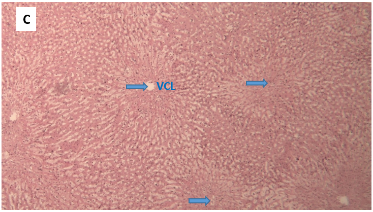
Figure 5. Microphotograph of a histological section of the liver of a rat from the paracetamol alone group, HE staining, transverse section, magnification × 100, blue arrows show tissue necrosis.
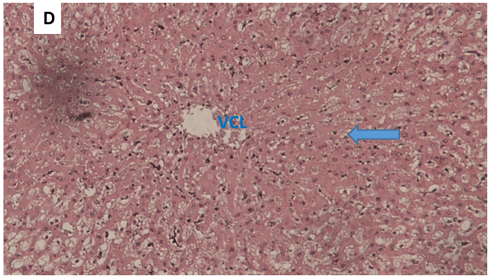
Figure 6. Microphotograph of a histological section of the liver of a rat in the paracetamol alone group, transverse section, HE staining, magnification × 400, the blue arrow indicates tissue necrosis.
Observation of microphotographs C and D: hepatocyte necrosis predominantly in the centrilobular region marked by a homogenization of the structure of the cytoplasm, a disappearance of the nuclei and cell boundaries. At a distance from the centrilobular vein, the structure of the hepatocytes remains relatively preserved.
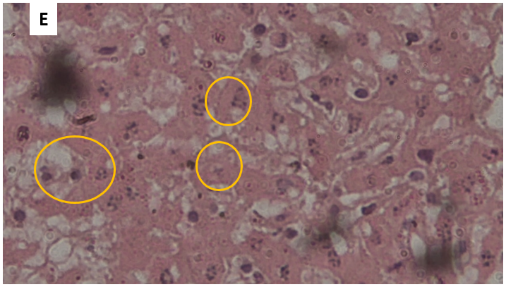
Figure 7. Microphotograph of a histological section of the liver of a rat from the paracetamol alone group, cross-section, HE staining, magnification × 1000.
Observation microphotograph E: the nuclei of the hepatocytes are at different stages of the necrosis process.
Histological images of rats from the silymarin group
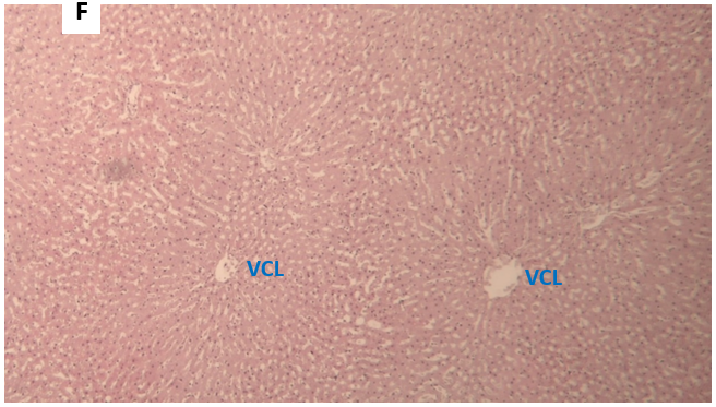
Figure 8. Photomicrograph of a histological section of the liver of a rat from the silymarin-treated group, transverse section, HE staining, magnification × 100.
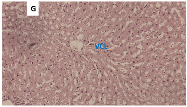
Figure 9. Microphotograph of a histological section of the liver of a rat from the silymarin-treated group, cross-section, HE staining, magnification × 400.
Observation of microphotographs F and G: Persistence of some signs of cellular damage limited to the first rows of hepatocytes closest to the centrilobular vein. The hepatic architectural structure is clearly recognizable.
Histological images of rats from the group extracted from the RCT
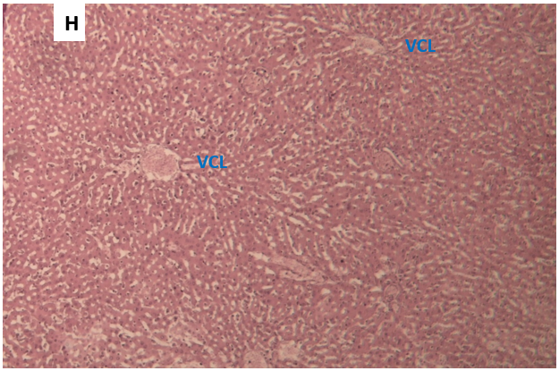
Figure 10. Photomicrograph of a histological section of the liver of a rat from the group treated with RCT extract, transverse section, HE staining, magnification × 100.
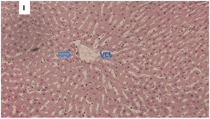
Figure 11. Microphotograph of a histological section of the liver of a rat from the group treated with RCT extract, transverse section, HE staining, magnification × 400.
Observation microphotographs H and I: Persistence of some signs of cellular suffering limited to the first rows of hepatocytes closest to the centrilobular vein. The hepatic architectural structure is well recognizable.
DISCUSSION
The main objective of this study is to study the hepatoprotective effects of the hydroethanolic extract of RCT in the wistar rat against PCM-induced toxicity. Previous studies have shown choleretic and hepatoprotective properties of the plant in vivo against carbon tetrachloride (CCl4) and aflatoxin B poisoning [14,19]. Literature searches did not find data on the protective effect of the liver against PCM-induced toxicity, despite its use against very popular liver diseases in West Africa. This motivated our study against liver damage due to PCM poisoning.
Yield of hydroethanolic extraction of cochlospermum tinctorium. A. Rich
Our yield of 13.47% with the hydroethanolic extraction of RCT is comparable to that of H. Abu et al in Nigeria [20] in 2012 who obtained a yield of 16.5% with a hydromethanolic extract. Furthermore, GUIGUEMDE Wendyam Armand in Burkina Faso [21] obtained in 2000 a yield of 13.47% by successive exhaustion with solvents of increasing polarity (petroleum ether, dichloromethane, dichloromethane-methanol, dichloromethanemethanol, methanol and water). This high yield probably illustrates the richness of this plant in secondary metabolites.
Phytochemical study of hydroethanolic extracts of RCT
Our hydroethanolic extract of RCT contains alkaloids, coumarins, tannins, flavonoids, triterpenes, anthracene derivatives, saponins and cardiac glycosides. The extract does not contain naphthoquinones according to the method used.
Our result is comparable to those of:
Nergard et al [22] who reported the presence in aqueous extracts of RCT of polyphenols, polysaccharides, gallotannins, ferullic acids. Collinlaw J. NDOUYANG et al [23] in Chad reported as secondary metabolites of RCT extracts tannins, phytates, oxalates, carotenoids, cyanides, alkaloids, flavonoids and phenols. Ahmed TS et al [8] in Nigeria in 2011, Diallo et al in 1989, 1991 and 1992 [24–26] in Mali reported the presence in different parts of the plant (leaves, stem, root) 3 families of bioactive molecules which are phenols/polyphenols, triterpenes and alkaloids. On the other hand, Akpemi Audu Musa in Nigeria reported the presence of anthraquinones (extracted by fractionation with 80% acetones) in addition to what was found [27]. Our data and those of the literature show the richness of this plant in secondary antioxidant, anti-inflammatory, immunomodulatory metabolites. Thus we can deduct from its properties a broad therapeutic possibility.
Animal mortality
We did not record any deaths in the control group and the silymarin-treated group. On the other hand, the paracetamol-intoxicated group showed a mortality of 66.66% (4 rats out of 6), which is higher than the group treated with RCT extracts with a mortality of 50% (3/6). This result is contrary to the results of other studies that have not reported deaths with CCl4 and Aflatoxin B poisoning [25,28–30].
Liver damage induced by different hepatotoxic agents is recognized as a major toxicological problem that leads to death. The toxicity of PCM is known, at high doses, it can induce fatal hepatic necrosis. PCM caused fatal damage to rats in groups 2 and 4. But despite the toxic effects, the silymarin-treated group did not experience any deaths. We can deduce that our extract had a protective effect but less compared to the batch treated with silymarin.
Effects of the extract on the evolution of the average body weight of rats
From the beginning to the end of the experiment, a significant decrease in the average weight of rats in the RCT extract batch was observed compared to the control batch. This observation is comparable to that of Collinlaw Joseph NDOUYANG and colleagues in Chad in 2021 for whom RCT flour slows down weight gain in young rats but without significant effect on growth [31,32]. RCT powder is widely used by the population of Northern Benin for culinary purposes without mention of nutritional data and the effects on weight growth [33].
Effect of the extract on biochemical and hematological parameters
At high doses, PCM is responsible for dose-dependent cytolysis and hepatocyte necrosis, initiated by the formation of its reactive metabolite, N-acetyl-p-benzoquinone imine (NAPQI), produced by cytochrome P450 2E1. Since the neutralization capacities of NAPQI by glutathione are exceeded, oxidative stress is produced, causing mitochondrial dysfunction, mediated by the activation of a cascade of cytosolic kinases and followed by DNA fragmentation. This is followed by an influx of inflammatory cells amplifying the production of cytokines and enzymes with cytolytic activity. These molecular mechanisms are at the origin of the release into the blood of early markers of liver toxicity. Among the markers of hepatic PMC intoxication sought, the elevations of γ-GT, transaminases, total bilirubin.
The non-significant increase in γ-GT and total bilirubin in the batch treated with RCT extracts and the batch treated with silymarin compared to the control batch shows that there was no major cholestasis. On the other hand, the significant increase in transaminases in batches No. 2, No. 3 and No. 4 indicates the presence of cytolysis in these groups. This cytolysis is more marked in batch 2 with in addition a significant increase in total bilirubin. The variations in the blood biochemical parameters of the rats show the protective effect of our RCT extract against PMC intoxication.
Our observations are similar to those of:
- Etuk E. U et al in 2009 in Nigeria who reported a significant decrease in transaminases and bilirubin compared to the group poisoned by carbon tetrachloride [34].
- B. Diallo and al in Mali in 1992 reported a dose-dependent decrease in transaminases of aqueous, hydro-ethanolic and ethanolic extracts of RCT against in-vivo CCl4 poisoning and in-vitro t-butyl-hydroperoxide poisoning [10].
- Mouzouvi R et al in Benin in 2014 reported a significant decrease in transaminases in patients with hepatitis B after treatment with a recipe combining Combretum micranthum G Don and cochlospermum tinctorium A Rich [35].
The results of the liver marker assays (ALAT, ASAT, total bilirubin) were in the same direction, since the reductions in the levels of the latter were better with the batch treated with the RCT extracts than with the batch treated with PCM alone.
Intoxication of rats with PCM resulted in a non-significant decrease in red blood cells, white blood cells and hemoglobin levels compared to the control group. On the other hand, platelets in the paracetamol-intoxicated group significantly decreased compared to the other groups. Treatment with the extracts did not result in significant changes in hematological parameters compared to the control group.
Effects of the extract on the histological architecture of the liver under optical microscope
The histological analysis of the liver of rats intoxicated by PMC correlated with the results of biochemical analyses. Indeed, the examination of liver sections showed that the poisoning of rats by PMC causes hepatic lesions, such as massive deformation of the architecture of the hepatic tissue and necrosis (Figures 1&2).
The liver of rats in the control group presents a normal lobular architecture marked by the presence of hepatocyte trabeculae arranged radially around a centrilobular vein. These trabeculae are separated by sinusoids (Figures 3-4).
In animals intoxicated with paracetamol, a mosaic aspect of the liver is noted, reflecting hepatic suffering, sinusoidal congestion, an inflammatory portal infiltrate, clarified and ballooned or necrotic hepatocytes with a predominance around the centrolobular vein, as well as microvacuolar steatosis and very significant vascular congestion (Figures 5-7).
For rats that received silymarin and RCT extracts, the liver lesions are less significant: the hepatic architecture remains recognizable but the congestion persists. Around the centrolobular veins, the hepatocytes are almost normal (Figures 8-11).
Treatment with RCT extracts reduces lesions and significantly protects hepatocyte morphology. This hepatoprotective effect of RCT extracts could be due to the antioxidant and anti-inflammatory properties. Indeed, several studies have reported that RCT extracts have antioxidant, anti-inflammatory and immunomodulatory effects [2,8,33,36,37]. All the hepatoprotective effects reported in this work may be due to the presence of bioactive compounds in the extracts studied. Indeed, phytochemical analysis revealed the presence of phenolic and polyphenolic compounds in the active fraction [36,37]. Several studies have demonstrated the hepatoprotective effect of the plant:
- The tannin content of RCT has shown remarkable anti-hepatotoxic activity and gallic acid in particular inhibits the production of oxygen free radicals [38].
- The polymers contained in the aqueous extract have been described as having antiulcer, immunomodulatory and antioxidant activities [22].
All of these molecules contained in RCT extracts increase the level of hepatic antioxidant enzymes such as catalase, Superoxide Dismutase (SOD), Glutathione Peroxidase (GPX), glutathione-S-transferase and decrease lipid peroxidation.
CONCLUSION
Hydroethanolic extracts of RCT protect the liver against PCM-induced aggressions. This activity could be attributed to the antioxidant and anti-inflammatory properties of this plant. The functional disorders and destruction of the hepatic parenchyma obtained after exposure to PCM are due to the induction of the generation of free radicals and the initiation of the inflammatory reaction.
The phytochemical study of RCT extracts revealed the presence of several phenolic compounds, terpenoids and steroids. These secondary metabolites, in the presence of other compounds present in the extracts, can improve the functions and condition of the hepatic parenchyma.
Several studies of the in vivo antioxidant activity of this plant have shown its ability to detoxify and protect liver tissue. The molecular mechanisms of liver protection by its constituents are not yet well studied. Indeed, research has shown its free radical scavenging abilities, inhibition of lipid peroxidation, protective effect against protein and DNA oxidation, anti-inflammatory effect and immunomodulatory effect.
The improvement of the histological structure of the liver, the restoration of liver enzyme levels and other biochemical indicators reflect the hepatoprotective effect of the studied plant against paracetamol toxicity, this effect would probably be due to the antioxidant and anti-inflammatory activities of these compounds. The limitations of our work are the short duration of the study and the small group of rats (6 per group) used due to the high mortality of paracetamol intoxication. There is great interest in studying the hepatoprotective effect of other types of RCT extracts and in conducting studies in a chronic intoxication model.
ACKNOWLEDGEMENTS
None.
CONFLICTS OF INTEREST
The authors declare that there are no conflicts of interest.
REFERENCES
- Telefo PB, Lemfack MC, Bayala B, Lienou LL, Goka CS, Yemélé D, et al. (2012). Enquête éthnopharmacologique des plantes utilisées dans le traitement de l’infertilité féminine dans les localités de Fossong-Wentcheng et Foto (Cameroun). Phytothérapie. 10(1). DOI : 10.1007/s10298-011-0678-6.
- Organisation Ouest Africaine de la Santé (OOAS). (2020). Pharmacopée de l’Afrique de l’Ouest. Burkina Faso : OOAS. 308 p [cité 22 déc 2024]. Disponible sur: https://www.wahooas.org/web-ooas/fr
- Adjanohoun E, Adjakidjè V, Ahyi MA, et al. (1989). Contribution Aux Etudes Ethnobotaniques et Floristiques en République Du Bénin,” Agence de Coopaeration Culturelle et Technique, Niamey. p. 895. ISBN: 92-9028-152-9.
- Dossa AK, Klotoe JE, Agbodjento E, Dougnon V, Sinkou JS, Loko F. (2021). Usages de poudres à base de rhizomes de Cochlospermum tinctorium Perrier ex A. Rich au Bénin : fréquence, formes et indications. Int J Biol Chem Sci. 15(4):1511-1523.
- Bouquet A, Debray M. (1974). Plantes Medicinales de Cote d’Ivoire. O.R.S.T.O.M. Office de la recherche scientifique et technique outre-mer, Paris. 231 p.
- Kerharo J, Bouquet A. (1950). Plante médicinales et toxiques de la Côte d’Ivoire-Haute Volta. Office de la Recherche Scientifique Outre-Mer. Paris : Plante médicinales et toxiques de la Côte d’Ivoire-Haute Volta. 295 p.
- Adjanohoun EJ. (1985). Contribution aux études ethnobotaniques et floristiques au Mali. ACCT. Paris : Agence de coopération culturelle et technique. 249 p. ISBN: 92-9028-016-6.
- Ahmed Ts, Magaji M, Yaro A, Musa A, Adamu A. (2011). Aqueous Methanol Extracts of Cochlospermum tinctorium (A. Rich) Possess Analgesic and Anti-inflammatory Activities. J Young Pharm. 3(3):237-242.
- Dalvi RR, Séré A. (1988). Protective Effect of Cochlospermum tinctorium A. Rich Extract versus Aflatoxin B Induced Liver Damage in Rats. International Journal of Crude Drug Research. 26(2):117‑120.
- Diallo B, Vanhaelen-Fastre R, Vanhaelen M, Fiegel C, Joyeux M, Roland A, et al. (1992). Further studies on the hepatoprotective effects of Cochlospermum tinctorium rhizomes. J Ethnopharmacol. 36(2):137-142.
- Islam MT, Quispe C, Islam MA, Ali ES, Saha S, Asha UH, et al. (2021). Effects of nerol on paracetamol-induced liver damage in Wistar albino rats. Biomed Pharmacother. 140:111732.
- Senthilkumar R, Chandran R, Parimelazhagan T. (2014). Hepatoprotective effect of Rhodiola imbricata rhizome against paracetamol-induced liver toxicity in rats. Saudi J Biol Sci. 21(5):409-416.
- Mégarbane B. (2016). Intoxication par le paracétamol : mécanismes de toxicité, facteurs prédictifs et modalités de prise en charge. Toxicol Anal Clin. 28(3):240.
- Dalvi RR, Séré A. (1988). Protective Effect of Cochlospermum tinctorium A. Rich Extract versus Aflatoxin B Induced Liver Damage in Rats. Int J Crude Drug Res. 26(2):117‑120.
- Gillessen A, Schmidt HH. (2020). Silymarin as Supportive Treatment in Liver Diseases: A Narrative Review. Adv Ther. 37(4):1279-1301.
- Gür FM, Bilgiç S. (2023). Silymarin, an antioxidant flavonoid, protects the liver from the toxicity of the anticancer drug paclitaxel. Tissue Cell. 83:102158.
- Wagner H, Bladt S. (1996). Plant Drug Analysis: A Thin Layer Chromatography Atlas. Springer Science & Business Media. 373 p.
- Diallo D, Sanogo R, Yasambou H, Traoré A, Coulibaly K, Maïga A. (2004). Étude des constituants des feuilles de Ziziphus mauritiana Lam. (Rhamnaceae), utilisées traditionnellement dans le traitement du diabète au Mali. Comptes Rendus Chim. 7(10):1073‑1080.
- Sohn DH, Kim YC, Oh SH, Park EJ, Li X, Lee BH. (2003). Hepatoprotective and free radical scavenging effects of Nelumbo nucifera. Phytomedicine Int J Phytother Phytopharm. 10(2‑3):165‑169.
- Abu AH. (2012). Aqueous ethanolic extract of Cochlospermum planchonii rhizome enhances spermatogenesis in male albino rats. Afr J Biotechnol. 11(53).
- Guiguemde WA. (2000). Contribution à l'identification des principes antiplasmodiques de Cochlospermum tinctorium a. Rich (cochlospermaceae) (Thèse). Ouagadougou : Faculté des sciences de la santé (FSS) de l’Université de Ouagadougou. 59 p. Available at: https://beep.ird.fr/collect/uouaga/index/assoc/M08498.dir/M08498.pdf
- Nergard CS, Diallo D, Inngjerdingen K, Michaelsen TE, Matsumoto T, Kiyohara H, et al. (2005). Medicinal use of Cochlospermum tinctorium in Mali Anti-ulcer-, radical scavenging- and immunomodulating activities of polymers in the aqueous extract of the roots. J Ethnopharmacol. 96(1‑2):255‑269.
- Ndouyang CJ, Himeda M, Nguimbou RM. (2012). Antinutriments et propriétés nutritionnelles in vivo de Cochlospermumtinctorium A. Rich. (Bixaceae) chez les jeunes rats (Rattusnorvegicus L.). Int J Biol Chem Sci. 12(2):884-901.
- Diallo B, Vanhaelen M, Vanhaelen-Fastré R, Konoshima T, Kozuka M, Tokuda H. (1989). Studies on inhibitors of skin-tumor promotion. Inhibitory effects of triterpenes from Cochlospermum tinctorium on Epstein-Barr virus activation. J Nat Prod. 52(4):879-881.
- Diallo B, Vanhaelen-Fastre R, Vanhaelen M, Fiegel C, Joyeux M, Roland A, et al. (1992). Further studies on the hepatoprotective effects of Cochlospermum tinctorium rhizomes. J Ethnopharmacol. 36(2):137‑142.
- Diallo B, Vanhaelen-Fastré R, Vanhaelen M. (1991). Triacylbenzenes and long-chain volatile ketones from Cochlospermum tinctorium rhizome. Phytochemistry. 30(12):4153‑4156.
- Musa AK, Musa AS. (2012). Cytotoxicity Activity and Phytochemical Screening of Cochlospermum tinctorium Perr Ex A. Rich Rhizome. Journal of applied pharmaceutical science. 02(07):155-159.
- Dalvi RR, Séré A. (1988). Protective Effect of Cuchlospermum tinctorium A. Rich Extract versus Aflatoxin B1 induced Liver Damage in Rats. Int J Crude Drug Res. 26(2):117-120.
- Etuk EU, Agaie BM, Ladan MJ, Garba I. (2009). The modulatory effect of Cochlospermum tinctorium a rich aqueous root extract on liver damage induced by carbon tetrachloride in rats. African Journal of Pharmacy and Pharmacology. 3(4):151-157.
- Kecira M, Zitouni M, Bekkouche A, Kebsa wided (Encadreur). (2012). Evaluation de l’effet hépatoprotecteur de l’extrait brut de la propolis contre la toxicité de l’aflatoxine b1 chez les rats. université de Jijel. Available at: http://dspace.univ-jijel.dz:8080/xmlui/handle/123456789/6049
- Ndouyang CJ, Himeda M, Nguimbou RM. (2018). Antinutriments et propriétés nutritionnelles in vivo de Cochlospermum tinctorium A. Rich. (Bixaceae) chez les jeunes rats (Rattus norvegicus L.). Int J Biol Chem Sci. 12(2):884.
- Ndouyang CJ, Gaiani C, Scher J. (2021). Bouillie thérapeutique infantile à base de Tacca leontopetaloides (L.) Kuntze (Taccaceae) et de Cochlospermum tinctorium A. Rich. (Bixaceae).
- Dossa AK, Klotoe JR, Agbodjento E, Dougnon V, Sinkou JS, Loko F. (2021). Usages de poudres à base de rhizomes de Cochlospermum tinctorium Perrier ex A.Rich au Bénin : fréquence, formes et indications. Int J Biol Chem Sci. 15(4):1511‑1523.
- Etuk EU, Agaie BM, Ladan MJ, Garba I. The modulatory effect of Cochlospermum tinctorium a rich aqueous root extract on liver damage induced by carbon tetrachloride in rats.
- Mouzouvi R, Djègo JG, Sehonou J, Lalèyè A, Priuli F, Bigot A. (2014). Effet de l’association Combretummicranthum G Don (Combretaceae) et Cochlospermum tinctorium A Rich (Cochlospermaceae) dans la prise en charge de l’hépatite virale B. Série Pharm Méd Trad Afr. 17(1):10-14.
- Gnimansou AF, Gbèwonmèdéa D, Aristide CA, Adandé BF, et al. (2020). Cochlospermum planchonii Hook.f. ex Planch. and Cochlospermum tinctorium Perrier ex A. Rich.: extent of knowledge and prospects for sustainable use in West Africa. Genet Resour Crop Evol. 68:25-44.
- Ahmad MH, Jatau AI, Khalid GM, Alshargi OY. (2021). Traditional uses, phytochemistry, and pharmacological activities of Cochlospermum tinctorium A. Rich (Cochlospermaceae): a review. Future J Pharm Sci. 7(1):20.
- Ballin NZ, Traore M, Tinto H, Sittie A, Mølgaard P, Olsen CE, et al. (2002). Antiplasmodial compounds from Cochlospermum tinctorium. J Nat Prod. 65(9):1325‑1327.