Information Links
Related Conferences
Current Issue Volume 9, Issue 1 - 2026
Resolving Canal Obstruction: Excision of a Pedunculated External Auditory Canal (EAC) Osteoma
Pankaj Goyal1,*, Kishan Kumawat1, Manisha Chouhan2
1Apollo ENT Hospital, Pal road, Jodhpur, Rajasthan, India
2Medipulse Hospital, Jodhpur, Rajasthan, India
*Corresponding Author: Pankaj Goyal, Apollo E.N.T. Hospital, pal road, Jodhpur, Rajasthan, India, ORCID: 0000-0003-4098-7308, Email: [email protected]
Received Date: January 19, 2026
Published Date: February 26, 2026
Citation: Goyal P, et al. (2026). Resolving Canal Obstruction: Excision of a Pedunculated External Auditory Canal (EAC) Osteoma. Mathews J Surg. 9(1):42.
Copyrights: Goyal P, et al. © (2026).
ABSTRACT
Osteoma of the external auditory canal is a rare, benign, slow-growing osseous tumor that is usually asymptomatic. Progressive enlargement may cause canal obstruction, conductive hearing loss, or recurrent otitis externa. We report a case of a middle-aged patient presenting with ear blockage and hearing impairment. Otoscopic examination showed a solitary, hard, pedunculated mass arising from the bony external auditory canal. High-resolution computed tomography of the temporal bone revealed a well-defined hyperdense lesion suggestive of osteoma. Complete surgical excision was performed via a transcanal approach. Excellent prognosis and prevention of increasing canal obstruction are provided by surgical removal.
Keywords: Osteoma, External Auditory Canal, Benign Tumors, Hearing Loss, Surgical Excision.
INTRODUCTION
Osteomas of the EAC are benign, slow-growing osseous neoplasms that typically originate from the tympanosquamous or tympanomastoid suture lines [1]. With an estimated prevalence of less than 0.05% in otologic practice, they are among the rarest benign neoplasms of the EAC [2]. In contrast to exostoses, which are often multiple,bilateral, broad-based lesions strongly linked to recurrent exposure to cold water, EAC osteomas are usually solitary, unilateral, well-circumscribed, and pedunculated [2,3]. Though suggestions about congenital origins, persistent irritation, trauma, and developmental abnormalities have been put up, none of them have been proven to be the cause of osteomas [4].
Because of their slower growth process, EAC osteomas are frequently asymptomatic in clinical settings [5]. During a normal otologic examination, many cases are unintentionally found. But when the lesion grows, it could block the ear canal, resulting in symptoms like conductive hearing loss, water trapping, recurrent otitis externa, or cerumen impaction [6]. Additionally, obstruction disrupts the external canal's natural self-cleaning process, putting patients at risk for persistent irritation and debris buildup [7]. Therefore, rather than the tumor itself being present, clinical manifestation typically corresponds with progressive luminal narrowing [8].
Because it clearly shows the lesion's density, attachment site, and extent within the canal, high-resolution computed tomography (HRCT) of the temporal bone has become crucial for assessing EAC osteomas [9]. In order to distinguish osteomas from other bony lesions such exostoses or osteosclerotic diseases, HRCT findings usually reveal a dense, pedunculated mass emerging from a focal site on the canal wall [10]. Surgical decision-making is aided by early imaging, especially when there is a substantial obstruction or the lesion is near the tympanic membrane or temporomandibular joint [11].
The size of the lesion and its symptoms determine the management approach. Surgical removal is the preferred course of treatment when the mass results in functional impairment, recurring infections, or diagnostic ambiguity, even if small, asymptomatic osteomas may be seen with routine follow-up [12]. Because the pedicle is usually thin and well-defined, surgical removal—most frequently using a transcanal technique for tiny pedunculated lesions—offers excellent results with minimal recurrence rates [13]. Because osteomas exhibit compact, lamellar bone architecture that sets them apart from other benign or inflammatory bone diseases, histopathological confirmation is crucial [14].
CASE PRESENTATION
A 32-year-old man had been experiencing progressive conductive hearing loss and right ear obstruction for the past eight months. The patient's condition was consistent with the fact that osteomas usually only cause symptoms when they restrict the canal lumen.
Otoscopy (Figure 1a and 1b) showed a solitary pedunculated bony mass arising from the anterior wall of the external auditory canal (Figure 1), consistent with typical osteoma morphology described in literature [1]. Audiometry revealed a 20 dB air–bone gap.
A distinct, single, pedunculated osseous mass originating from the lateral bony canal—typically near the tympanomastoid suture line—was visible on High resolution Computed Tomography (HRCT) of the temporal bone (Figure 2a and 2b). The lesion is a dense osseous lesion connected by a thin pedicle, appearing as a uniform, hyperdense core of compact bone with smooth, well-corticated borders. Imaging shows that the middle and inner ear structures are normal, and there is no bony erosion, invasive behavior, or soft-tissue component.
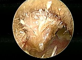
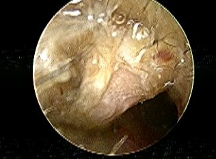
Figure 1a Figure 1b
Figure 1(a & b). Clinical picture of Right External Auditory Canal(E.A.C.) osteoma.
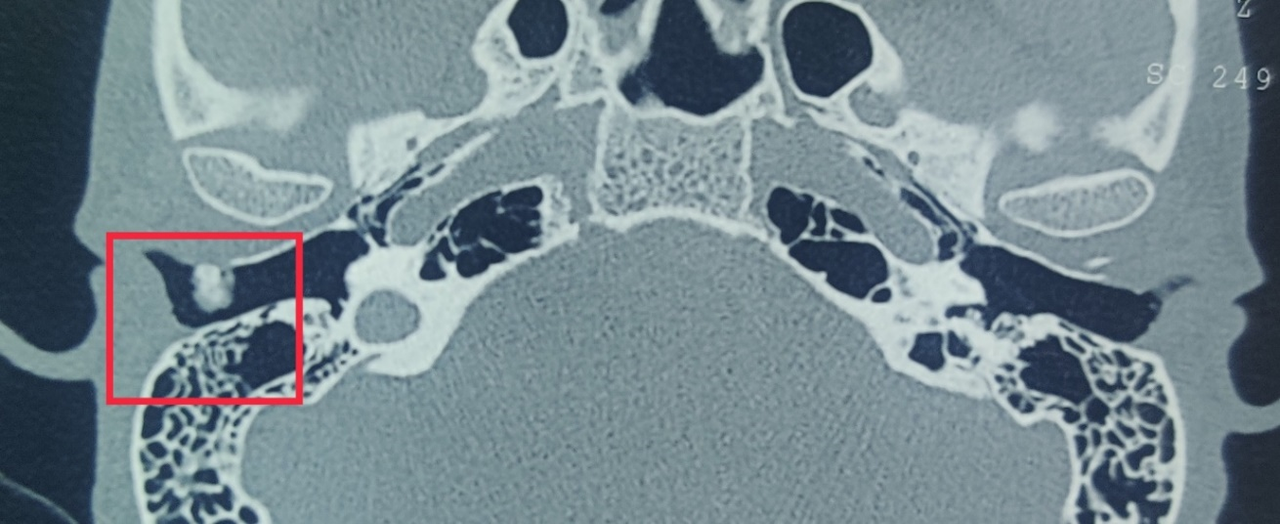
Figure 2a
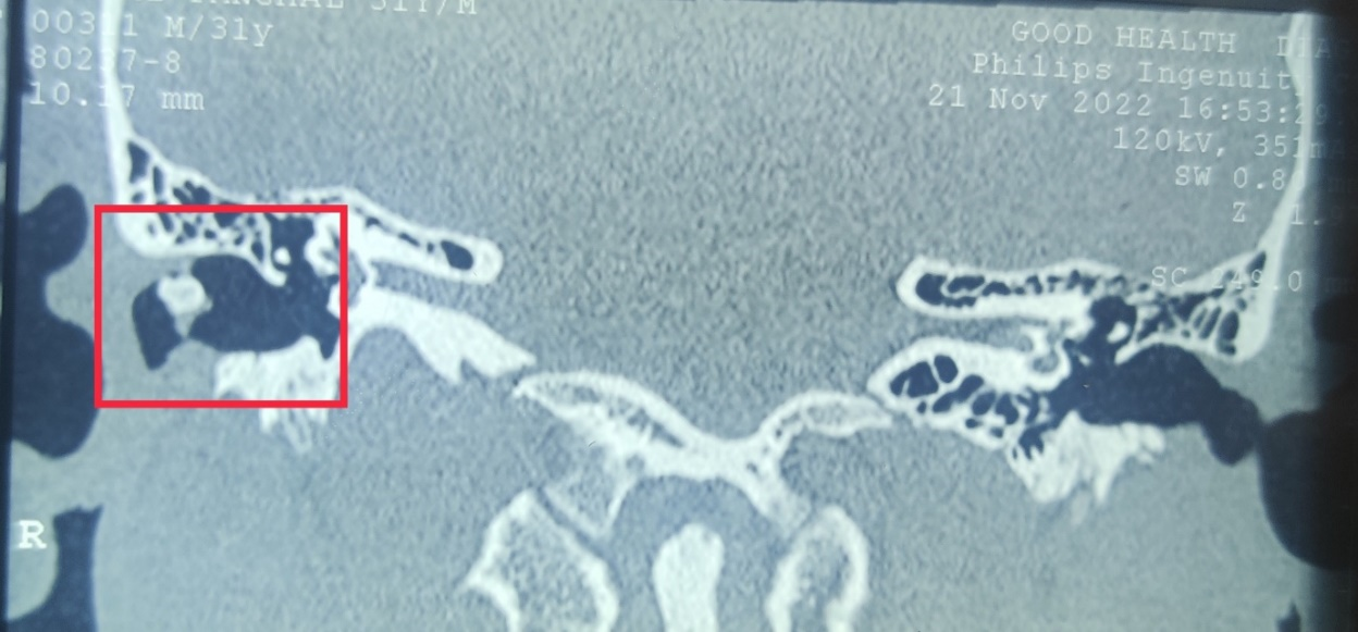
Figure 2b
Figure 2(a & b). HRCT temporal bone showing right EAC osteoma.
Management
When the osteoma starts to produce symptoms, recurrent infections, or canal obstruction, surgical removal is advised. For this patient, a transcanal excision was scheduled in accordance with this idea. An osteotome was used to remove the osteoma after the canal skin was raised circumferentially while under local anesthetic. The osteoma was completely removed and sent for histopathology (Figure 3). The underlying bone was smoothed with a diamond burr. Packing was put in place and the canal skin was moved.
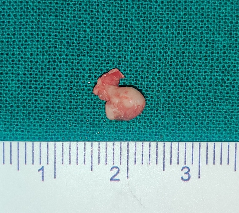
Figure 3. Main histopathological specimen.
Outcome
The postoperative course was uneventful, with early recovery, complete resolution of symptoms, and no complications or recurrence on follow-up. Following the restoration of canal patency, audiometry revealed that the conductive gap had closed. At six months, there was no recurrence, which is in line with research showing that recurrence following total pedicle removal is incredibly uncommon. In line with earlier documented occurrences, a histopathological investigation revealed solid lamellar bone with little fibrous tissue, confirming compact osteoma [1].
DISCUSSION
External auditory canal (EAC) osteomas are uncommon, benign osseous tumors that grow slowly from the tympanosquamous or tympanomastoid suture lines [1]. In contrast to exostoses, which are many, bilateral, broad-based, and strongly linked to repeated exposure to cold water (also known as "surfer's ear"), they are usually solitary, unilateral, and pedunculated lesions. Two. While the precise cause of EAC osteomas is still unknown, several theories have been put forth, including developmental abnormalities, trauma, persistent irritation, and congenital factors [4].
Clinically, because EAC osteomas grow slowly, they are frequently asymptomatic in the early stages. Significant canal obstruction caused by the osteoma typically results in cerumen impaction, water trapping, recurrent otitis externa, or conductive hearing loss [6]. As the bone mass grows, it could make it harder to see the tympanic membrane or disrupt the EAC 's self-cleaning systems [5]. In line with earlier reports highlighting functional impairment as the primary justification for surgical intervention, our patient's 70% canal obstruction led to a discernible air–bone gap and episodic occlusion [7].
The preferred imaging technique for osteoma diagnosis is high-resolution computed tomography (HRCT). A well-defined, hyperdense lesion connected to the canal wall by a thin pedicle is consistently shown by HRCT [10]. Osteomas can be distinguished from exostoses, which manifest as numerous dense outgrowths with wide bases, thanks to this radiological clarity [2]. Additionally, by pinpointing the precise location of the lesion's attachment and its closeness to the tympanic membrane or temporomandibular joint, CT imaging is essential for preoperative planning [10].
The severity of symptoms and canal obstruction determine how EAC osteomas are managed. Periodic follow-up can detect asymptomatic lesions; nevertheless, surgical excision is advised for lesions that are symptomatic, obstructive, or cosmetically problematic [12]. Because it offers direct access with little morbidity, a transcanal technique is perfect for tiny or pedunculated osteomas [13]. Endaural or postauricular techniques may be necessary for the full excision of larger or broad-based osteomas [15]. Because of the tiny pedicle origin in this instance, the transcanal approach permitted total excision.
To avoid canal stenosis or damage to nearby structures, it is crucial to carefully elevate the canal skin and remove the bony pedicle during surgery [16]. In order to avoid lingering imperfections that could result in long-term discomfort, many authors advise utilizing small osteotomes for detachment and diamond burrs to smooth the canal wall afterwards [17]. This typical approach was followed by our surgical procedure, which produced good functional and cosmetic results.
The diagnosis of osteoma is usually confirmed by histopathological investigation, which shows dense lamellar or compact bone with little fibrovascular stroma [14]. Because osteomas are pedunculated and originate from distinct attachment locations, recurrence following total surgical removal is extremely rare [18]. Long-term results following surgery are outstanding in published series, with the majority of patients regaining normal canal patency and hearing function [19]. This is consistent with our findings because, at six months, the patient had fully recovered and had not experienced a recurrence.
CONCLUSION
Even though these tumors are rare, EAC osteomas should be taken into account in individuals who have prolonged ear obstruction feelings, chronic cerumen impaction, or unexplained conductive hearing loss. While thorough excision using the proper technique guarantees great long-term results, HRCT is essential for diagnosis and surgical planning.
COMPLIANCE WITH ETHICAL STANDARDS
The procedure performed in this case report was in accordance with the ethical standards of the institutional and/or national research committee and with the 1964 Helsinki declaration and its later amendments or comparable ethical standards.”
FUNDING
This study is not funded by any resources.
CONFLICT OF INTEREST
The author(s) declare no potential conflicts of interest with respect to the research, authorship, and/or publication of this paper.
ETHICAL APPROVAL
The study was published with written consent of the patient.
REFERENCES
- Swartz JD, Hagiwara M. (2006). Lesions of the external auditory canal. Radiol Clin North Am. 44(5):743-754.
- Hetzler D. (2007). Differentiating exostoses from osteomas: Radiologic and clinical pearls. Otol Neurotol. 28(6):758-763.
- House JW, Wilkinson EP. (2003). External auditory exostoses: Evaluation and treatment. Otolaryngol Clin North Am. 36(2):391-402.
- Magliulo G. (2000). Osteoma of the external auditory canal: Etiology and clinical features. J Otolaryngol. 29(6):360-365.
- Van Caulaert J, Somers T. (2011). External auditory canal osteoma and its impact on ear physiology. Acta Otolaryngol. 131(9):997-1002.
- Albright JT, Lesperance MM. (2007). External auditory canal osteomas: Clinical presentation and surgical results. Otolaryngol Head Neck Surg. 137(4):611-615.
- Prasad SC, Meyers AD. (2014). Indications for removal of external auditory canal osteomas. Ear Nose Throat J. 93(3):92-96.
- Choi JS, Lee S. (2017). Long-term outcomes of external auditory canal osteomas. J Laryngol Otol. 131(10):895-899.
- Iwasaki S, Sano H. (2010). CT evaluation of external auditory canal osteomas and exostoses. Auris Nasus Larynx. 37(3):348-353.
- Zhen Y, Li X. (2015). Role of CT in surgical planning for EAC osteomas. Clin Imaging. 39(2):338-342.
- Goh EK, Park CI. (2001). Radiologic assessment for EAC tumors: Implications for surgery. Am J Otolaryngol. 22(5):294-300.
- Blevins NH, Owens RM. (2002). Management of symptomatic osteomas of the external auditory canal. Laryngoscope. 112(6):1115-1120.
- Kim CW, Lee HK. (2013). Transcanal excision of external auditory canal osteomas: Outcomes and techniques. Clin Exp Otorhinolaryngol. 6(4):233-237.
- Dahl I. (1977). Histopathologic features of external auditory canal osteoma. Arch Otolaryngol. 103(3):152-155.
- Goh EK, Park CI. (2000). Postauricular approach for large EAC osteomas. Am J Otolaryngol. 21(4):263-267.
- Kania RE. (2001). Surgical considerations in EAC osteoma excision. Otolaryngol Head Neck Surg. 125(6):627-631.
- Naim R, Linthicum FH. (2005). Canaloplasty techniques in osteoma surgery. Otol Neurotol. 26(4):626-631.
- Somers T, Casselman J. (2010). Recurrence of EAC osteoma after surgical excision: A long-term review. Eur Arch Otorhinolaryngol. 267(1):37-43.
- Choi JS, Lee S. (2017). Long-term outcomes following surgical excision of EAC osteomas. J Laryngol Otol. 131(10):895-899.
