Information Links
Related Conferences
Previous Issues Volume 7, Issue 4 - 2023
Progressive Cutaneous Angiomatosis in a Persian Cat: Case Report
Daniela de Alcantara Leite dos Reis1, André Rinaldi Fukushima2,3,4*, Simone Rodrigues Ambrosio1, Priscyla Taboada Dias da Silva1,5, Mariane Borges da Silva1, Larissa da Silva Costa1, Rodrigo Casemiro Pinto Monteiro1,6, Jordana Casemiro Pinto Monteiro1,6
1Médica Veterinária, Brazil
2Universidade de São Paulo Faculdade de Medicina Veterinária e Zootecnia, Brazil
3Centro Universitário das Américas–FAM, Brazil
4Faculdade de Ciências da Saúde–IGESP, Brazil
5Histopet, Brazil
6Universidade São Judas Tadeu-USJT, Brazil
*Corresponding author: André Rinaldi Fukushima, School of Veterinary Medicine and Animal Science, University of São Paulo, São Paulo, Brazil, Tel: (11) 98133-7311, ORCID: 0000-0001-6026-3054; E-mail: [email protected].
Received Date: October 12, 2023
Published Date: November 02, 2023
Citation: Reis DDALD, et al. (2023). Progressive Cutaneous Angiomatosis in a Persian Cat: Case Report. Mathews J Vet Sci. 7(4):27.
Copyrights: Reis DDALD, et al. © (2023).
ABSTRACT
Angiomatosis is a vascular disorder characterized by proliferative granulation tissue formation, with the growth of angioblastic cells surpassing that of fibroblasts, resulting in inflammatory connective tissue with a tendency for spontaneous bleeding. It is a rare pathology in veterinary medicine, and its pathogenesis remains poorly understood. Therefore, the objective of this study is to report a case of angiomatosis where the diagnostic confirmation was achieved through histopathological examination. A female Persian cat, one year old, was presented with lameness, marked edema, and active bleeding in the distal region of the left thoracic limb, with symptoms persisting for several months. Physical examination revealed ulcerated skin lesions with bloody crusts, areas of hyperpigmentation, and extensive hematomas. An incisional biopsy was performed, and the samples were sent for histopathological examination. Microscopic evaluation revealed a low-grade vascular proliferation involving the superficial, perianexial, and deep dermis. The cells exhibited oval nuclei with mild anisocaryosis, stippled chromatin, and an absence of mitotic figures, consistent with the morphological appearance of progressive cutaneous angiomatosis. Treatment involved limb amputation since the patient had previously been treated unsuccessfully with corticosteroid anti-inflammatories and antibiotics. In conclusion, due to the rarity of this disease in the veterinary field, only histopathological analysis can lead to an accurate diagnosis. The treatment employed in this case was deemed satisfactory, with no disease recurrence in the patient thus far.
Keywords: Progressive Angiomatosis, Skin, Endothelium.
INTRODUCTION
Angiomatosis and vascular tumors, such as hemangiosarcoma, lymphangiosarcoma, hemangioma, and vascular hematomas, are characterized by the proliferation of endothelial cells [1]. Angiomatosis is an alteration in vascular tissue resulting from neovascularization caused by angiogenic growth factors or vascular anomalies, inflammatory reactions, viral or bacterial infection, hyperplasia, and lacks neoplastic characteristics [1]. In angiomatosis, there can be the formation of proliferative granulation tissue, with the growth of angioblastic cells surpassing that of fibroblasts. It is an inflammatory connective tissue with a tendency for spontaneous bleeding, which may or may not have an associated secondary infection [2,3].
In Brazil, there are still few reported cases in animals; however, cases have been described in dogs and a captive llama. Worldwide, there are also reports in cats and cattle [3]. Angiomatosis is a rare pathology in veterinary medicine, and its pathogenesis is not yet well understood [4].
In reports, angiomatosis lesions can occur in the skin, intestines, viscera, vertebrae, ovaries, and meninges [5], often associated with pain and bleeding [6].
The best diagnostic method is histopathological examination, due to its ability to differentiate cells, especially to distinguish them from neoplastic cells. Therapeutic options are limited, but there are reports of treatments with laser therapy and surgery [6,7].
The scenario in which angiomatosis presents itself as a pathology rarely addressed in veterinary medicine and with aspects still not fully understood, this report seeks to expand the understanding of such a condition. The main objective of this study is to present a clinical case of progressive cutaneous angiomatosis in a Persian cat, emphasizing its clinical manifestation, diagnostic challenges, and the therapeutic approach adopted, in order to contribute to a better recognition and management of the disease in future veterinary practices.
CASE REPORT
Physical Examination
A Persian cat, approximately one year old, weighing 3.8 kg, was referred for evaluation. The owner reported injuries to the left forelimb in the metacarpal and phalangeal regions, accompanied by progressive bleeding and lameness. The owner mentioned a persistent increase in the volume of the soft tissue adjacent to the metacarpal and phalangeal areas and bleeding from the paw pads and digits, intensified over the last two months. The patient had previously been treated by other veterinarians with non-steroidal anti-inflammatory drugs and antimicrobials, but with little response to treatment. On physical examination, the animal presented edema in the distal region of the limb with continuous bleeding from the metacarpal, phalangeal, and paw pad areas showed in a figure 1a, 1b, 1c, and 1d the palmar and dorsal views of the left forelimb at the time of physical examination and after shaving.
.png)
Figure 1. Palmar view (a) and dorsal view (b) of the left thoracic limb at the time of the physical examination, showing active bleeding, soft tissue edema, and bloody crusts adhered to the fur. The same limb, after shaving, in the palmar view (c) and dorsal view (d), revealing areas of hyperpigmentation and extensive hematomas (arrows).
Source: Author's Own
X-ray Findings
Radiography of the region, including the carpal joint and phalanges, showed only an increase in the volume of the soft tissue, suggesting an inflammatory process, with no observed bone abnormalities showed in the figure 2, the dorsopalmar projection radiography of the carpal, metacarpal, and phalangeal region.
.png)
Figure 2. Radiography in the dorsopalmar projection of the carpal, metacarpal, and phalangeal region reveals a pronounced increase in soft tissue volume adjacent to the metacarpal and phalangeal regions (arrows), with no evidence of radiographic changes in the bone structures.
Source: Own authorship
Histological Studies
Due to the high number of cases in this region, the initial clinical suspicion was sporotrichosis, with a differential diagnosis of possible neoplasia. Cytology samples were collected, tests for FeLV and FIV were also performed, as well as complete blood count and blood biochemistry.
The patient was referred for incisional biopsy of the skin and subcutaneous tissue. Subsequently, the collected fragments were sent for histopathological examination, bacterial and fungal culture, showed in the figure 3 and 4 Photomicrographs of sections from the biopsied sample.
.png)
Figure 3. Photomicrographs of sections from the biopsied tissue sample, (a) proliferation of vessels of various calibers located in the dermis and hypodermis (arrows), (b) proliferation of vessels of various calibers without signs of atypia located in the hypodermis (arrows). Hematoxylin & Eosin staining. Objective 40x.
Source: Histopet
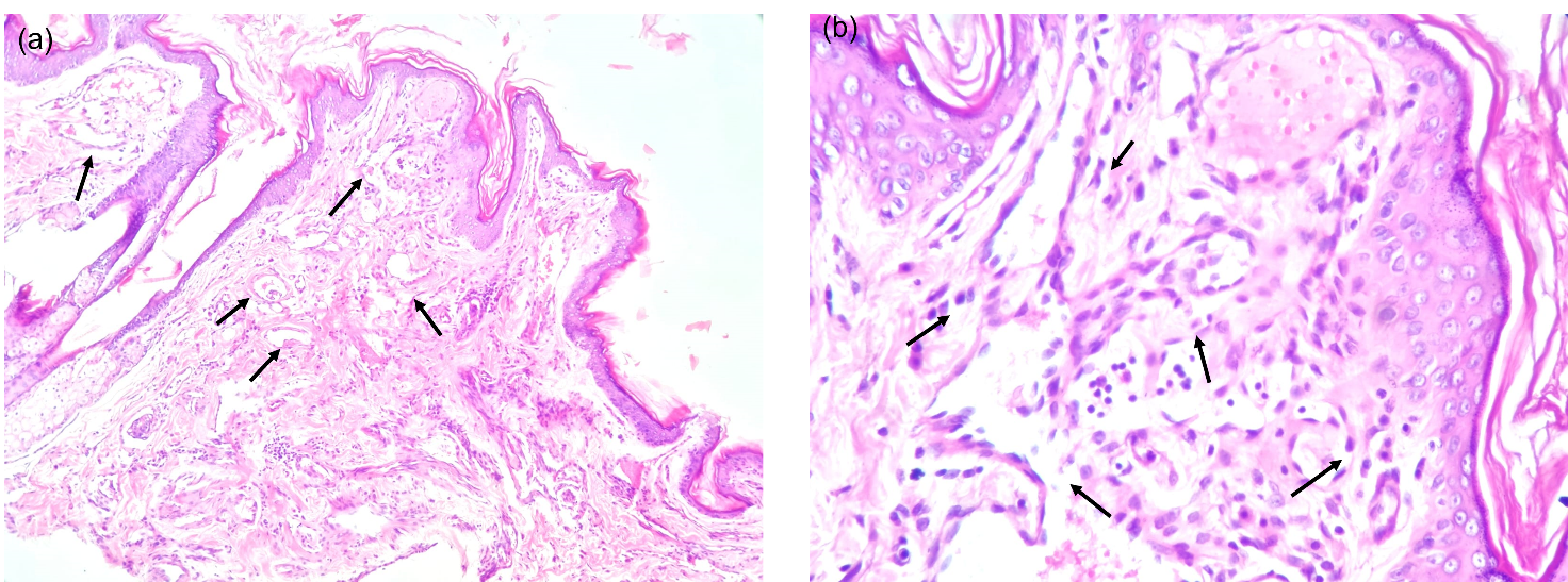
Figure 4 Photomicrographs of sections from the biopsied tissue sample, (a) proliferation of vessels of varying calibers located in the superficial dermis and perianexial region (arrows), (b) proliferation of vessels in the superficial dermis without signs of atypia (arrows). Hematoxylin & Eosin staining. Objective 10x (a) and 40x (b).
Source: Histopet
Based on the physical evaluation, radiography, and histopathological findings, the patient was diagnosed with angiomatosis.
The animal was scheduled for the surgical procedure of amputation of the left forelimb. Post-surgical medication included various drugs, and the animal returned daily for post-operative monitoring and bandage changes.
One day after the procedure, the patient returned showing signs of pain. Ten days after the amputation surgery, the skin sutures were removed, and the patient resumed her activities.
DISCUSSION
Cutaneous angiomatosis is characterized as a progressive proliferative vascular disorder that affects the dermis and subcutaneous layers. This pathology can be observed in both dogs and cats, being more prevalent in young individuals [8-10]. In the context of veterinary medicine, this disease is of rare occurrence, with limited data on its clinical behavior and available therapeutic options.
The term "angiomatosis" is used to designate a heterogeneous group of non-neoplastic vascular diseases that can affect cutaneous and visceral tissues in a variety of species [3,11].
The clinical signs exhibited can vary widely, depending on the affected organ, ranging from weakness, collapse, seizures to hemorrhages or the presentation of palpable masses.
In the young feline patient described in this report, angiomatosis was associated with persistent edema and recurrent bleeding of the affected limb, a scenario like what has been reported in other studies involving the same species [7].
The definitive diagnosis of cutaneous angiomatosis was established through histological examination of a biopsy sample from the affected limb. The histopathological findings demonstrated a proliferation of blood vessels of different calibers, including arterial and capillary vessels. These vessels were lined by endothelial cells without signs of atypia, with some vessels containing red blood cells within, corroborating with previous observations in the literature [7,12].
Due to financial constraints, angiography, often recommended as a preoperative assessment to identify anomalous vessels and the extent of vascular lesions, was not performed. However, even though it is a reliable tool for evaluating angiomatosis, the progressive nature of the disease can lead to recurrences [7,12].
Laser treatments were considered as a therapeutic alternative; however, in the cases reviewed, there was no regression of the lesions nor cessation of bleeding, with recorded recurrences [12]
Although the complete surgical excision of the affected tissue area may be curative in some animals, the lesions can be progressive and spread through tissues, mimicking the behavior of a low-grade malignant tumor. Thus, in specific cases, amputation is suggested as a treatment [7].
In this case, the decision was made to amputate the affected limb due to lesion recurrence. The patient showed a good post-surgical recovery, adapting well after the amputation, and showed no signs of angiomatosis recurrence in other regions. This case report highlights that the recurrence of cutaneous angiomatosis lesions can occur within a short time frame, and the amputation of the compromised limb emerges as a viable therapeutic option.
CONCLUSIONS
Thus, it is evident that surgical treatment involving limb amputation, as employed in this case, was the best choice due to the recurrences observed in previous clinical treatments. Limb amputation is justified in the face of surgery involving resection of the affected areas, as even though it is not a neoplasm, the proliferative process is not circumscribed, and the disease's behavior may resemble that of a low-grade sarcoma. Performing surgery with wide margins is not feasible due to the lack of delineation and localization of the lesions. It is worth noting that histopathological examination for cellular differentiation is of utmost importance, as only after obtaining the results can a diagnostic conclusion be reached.
REFERENCES
- Gross TL, Ihrke PJ, Walder EJ, Affolter VK. (2008). Skin Diseases of the Dog and Cat: Clinical and Histopathologic Diagnosis. United States: John Wiley & Sons.
- Cotchin E, Swarbrick O. (1963). Bovine cutaneous angiomatosis: a lesion resembling human “pyogenic granuloma” (“granuloma telangiectaticum”). Vet Rec. 75:437-444.
- Benetti AH, Maruyama S, Taboada P, Amude AM, Santos CF, Igarashi M. (2015). Angiomatose cutânea em cão jovem: primeiro relato brasileiro. Revista de Educação Continuada em Medicina Veterinária e Zootecnia do CRMV-SP. 13(3):61-61.
- Frizzi M, Ottolini N, Spigolon C, Bertolini G. (2017). Feline vertebral angiomatosis: two cases. JFMS Open Rep. 3(2):2055116917744127.
- Luppi MM, Malta MC, Ocarino NM, França SA, Serakides R. (2010). Cutaneous angiomatosis in a llama (Lama glama). J Comp Pathol. 142(2-3):223-227.
- Peavy GM, Walder EJ, Nelson JS, Rosenberg M. (2001). Use of laser photocoagulation for treatment of cutaneous angiomatosis in one dog and two cats. J Am Vet Med Assoc. 219(8):1094-1097.
- Baron CP, Puntel FC, Fukushima FB, da Cunha O. (2020). Progressive cutaneous angiomatosis in the metatarsal region of a cat. J Am Vet Med Assoc. 256(2):226-229.
- Marr J, Miranda IC, Miller AD, Summers BA. (2021). A Review of Proliferative Vascular Disorders of the Central Nervous System of Animals. Vet Pathol. 58(5):864-880.
- Hendrick MJ. (2016). Mesenchymal tumors of the skin and soft tissues. Tumors in domestic animals. United States: John Wiley & Sons. p. 142-175.
- Raskin RE. (2015). Skin and subcutaneous tissue. Canine and feline cytology: a color atlas and interpretation guide. Netherlands: Elsevier. p. 34-90.
- Sergi CM, Sergi CM. (2020). Soft tissue. Pathology of Childhood and Adolescence: An Illustrated Guide. Germany: Springer. p. 1003-1094.
- Bulman-Fleming JC, Gibson TW, Kruth SA. (2009). Invasive cutaneous angiomatosis and thrombocytopenia in a cat. J Am Vet Med Assoc. 234(3):381-384.