Information Links
Related Conferences
Previous Issues Volume 5, Issue 1 - 2020
PICTURE IN A PICTURE
Anubhav Chauhan1*, Poonam Samyal2, Kulbhushan Prakash Chaudhary3
1Dept. of Ophthalmology, Shri Lal Bahadur Shastri Government Medical College and Hospital, Nerchowk, Distt. Mandi, Himachal Pradesh, India 2Kamla Nehru Hospital(Indira Gandhi Medical College), Shimla 171001, Himachal Pradesh, India 3Private Practitioner at Shimla.Former Director Medical Education, Himachal Pradesh, India
*Corresponding Author: Anubhav Chauhan (M.S Ophthalmology), Medical Officer(Specialist), Deptt. of Ophthalmology, Shri Lal Bahadur Shastri Government Medical College and Hospital, Nerchowk, Distt. Mandi, Himachal Pradesh, India, Tel: +91-9816991482; E-mail: [email protected]
Received Date: November 03, 2020 Published Date: December 21, 2020 Copyright: Chauhan A, et al. © 2020. Citation: Chauhan A, et al. (2020). Picture in Picture. Mathews J Ophthalmol. (5)1:25.
ABSTRACT
Amniotic band syndrome (ABS) is a rare syndrome having a constellation of congenital malformations. Choriostoma in ABS has rarely been reported. We report a rare case of ABS with choriostoma in which individual ocular parts in the left eye choriostoma have been highlighted by the authors.
KEYWORDS: Choriostoma; Amniotic band syndrome; Picture
INTRODUCTION
ABS is an uncommon congenital disorder causing fetal entrapment in strands of amniotic tissue leading to an array of abnormalities in various body parts. Prenatal ultrasound helps in diagnosing the condition, though karyotying and genetic studies have also been carried out. Choriostoma in ABS is a rarest of rare entity and our case highlights this aspect. Secondly, highlighting the various ocular structures in choriostoma involving left eye remanant is another important feature of our case.
CASE
A 15-year-old female was referred to us from the department of obstretics and gynaecology for routine ocular examination. She was a diagnosed case of ABS. She had no ocular symptoms. There was no other significant medical, surgical, family, traumatic or drug abuse history. Ocular examination was carried out and her visual acuity was 6/6 in the right eye while in the left eye remanant with choriostoma, it was no perception of light. Slit lamp, fundus and intraocular pressure examination of the right eye was within normal limits She had cleft lip, cleft left nose, cleft left upper eyelid, skull anomalies, updrawn shallow fornices with choriostoma involving left eye remanant and left forehead area (Figure 1). Choriostoma of the forehead area had a dense tuft of hair on it. The left eye was studied in great detail by the authors and a diagramatic representation of all the structures visualized was made (Figure 2a and 2b). To the best of our knowledge and after an intensive internet search, choriostoma in ABS is a rarest of rare association.
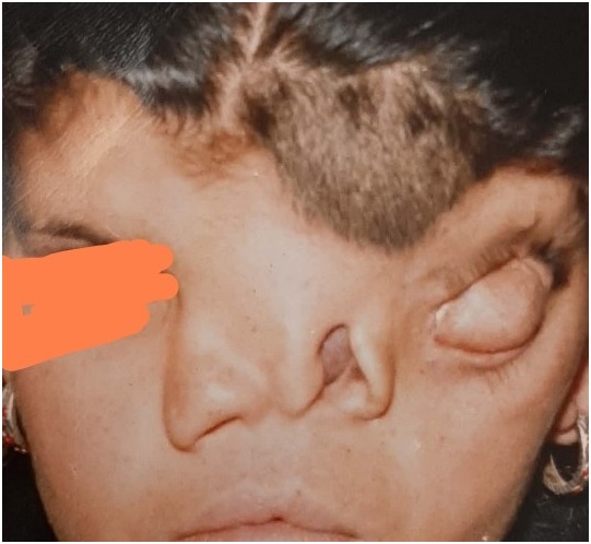
Figure 1: Choriostoma of the forehead area had a dense tuft of hair on it.
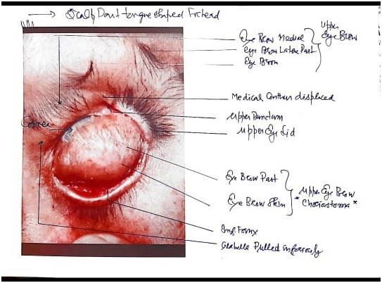
Figure 2A): Diagrammatic representation of all the structures.
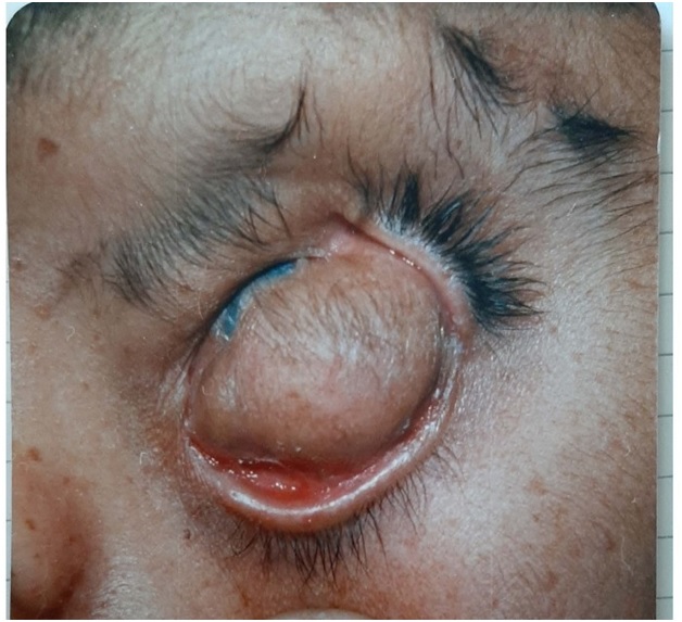
Figure 2B): Choristomas eye.
DISCUSSION
Choristomas are congenital lesions representing normal tissue in an abnormal location [1]. ABS is a group of rare congenital abnormalities caused by wrapping of parts of the foetus by fibrous amniotic bands during intrauterine life [2]. The condition is also known as amniotic band sequence, Streeter’s dysplasia, congenital constriction bands, and pseudoainhum. It is found approximately 1 in 1200 live births [3].
One of the widely accepted pathogenic mechanism theory of ABS states that once the amnion is ruptured, the fetus lies outside the amniotic cavity and bands extending from the chorionic side of the cavity entrap various parts of the fetus and disturb normal development. Common anomalies encountered are cleft lip and palate, club feet, syndactyly, cranial, cardiac, abdominal wall defect and abdominal organs extrophy, chest wall defect with heart extrophy. Associations with abdominal trauma, intrauterine devices, Ehlers Danols and epidermilosis bullosa have also been reported [4].
The diagnosis is based on ultrasound visualization of amniotic bands. Amniotic bands are now being successfully released fetoscopically through minimally invasive surgery. Extrauterine surgical intervention should be performed within weeks to months [5].
REFERENCE
1. Ponnudurai T, Louisraj S, Salman A. (2017). Epibulbar osseous choristoma: a case report. International Medical Case Reports Journal. 10:337–9. 2. Mitrović D, Vasić K, Ćirić D, Miletić E, Bogoslović M, et al. (2015). Amniotic band syndrome: Case report. Timočki medicinski glasnik. 40(3):171-5. 3. Shetty P, Menezes LT, Tauro LF, Diddigi KA. (2013). Amniotic Band Syndrome. Indian J Surg. 75(5):401–2. 4. Osama T. Abu-Salah. (2011). Amniotic band syndrome; a case report. RMJ. 36(2):159-161. 5. Rezai S, Faye J, Chadee A, Gottimukkala S, Upadhyay R, et al. (2016). Amniotic Band Syndrome, Perinatal Hospice, and Palliative Care versus Active Management. Case Reports in Obstetrics and Gynecology. Article ID 9756987. https://doi.org/10.1155/2016/9756987