Previous Issues Volume 3, Issue 1 - 2018
Massive Gastric Submucosal Hematoma: An Unusual Complication After Diagnostic Gastroscopy
Yoshihiko Kadowaki, Ayako Watanabe, Kohei Kuroda, Daisuke Shirasaka
Corresponding Author: Yoshihiko Kadowaki, Department of Surgery, Japanese Red Cross Kobe Hospital, 1-3-1, Wakinohamakaigandouri, Chuo-ku, Kobe 651-0073, Japan, E-mail: [email protected]
Received Date:05 Feb 2018 Accepted Date: 06 Feb 2018 Published Date: 07 Feb 2018
Copyright © 2017 Hoya Y
Citation: Kadowaki Y, Watanabe A, Kuroda K and Shirasaka D. (2018). Massive Gastric Submucosal Hematoma: An Unusual Complication After Diagnostic Gastroscopy. Mathews J Gastroenterol Hepatol 3(1): 010.
INTRODUCTION
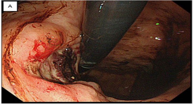
A previously healthy 81-year-old man was admitted to our hospital with weakness and fatigue caused by anemia. Laboratory studies were notable for a red-cell count of 1,580,000 per ml, a hemoglobin level of 4.8 g per deciliter, and hematocrit level of 15.1 %. Emergency endoscopic examination revealed an active gastric ulcer (Panel A).
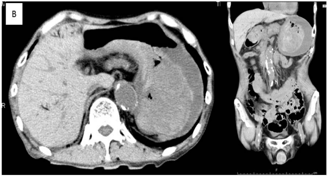
Two days later, mild epigastric tenderness was present. Computed tomography of the abdomen revealed hepatic portal venous gas and massive gastric submucosal hematoma (Panel B)
Two days later, mild epigastric tenderness was present. Computed tomography of the abdomen revealed hepatic portal venous gas and massive gastric submucosal hematoma (Panel B);
however, there was no evidence of perforation. Four days after conservative treatment, on follow-up computed tomography, neither gastric submucosal hematoma nor hepatic portal venous gas was observed (Panel C). Recovery was uneventful and the patient was well discharged on the 20th hospital day.