Information Links
Related Conferences
Current Issue Volume 9, Issue 1 - 2026
Laser Precision in Voice Preservation: A Case of True Vocal Fold Hemorrhagic Polyp Excision
Pankaj Goyal1,*, Kishan Kumawat2, Nirupama Kothari3, Manisha Chouhan4
1,2Apollo E.N.T. Hospital, Pal Road, Jodhpur, Rajasthan, India
3Sterling Accuris Diagnostics, Saras dairy road, Jodhpur, Rajasthan, India
4JIET Medical College and Hospital, Jodhpur, Rajasthan, India
*Corresponding author: Pankaj Goyal, Apollo E.N.T. Hospital, Pal Road, Jodhpur, Rajasthan, India, ORCID: 0000-0003-4098-7308, Email: [email protected]
Received Date: May 28, 2026
Published Date: June 11, 2026
Citation: Goyal P, et al. (2026). Laser Precision in Voice Preservation: A Case of True Vocal Fold Hemorrhagic Polyp Excision. Mathews J Surg. 9(1):44.
Copyrights: Goyal P, et al. © (2026).
ABSTRACT
Benign lesions known as hemorrhagic polyps of the actual vocal folds can seriously degrade voice quality, especially in professional voice users. When conservative therapy is unsuccessful, surgical excision is still the last resort. The use of CO2 laser microlaryngoscopic surgery for the removal of a hemorrhagic polyp is highlighted in this case study, with a focus on quick recovery after surgery and the best possible voice preservation. The case illustrates how laser-assisted methods can reduce tissue damage while maintaining vibratory performance.
Keywords: Hemorrhagic Polyp, True Vocal Fold, Benign Lesions, CO2 Laser, Microlaryngoscopic Surgery.
INTRODUCTION
The human voice is a finely tuned function that depends on the structural integrity and vibratory characteristics of the true vocal folds, particularly the superficial lamina propria (Reinke’s space), which plays a critical role in voice production and modulation [1]. One of the most frequent causes of dysphonia seen in clinical practice, particularly in those with high vocal demand, is benign lesions of the vocal folds, such as polyps, nodules, and cysts [2]. Of these, hemorrhagic vocal fold polyps are a unique clinical condition marked by vascular proliferation and subepithelial blood extravasation, frequently brought on by acute or chronic phonotrauma [3]. The mid-membranous region of the vocal fold, which experiences the most contact and vibratory stress during phonation, is where hemorrhagic polyps usually develop [4]. Patients typically exhibit persistent hoarseness, breathiness, decreased vocal endurance, and sporadic pitch breakdowns. These symptoms can seriously impede quality of life and professional performance, especially for public speakers, instructors, and singers [5]. Subepithelial capillary rupture is the pathophysiology, which results in localized blood accumulation, edema, and the eventual formation of a polypoid lesion [6]. Conservative measures including voice therapy, vocal hygiene education, and medical treatment of contributory causes like laryngopharyngeal reflux are frequently used in the initial management of vocal fold polyps [7]. However, compared to non-hemorrhagic lesions, hemorrhagic polyps have less likelihood of resolving on their own and sometimes necessitate surgical intervention for long-term management [8]. Complete lesion excision while maintaining the vocal fold's layered microstructure is the fundamental objective of surgical therapy in order to preserve ideal vibratory performanc [9]. Although cold-instrument microlaryngoscopic excision has long been the accepted surgical method, technological developments have made laser-assisted phonosurgery a better option in certain situations [10]. Because of its capacity for accurate tissue ablation, excellent hemostasis, and minimal adjacent tissue damage, the CO2 laser is widely used [11]. Super-pulse and ultra-pulse laser energy delivery modes provide controlled penetration depth and lower the possibility of thermal damage to the vocal ligament and deeper tissue [12]. Compared to traditional approaches, laser-assisted techniques have a number of advantages, such as better visibility because of a bloodless surgical field, less need to grasp and manipulate fragile vocal fold tissue, and better mucosal wave preservation [13]. These advantages are especially crucial for individuals whose voice quality is crucial. Additionally, research has shown that, in comparison to conventional cold steel procedures, laser excision improves phonatory outcomes, reduces scarring, and speeds up postoperative recovery [14]. Despite these benefits, the use of lasers in phonosurgery necessitates careful control of energy levels, careful case selection, and careful attention to detail in order to prevent problems such fibrosis, thermal damage, and voice impairment [15]. The purpose of this case study is to demonstrate the importance of laser accuracy in the removal of a genuine vocal fold hemorrhagic polyp, highlighting its efficacy in maintaining vocal function and promising postoperative voice results.
CASE REPORT
The patient, a 35-year-old woman, has been experiencing increasing hoarseness for five to seven months. The patient did not have dysphagia or dyspnea, but did report voice fatigue and sporadic throat pain. Overuse of the voice had a history. There were no past complaints of this kind. No past addiction history. No history of long-term drug use.
Upon closer inspection, the voice sounded gruff and breathy. A pedunculated hemorrhagic polyp situated at the intersection of the anterior and middle thirds of the vocal fold was confirmed by indirect laryngoscopy, which also showed a solitary reddish lesion on the right true vocal fold with phonatory gap. Routine blood tests show normal levels.
Investigations
- Indirect laryngoscopy: Reddish, vascular lesion on right vocal fold
- Routine blood investigations: Within normal limits
Management
Initially she patient was advised conservatively in form of strict voice rest for one week, followed by relative voice rest with avoidance of vocal abuse such as shouting, whispering, and prolonged speaking. Voice therapy was initiated with a speech-language pathologist to improve vocal technique and reduce strain. Adequate hydration was encouraged, including increased oral fluid intake and regular steam inhalation. Medical management included initiation of a proton pump inhibitor, namely Omeprazole, to address associated reflux symptoms, along with dietary and lifestyle modifications. The patient was counseled to avoid irritants such as smoking, alcohol, and environmental pollutants, and was educated regarding vocal hygiene practices. Due to lack of significant improvement after 6 weeks, surgical intervention was planned.
Surgical Procedure
After obtaining informed consent, the patient was brought to the operating room and placed in the supine position. General anesthesia was induced and the airway secured with a laser-resistant endotracheal tube. Adequate eye protection and standard laser safety precautions were implemented. Moist saline-soaked pledgets were placed around the tube cuff to minimize the risk of airway fire. A direct suspension laryngoscope was introduced transorally and suspended to provide optimal visualization of the larynx. The operating microscope was brought into position, and microlaryngoscopic examination was performed. A hemorrhagic polyp was identified on the vocal cord. The CO₂ laser was connected to the operating microscope and set at appropriate power settings in super-pulse/ultra-pulse mode. Under microscopic magnification, the lesion margins were carefully delineated. Using a focused CO₂ laser beam, a precise mucosal incision was made around the base of the polyp. The lesion was elevated and dissected meticulously from the underlying superficial lamina propria while preserving the surrounding normal vocal fold mucosa and vibratory tissue. The hemorrhagic polyp was completely excised and removed. Hemostasis was achieved with the CO₂ laser in a defocused mode. The surgical field was inspected thoroughly, confirming complete removal of the lesion and preservation of the vocal fold architecture. The excised specimen was sent for histopathological examination. The laryngoscope was removed carefully. The patient tolerated the procedure well and was extubated uneventfully before transfer to the recovery room in stable condition. Following surgery, patients are typically prescribed strict voice rest for a few days, followed by speech therapy and gradual voice use. Proton pump inhibitors, analgesics, fluids, and rarely corticosteroids are examples of medical management. Over the course of several weeks, healing entails re-epithelialization and mucosal wave repair. Although CO2 laser-assisted surgery has several benefits, such as reduced mechanical trauma, excellent vision due to low bleeding, and superior precision, inappropriate use can result in thermal damage and poor vocal results. In order to accomplish the best possible voice restoration, the essential premise of the procedure is to preserve the sensitive vibratory microstructure of the vocal fold.
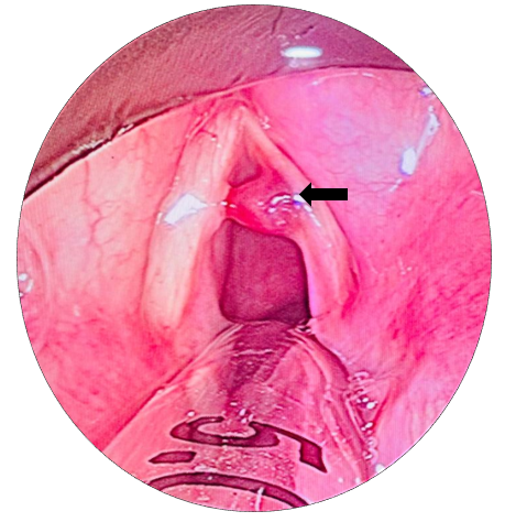
Figure 1. A hemorrhagic polyp with feeding vessel seen on the striking zone of the right vocal fold. A 5.5 endotracheal tube is seen in situ in the posterior larynx.
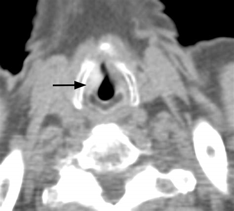
Figure 2. Computed tomography scan suggested mild focal thickening of the right true vocal cord.

Figure 3. An upward Bouchayer forcep holds the polyp and provide gentle traction.
Histopathology
Microscopic examination revealed:
Subepithelium is edematousand shows mild lymphoplasmacytic infiltration. Many dilated and congested blood vessels were presentalong with area of fibrinoid necrosis hemorrhage. No evidence of dysplasia or malignancy. Findings were consistent with a hemorrhagic vocal polyp.
Postoperative Care
- Absolute voice rest for 7 days
- Gradual reintroduction of speech
- Structured voice therapy
- Anti-reflux measures and lifestyle modification
Outcome and Follow-Up
At 4-week follow-up:
- Significant improvement in voice quality
- No residual lesion on laryngoscopic examination
The patient was advised to continue vocal hygiene practices.
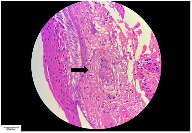
Figure 4a.
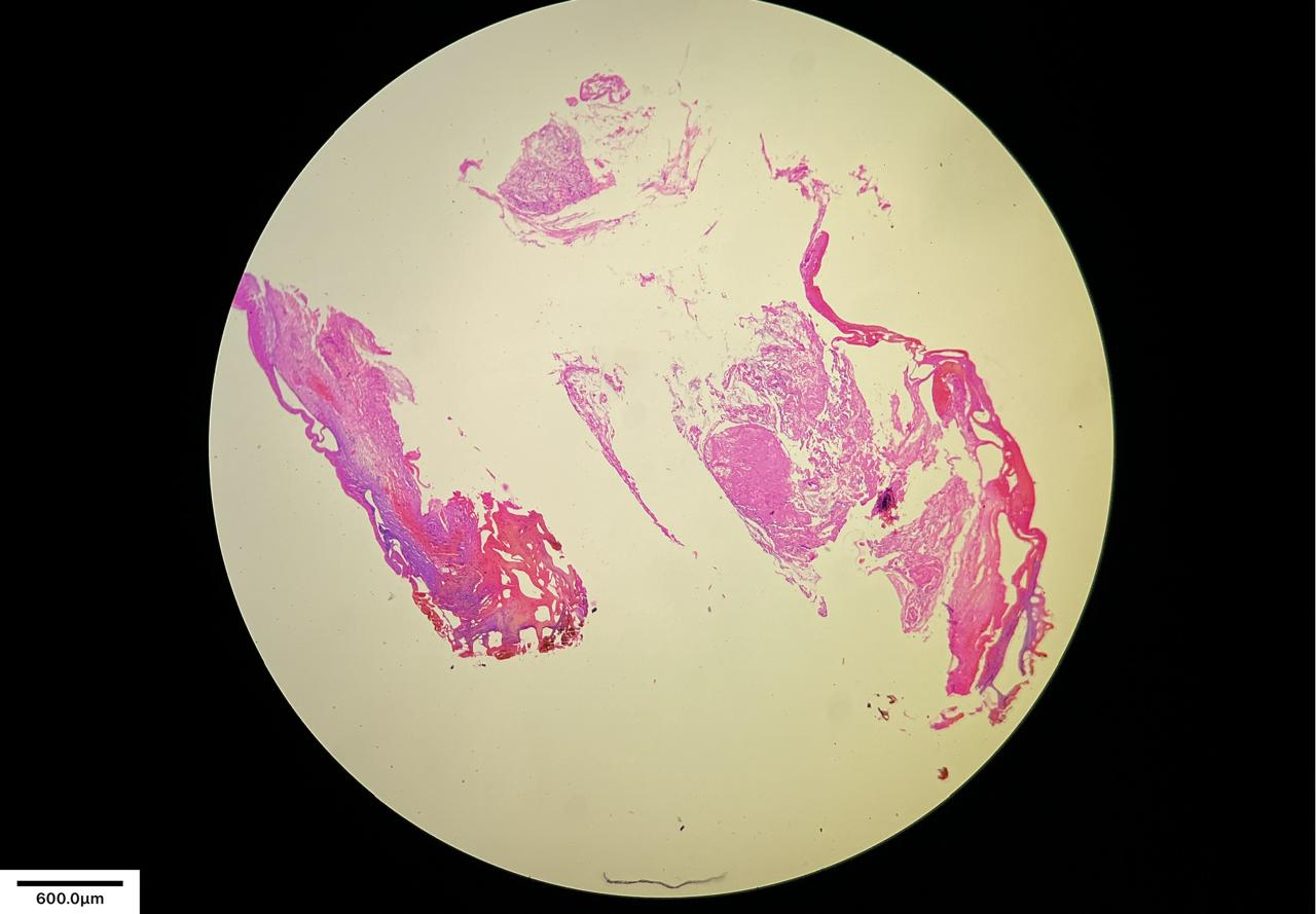
Figure 4b.
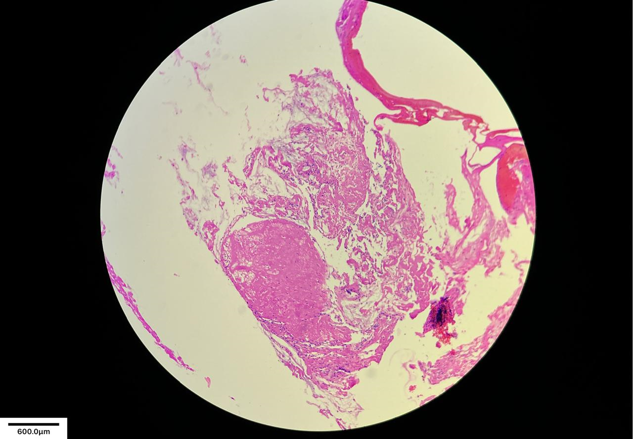
Figure 4c.
Figure 4 (a, b & c). Histopathology slide pictures.
DISCUSSION
A distinct subclass of benign laryngeal lesions, hemorrhagic polyps of the true vocal folds are defined by subepithelial accumulation and significant vascular components. They are usually caused by acute phonotrauma or recurrent vocal strain [3]. The most prevalent location for these lesions is the mid-membranous section of the vocal fold, which is especially susceptible to microvascular damage because it experiences the greatest amount of stress during phonation [4]. As a result, the mucosal wave is drastically altered by the rupture of the superficial lamina propria, which results in problems including breathiness, hoarseness, and decreased vocal effectiveness.
Restoring normal vibratory function while minimizing harm to the fragile vocal fold layers is the main objective in the treatment of hemorrhagic vocal fold polyps [9]. Because hemorrhagic polyps are organized vascular lesions, they are less likely to regress naturally and require surgical excision for definitive treatment [8]. However, conservative care, such as voice therapy and behavioral retraining, may be helpful in early or small lesions. Persistent dysphonia and compensatory maladaptive vocal actions might decrease voice quality if treatment is delayed or insufficient [5]. Although laser-assisted phonosurgery has greatly improved surgical results, microlaryngoscopic excision has long been regarded as the gold standard for treating such lesions [10]. In laryngeal microsurgery, the CO₂ laser offers unmatched precision, enabling surgeons to target diseased tissue specifically while maintaining the surrounding superficial lamina propria and epithelium [12]. This accuracy is especially important in phonosurgery, where voice quality can be significantly impacted by even little disruptions. The potential of lasers to provide hemostasis, particularly in hemorrhagic lesions, is one of its advantages [11]. By improving visibility, the laser-free surgical field reduces the requirement for mechanical vocal fold manipulation and allows for more precise lesion excision [13]. Reduced handling preserves the vibratory qualities necessary for regular phonation by minimizing damage to the mucosal layer [14]. Surgeons can also restrict the amount of heat that spreads to nearby tissues by precisely controlling the laser's power, duration, and pulse length [16]. In order to reduce collateral thermal damage and avoid fibrosis or scarring, techniques like super-pulse or ultra-pulse modes assist in delivering high energy in brief bursts [17]. Maintaining the mucosal wave and attaining the best postoperative voice results depend on the preservation of the layered microstructure, especially the superficial lamina propria [18]. According to comparative studies, laser-assisted excision has a number of advantages over traditional cold steel procedures, such as lower intraoperative bleeding, shorter operating times, less postoperative edema, and quicker voice recovery [2]. Additionally, especially in professional voice users, patients who undergo laser phonosurgery frequently show improved results in terms of phonatory efficiency, stability, and quality [19]. These results highlight how crucial it is for modern laryngology to use minimally invasive and precision-based surgical techniques. Nevertheless, laser surgery has certain drawbacks despite its benefits. Complications include thermal damage, delayed healing, scarring, and dysphonia can result from improper laser use [20]. Strict commitment to surgical principles, sufficient training, and good results are crucial [21]. Patient selection is particularly crucial because, depending on the lesion's size, depth, and underlying pathology, not all lesions are eligible for laser excision [2]. When it comes to long-term outcomes, postoperative care is equally important. Recurrence can be avoided and normal phonatory patterns can be restored with voice rest and organized voice therapy [22]. Improved outcomes and overall satisfaction are greatly influenced by early intervention and patient compliance.
CONCLUSION
In this instance, the hemorrhagic polyp was precisely removed with little damage to the surrounding tissues thanks to laser-assisted microsurgery, which produced a very good functional recovery. The success of this strategy is demonstrated by the restoration of the mucosal wave and near-normal voice quality seen during follow-up. Overall, this case supports the increasing amount of data that laser accuracy in phonosurgery is an excellent method for treating hemorrhagic vocal fold polyps, especially in patients when voice preservation is crucial.
CONSENT
Written informed consent was obtained from the patient for the publication of this case report and any accompanying images.
ACKNOWLEDGMENTS
We would like to acknowledge the patient for providing consent for the publication of this case report.
CONFLICT OF INTEREST
The authors declare no conflicts of interest.
FUNDING
No funding was received for the publication of this case report.
AUTHORS' CONTRIBUTIONS
All authors contributed to the conception, drafting, and critical revision of the manuscript. All authors have approved the final version of the manuscript for submission.
REFERENCES
- Hirano M. (1981). Structure of the vocal fold in normal and disease states. J Voice. 1(1):7-20.
- Sataloff RT. (2017). Professional Voice: The Science and Art of Clinical Care. 4th ed. San Diego: Plural Publishing.
- Johns MM. (2003). Update on the etiology, diagnosis, and treatment of vocal fold nodules, polyps, and cysts. Curr Opin Otolaryngol Head Neck Surg. 11(6):456-461.
- Gray SD. (2000). Cellular physiology of the vocal folds. Otolaryngol Clin North Am. 33(4):679-698.
- Roy N, Merrill RM, Thibeault S, Parsa RA, Gray SD, Smith EM. (2004). Prevalence of voice disorders in teachers. J Speech Lang Hear Res. 47(2):281-293.
- Pontes P, Behlau M, Gonçalves J. (1994). Alterations of the vocal folds due to voice misuse. J Voice. 8(3):257-264.
- Behrman A. (2005). Common practices of voice therapists in the evaluation of patients. J Voice. 19(3):454-269.
- Bouchayer M, Cornut G. (1988). Microsurgery for benign lesions of the vocal folds. Ear Nose Throat J. 67(6):446-466.
- Zeitels SM. (2002). Phonosurgery: past, present, and future. J Voice. 16(4):457-464.
- Remacle M, Eckel HE, Antonelli A, Brasnu D, Chevalier D, Friedrich G, et al. (2000). Endoscopic cordectomy with CO₂ laser. Eur Arch Otorhinolaryngol. 257(6):297-301.
- Steiner W. (1993). Results of curative laser microsurgery of laryngeal carcinomas. Am J Otolaryngol. 14(2):116-121.
- Mallur PS, Johns MM. (2013). Laser applications in laryngeal surgery. Otolaryngol Clin North Am. 46(1):131-145.
- Zeitels SM, Burns JA. (2007). Laser applications in laryngology. J Voice. 21(6):732-742.
- Franco RA, Andrus JG. (2007). Common diagnoses and treatments in professional voice users. Otolaryngol Clin North Am. 40(5):1025-1061.
- Benninger MS, Jacobson BH, Johnson AF. (1994). Vocal Arts Medicine. New York: Thieme.
- Strong MS, Jako GJ. (1972). Laser surgery in the larynx. Ann Otol Rhinol Laryngol. 81(6):791-798.
- Remacle M, Lawson G. (2001). CO₂ laser in phonosurgery. Curr Opin Otolaryngol Head Neck Surg. 9(6):386-393.
- Hirano M, Kakita Y. (1985). Cover-body theory of vocal fold vibration. In: Speech Science. Tokyo: University of Tokyo Press.
- Sataloff RT. (2017). Professional voice care and outcomes. J Voice. 31(2):145-152.
- Garrett CG, Ossoff RH. (2006). Risks of laser surgery in laryngology. Otolaryngol Clin North Am. 39(1):211-223.
- Zeitels SM, Healy GB. (19910. Laryngeal laser surgery: complications and prevention. Ann Otol Rhinol Laryngol. 100(6):481-486.
- Rosen CA, Simpson CB. (2008). Operative Techniques in Laryngology. Berlin: Springer.
