Information Links
Related Conferences
Previous Issues Volume 7, Issue 2 - 2023
In Vivo Studies on Mortality and Histopathological Indices of Phragmenthera capitata (Mistletoes) on Clarias gariepinus Fingerglings in Aquarium
George UU1, Mbong EO2, Abiaobo NO3, Akpan II3
1Department of Fisheries and Aquaculture, Faculty of Agriculture, Akwa Ibom State University, Nigeria
2Department of Biological Sciences, Ritman University, Nigeria
3Department of Zoology, Akwa Ibom State University, Nigeria
*Corresponding author: Dr. UU George, Department of Fisheries and Aquaculture, Faculty of Agriculture, Akwa Ibom State University, P. M. B. 1167, Ikot Akpaden, ORCID ID: 0000-0003-1773-5057, Tel: +2348032625310; Email: [email protected].
Received Date: October 18, 2023
Published Date: November 22, 2023
Citation: George UU, et al. (2023). In Vivo Studies on Mortality and Histopathological Indices of Phragmenthera capitata (Mistletoes) on Clarias gariepinus Fingerglings in Aquarium. Mathews J Cytol Histol. 7(2):25.
Copyrights: George UU, et al. © (2023).
ABSTRACT
Background: The effects of ethanolic extracts of Phragmenthera capitata on the histopathology of gills of Clarias gariepinus fingerlings were investigated under laboratory conditions to understand its toxicity with a view of adopting the plant as an indigenous fish feed additive. Methods: Five concentrations ranging from 0 mg/l, 20 mg/l, 30 mg/l, 40 mg/l and 50 mg/l were prepared for mortality and histopathological examination over a 96-hours in two batches (A and B) using aquarium. The mortality of C. gariepinus in the ethanolic extract (EE) of Phragmenthera capitata was estimated in percentage. Two-way analysis of variance (ANOVA) was used to test for significant (p<0.05) difference considering the extract concentration and mortality time. Also, the LC50 was determined using Probit analysis. Results: The results revealed a concentration-dependent mortality rate. The 96 hours LC50 was 37.653 mg/l representing a log concentration of 1.576 for both batches (A and B). However, the pathological effects on gill lamellae of Clarias gariepinus fingerlings were significant (P<0.05) at 40 mg/l and 50 mg/l concentrations of the EE when compared to the control group. At 40 mg/l and 50 mg/l concentrations there were evidences of diffused epithelial degeneration of the lamella. Hence, the histological changes observed in the present study were adjudged as being concentration-dependent with severe alterations associated with the higher concentrations. Conversely, there were no pathological changes on the gills of the control group, 20 mg/l and 30 mg/l respectively in both batches. Conclusion: Based on the results obtained from the present study, further research on plant nutraceutical potentials and its suitability as an additive for fish feed formulation at different inclusion level to ascertain the concentration that may promote fish growth is recommended.
Keywords: Histopathology, Phragmenthera capitata, Mortality, Gills, Fingerlings.
INTRODUCTION
In Nigeria, mistletoe is used as a remedy for several human and animal ailments that include stomach ache, diarrhoea, dysentery, wound and cancer. Ruminants and local fowls do relish it without any reported digestive disorder [1]. Mistletoe has been analyzed and observed to contain lecithin, viscotoxin, polysaccharides and many phytochemicals as an active ingredient [2]. It has been reported to have hypoglycaemic properties; since it decreases the blood glucose level and has effects in controlling the loss of body weight which occurs in person with diabetes mellitus [3]. Mistletoe has also been used in the treatment of problems, such as epilepsy, infertility in men and women, menopausal syndrome and rheumatism [4].
Aquaculture production has been supplementing the output of capture fisheries for the last 6 decades, thus, helping to sustain humanity’s demand for aquatic products. This is due to constant improvement in the techniques employed in raising aquatic animals and plants. Reports shows that aquaculture marginally supplied 7% of fish for human consumption in 1974, increasing to 26% in 1994, 39% in 2004, and 44.1% in 2014 [5]. The rearing of Clarias gariepinus started in the early 70s in central and western African countries. It received wide acceptance when it was realized to be a very suitable species for aquaculture and of high economic value. It has since been the most widely cultured fish in Nigeria and even in Africa [6]. The fish matures quickly and has a wide range of tolerance to climatic conditions [7].
Histopathological changes have been widely used as biomarkers in the evaluation of the health status of organisms exposed to contaminants in laboratory animal and wild specimen [8]. One of the great advantages of using histopathological biomarkers in environmental studies is that this category of biomarkers allows for examination of the target organs [9]. Furthermore, the alterations found in these organs are clearer than functional ones. It also reveals overall signs of damage to animal health. The gills being a respiratory organ of fish are frequently in contact with external environment and thus vulnerable to aquatic toxicants [10,11].
Massive unemployment and economic realities in the country have pushed a large part of the populace into commercial and subsistence fish farming. This trend is limited by the high cost of commercial fish feed brands which are imported from foreign countries and sold within our country. This bring the task of embarking on researches to identify indigenous plants that are common which may be adopted as an essential ingredient for the formulation of locally source fish feed with results comparable with imported feed brands. The adoption of plants as known feed additives proceeds from toxicological assay to determine species tolerance limits and evaluate the presence of undesired growth and metabolic effects in the plant. Therefore, the present study is carried out to elucidate the tolerance limits and toxicological response of C. gariepinus fingerlings to Phramenthera capitata ethanolic extract in aquarium with a view of adopting the plant as an indigenous fish feed additive.
MATERIALS AND METHODS
Collection of Test Organism
Fingerlings of Clarias gariepinus were collected from Akwa Ibom State University fish farm, Obio Akpa Akwa Ibom State, Nigeria located within 40 57’52” N and 7045’29’’ E. The climate of the area is tropical and is characterized by distinct wet and dry seasons. The vegetation of the study area is generally rainforest close to the mangrove belt. Human activities in the area include farming, hunting, boat building and sand mining. A total of two hundred (200) fingerling were collected and used for the study.
Acclimatization of Specimen's
The fingerlings where acclimated in a re-circulatory glass aquaria measuring 96 x 50 x 29 cm containing habitat water for 24hours in the fisheries and aquaculture laboratory of Akwa Ibom State fish farm. This enhanced the stability of the fingerlings from stress of collection and transportation [12].
Collection of Plant Sample
Fresh leaves of mistletoe (Phragmanthera Capitata) growing on Avocado Leaf (Persea americana) were collected for the study. The collection site of the plant was Ikot Udota in Afaha Eket Local Government Area, Akwa Ibom State. The date of collection was 20th January, 2023. The plants material was taken for identification and authentication by a plant systematics at the Department of Botany Herbarium, Akwa Ibom State University, Ikot Akpaden, Mkpat Enin Local Government Area.
Preparation of Plant Material
After the identification, the leaves were washed and sun dried. The leaves were shredded and spread on cellophane and allowed to dry for 72 hours under room temperature. The dried leaves were pulverized (grounded) into fine powder using wooden pestle and mortar.
Preparation of Ethanolic Extract (Maceration and Extraction)
Cold extraction method (Maceration) was used in this research according to [13]. In the extraction procedure, 1000ml of 99% Concentrated Ethanol was used to Macerate 240g of the plant materials in an airtight container and kept in the laboratory under room temperature for 72 hours (3 days). In the due date of filtration, the mixture was filtered with Muslim cloth to acquire the filtrate. The extract was stored in 250ml conical flasks. The conical flask was well labelled, the mouth of the conical flask was covered with foil paper and masking tape rapped around the mouth to ensure that it is tightly covered.
Preparation of Experimental Aquaria
Ten (10) rectangular plastic aquaria measuring 25 ×10 × 15 cm were thoroughly washed with tap water and properly rinsed with fresh water of similar salinity and allowed to drain dry for 24 hours on the laboratory bench based on [14].
Stocking of Specimen
Each of the Ten (10) plastic aquaria was filled with two liters of water and 10 Clarias gariepinus fingerlings were stocked in each aquarium. The ethanolic extract of mistletoe (Phragmanthera capitata) with varying concentrations was added to each stocked aquaria and allowed to stand for 96 hours for mortality examination. Each of the aquariums had a replicate to ensure accuracy (Batch A and Batch B).
Toxicant Concentration for the Bioassay
Prior to commencement of the bioassay, a preliminary test was conducted to give the actual variations in concentration to be used for the bioassay.
Monitoring of Water Quality
Water quality parameters were monitored prior to commencement of the experiment and also periodically according to Standard Method [15]. Parameters that were monitored include dissolved Oxygen (DO), pH, And Temperature (0C). Temperature and pH were measured using portable pH /Ec/ TDS/ Temperature HANNA, H1 991301 model instrument while oxygen was measured using digital portable analyser JPB - 607A from "Search Tech Instrument".
Monitoring of Specimen for Mortality
The effects of the various concentrations of the ethanolic extract of mistletoe (Phragmanthera capitata) on the fingerlings was monitored on a 24-hour basis for 96hours as recommended by [12] and [16].
Determination of Mortality and Survival Rates of Fingerlings
The percentage mortality and survival rates of the fingerlings in the different concentrations of the ethanolic extract of Phragmanthera capitata during the period of study was determine using the formula;
% mortality =n/N × 100 [17].
Where;
n = number of dead fish per aquarium per concentration
N = Total individuals stocked
The difference between dead fish and survivors will give the percentage survival of the fingerlings at the end of the experiment (96 hours) [12].
Determination of Mortality and Lethal Median Concentration (96 hours LC50)
The effects of the various concentrations of the ethanolic extract of plant (Phragmanthera capitata) on the fingerlings of C. gariepinus was determined by graphical method (Probit Level Determination as recommended by [12,16,18,19]. At Lethal Median Concentration LC50, after 96 hours of test, the number of fingerlings that were expected to die was determined from the graph. Similarly, the concentration that killed 50 % of the stocked fingerlings at the end of the test (96 hours) was determined at the probit level [12,16,18,19].
Collection of Sample for Histopathology Examination
The gill tissues were isolated from the test animal and fixed in formalin-saline for 48 hours. The fixed tissue was processed manually through graded ethanol, cleared in xylene impregnated and embedded in paraffin wax, sections of the tissue sample were cut with a rotary microtome, stained by hematoxylin and eosin technique, prepared tissues were finally observed using a microscope for pathological changes at x100 and x400 magnification.
Data Analysis
The results of the respective concentration effects of the ethanolic extract of Phragmanthera capitata was presented in tables. Two-way analysis of variance (ANOVA) was used to test for significant (p<0.05) difference considering the extract concentration and mortality time. Also, the LC50 was determined using Probit analysis. All statistics were carried out using SPSS version 20.0.
RESULTS
Initial Water Quality Parameters
The initial water quality parameters prior to stocking are shown in Table 1. Dissolved oxygen had a value of 5.2 mg/l, with a value of 29.8 °C for Temperature and 6.77 for pH.
Table 1. Initial Physico-chemical parameters of the test water prior to stocking of test organism
|
Fish Species |
Initial physico-chemical parameters prior to stocking |
||
|
DO (mg/l) |
Temp (°C) |
pH |
|
|
Clarias gariepinus |
5.2 |
29.8 |
6.77 |
The percentage mortality and survivors of C. gariepinus at the end of the test period in each of the concentrations are shown in Table 2 for the two batches of the experiment.
In the 0 mg/l concentration of the extract, no mortality was recorded throughout the test period in both batches A and B. Similar observations were made for 20 and 30 mg/l concentration of the extract in both bathes. In the 40 mg/l concentration of the extract, 30 % mortality and 70 % survivors were recorded while in the 50 mg/l concentration of the extract all the test organisms were observed dead leaving 0 % survivors in both batches (Table 3). Statistical Analysis using Anova (SPSS 20.0) showed that there was no significant difference (p>0.05) in mortality between the two batches in the different concentration and time.
Table 2. Summary of the Percentage Mortality and survivors of C. gariepinus in the different concentrations of the ethanoic extract of Phragmenthera capitata at the end of the experiment (96 hrs)
|
Conc. of extract (mg/l) |
BATCH A |
BATCH B |
||||||
|
|
Mortality (M) |
% M |
Survivors (S) |
% S |
Mortality (M) |
% M |
Survivors (S) |
% S |
|
0 |
0 |
0 |
10 |
100 |
0 |
0 |
10 |
100 |
|
20 |
0 |
0 |
10 |
100 |
0 |
0 |
10 |
100 |
|
30 |
0 |
0 |
10 |
100 |
0 |
0 |
10 |
100 |
|
40 |
3 |
30 |
7 |
70 |
3 |
30 |
7 |
70 |
|
50 |
10 |
100 |
0 |
0 |
10 |
100 |
0 |
0 |
The 96 hours LC50 for C. gariepinus exposed to the different concentrations of the ethanoic extract of Phragmenthera capitata is shown in Table 3 for both batches. The 96 hours LC50 is given at 37.653 mg/l representing a log transformed concentration of 1.576 mg/l a point where 50 % of the test organisms are expected to die at the end of the experiment.
Table 3. LC50 determination for C. gariepinus at the end of the 96-hours bioassay
|
Plant |
Species |
Probit |
S.E |
LC50 (mg/l) |
Log Con. (mg/l) |
|
Phragmenthera capitata |
Clarias gariepinus |
P= -60.749 + 38.551X |
729.203 |
37.653 |
1.576 |
The histological examination of gills in the control and the four experimental groups revealed distinct changes in their gill’s epithelial tissues (Figure 1). The control (0 mg/l) group showed a normal primary lamellar epithelium with no apparent degenerative or pathological alterations. The experimental, groups 2 (20 mg/l) and 3 (30 mg/l) also exhibited a normal epithelium of the filament, indicating that their respiratory surfaces were intact and functional. However, groups 4 (40 mg/l) and 5 (50 mg/l) displayed a diffused degeneration of both the primary and secondary lamellar epithelium, which could compromise their gas exchange capabilities. The degenerative changes observed in these groups are indicative of exposure to higher dose of the ethanolic extract of the mistletoes (Phragmenthera capitata) which could damage the delicate gill tissues. Overall, these findings suggest that exposure to the ethanolic extract of P. capitata at higher dose can cause histological changes in the gills of the fish, particularly in the lamella epithelium. These changes may have implications for the respiratory function of the gills and overall health of the fish.
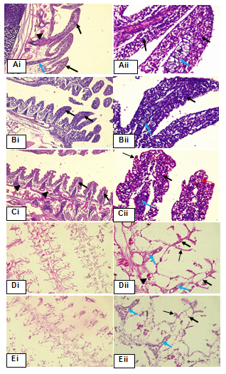
Figure 1. Phtotomicrograph of Gill arch tissue sections of Group 1-5. Haematoxylin and Eosin (H&E) stain. Each group was shown x100 and x400 magnification
Control Group (Ai&Aii) showed gills with normal filament epithelium (thick black arrow), the supporting cartilage epithelium (thick blue arrow), muscle fibre (arrowhead) and the capillaries (thin red arrow). (Bi&Bii) and (Ci&Cii) showed normal filament (lamella) epithelium, no pathological changes seen. (Di&Dii) and (Ei&Eii) depicted diffused epithelium degeneration of both primary (thick black arrow) and secondary lamellar (thin black arrow).
DISCUSSION
Initial values of the phsico-chemical parameters of the experimental water
Three basic physicochemical parameters were taken prior to the stocking of the fish species. Dissolved oxygen had a value of 5.2 mg/l with a value of 29.8 °C recorded for temperature and a value of 6.77 was recorded for ph.
In aquaculture operations, standard ranges of values for these parameters are known. For dissolved oxygen a range of between 4.0–6.0 mg/l is suitable, 6.7–8.6 for pH and 25.0–30.0 °C for temperature are recommended values for smooth operation of aquaculture [20-25].
The ranges of the physicochemical parameters of the experimental water were found to fall within the acceptable limits prior to the commencement of the experiment as previously reported by the authors under reference.
The maintenance of the normal values of the physicochemical parameters by the experimental water prior to the commencement of the experiment might have been as a result of absence of impurities or the toxicant and the organisms themselves [26-30]. Impurities, pollutants and toxicants are known to play to elevate or reduce the different physico-chemical parameters in aquatic environment [26,27,30].
Percentage Mortality and Survivors of Clarias gariepinus
The percentage mortality of C. gariepinus in the ethanolic extract of Phragmanthera capitata ranged from 0–100 % in both batches A and B at the end of the 96-hours bioassay. No mortality was recorded in the 0–30 mg/l concentration of the toxicant. However, 70 % mortality was recorded in the 40 mg/l concentration in each of the batches while 100 % mortality was recorded in the 50 mg/l concentration of the toxicant. The results of the present findings are in consonance with the earlier reports by George et.al. [22] While reporting the effect of lethal concentrations of rubber extract (Hevea brasiliensis) on Clarias gariepinus fingerlings survival under laboratory condition and synchronize with earlier assertions made by [31] while investigating on the toxicity of Qua Iboe light crude oil on the gills of Clarias gariepinus juveniles.
In the present study, percentage mortality was concentration dependent. Similar results have been reported by [32,33] and [34] while investigating on the toxicity of different plant extracts on Clarias gariepinus. Calta and Ural, (2004) also reported a dose-dependent toxic response of Cyprinus carpio to synthetic pesticides [35]. Noted that Clarias gariepinus generally tolerate and adapt to environments with rapid changes in water chemistry and increasing contaminants concentration. This explains the response of the species to 0 mg/l, 20 mg/l, and 30 mg/l concentration. However, 70% and 100% mortality of species at 40 mg/l and 50 mg/l over the 96 hours’ period, vindicates Shellford’s law of tolerance [36]. Noted that selective toxicity of toxicants for various fish species is dependent on different inhibition rates of acetylcholinesterase, different detoxification rates and differential absorption rates. These may have been responsible for the different toxic reactions showed by the fish at the studied concentrations and time. The reactions are usually more pronounced at higher concentrations due to increased inhibition of acetylcholinesterase [36] which eventually results in the death of the test organisms [33,35].
96 Hours LC50
The 96 hours LC50 of any toxicant is the dose or concentration which kills 50 % of the stocked organisms at the end of the experimental period of 96 hours (4 days) [37,21-24]. The 96 hours LC50 is known to vary from toxicant (APHA, 1998; Samabaswa and Rao, 1985) and from concentration to concentration of the toxicant [38].
In the present study the 96 hours LC50 was 37.653 mg/l representing a log concentration of 1.576 for both batches (A and B). The 96 hours LC50 of toxicants are known to vary as previously reported by the authors earlier sited above. For instance, [34] reported 96 hours LC50 of 0.0166 mg/l and 0.0038 mg/l for Clarias gariepinus fingerlings under the toxicity effects of detergent effluents, 96 hours LC50 of 0.1 mg/l and 0.03 mg/l was reported by [39] when working on the effects of soap and detergent effluents on Clarias gariepinus fingerlings. Again, [35] reported the 96 hours LC50 of 0.033–0.33 mg/l on Clarias gariepinus adults using Carica papaya extract. In this study the 96 hours LC50 of 1.576 mg/l obtained for both batch A and B is higher than those of the authors under reference. This might have depended on the ranges of the concentration of toxicant finally used for the bioassay after conducting a preliminary test. However, the toxic nature of the toxicants previously reported by the authors under references was observed to be more toxic than P. capitata based on the values of LC50 reported.
Effects of the Extract on the gills of the test Organisms
The effects of the ethanolic extract of Phragmenthera capitata showed pathological effects on the gill lamellae of Clarias gariepinus fingerlings. However, the gill lamellae in the control (0 mg/l), 20 mg/l and 30 mg/l were not affected. Pathological effects were pronounced at 40 mg/l and 50 mg/l concentration of the extract which shows evidence of diffused epithelial degeneration of the lamella. Similar results were reported by [40] when reporting on studies on mortality and histopathological alterations on the gills of Oreochromis niloticus juveniles following exposure to ethanolic extract of Phramenthera capitata under laboratory conditions.
Gill lamellae cell disintegration has been reported by [41] in Carassius auratus gibelio when investigating biochemical and histological effects of deltamethrim on the species with different effects such lamellae cells hypertrophy and nuclear pycnosis in the basal cells.
Hypertrophy, necrotic, atrophy and dystrophy of secondary lamellae have also been reported by [42] in Clarias gariepinus exposed to refined petroleum oil and kerosene under laboratory conditions.
The histological changes observed in the present study were concentration dependent with severe alteration being pronounced at higher concentrations. The results of this findings are similar to earlier assertions reported by [21] when reporting on the acute toxic effects of Hevea brasiliensis on the gills of hatchery reared Oreochromis niloticus fingerlings and observed histological changes in the gills of the exposed organisms which were concentration dependent, [24] when investigating on the acute toxic effect of Qua Iboe light crude oil on the gills of Clarias gariepinus juveniles; [43] when studying the effect of Euphorbia hirta leaf extract on histopathology of juveniles Clarias gariepinus and [31] when reporting on the histopathological alterations in gills of fingerlings of Clarias gariepinus following sub-lethal acute exposure to Hevea brasiliensis.
The changes in the gills of the test organisms following exposure to the extract fall within the general responses for fish organs to environmental pollutants [5]. Observed that fish gills are the prime target organ of all pollutants due to their extensive surface in contact with the external medium and the reduced distance between the external medium and gill morphology. This assertion makes the gills an important biomarker in providing a rapid method of detection of the effect of pollutant [42,44,45].
SUMMARY AND CONCLUSION
Effects of ethanolic extract of Phragmenthera capitata (mistletoes) on the survival and histopathology of the gills of Clarias gariepinus fingerlings were investigated using static bioassay under laboratory condition. Prior to the toxicity test of the extract on the fish species physico-chemical parameters were taken before stocking of the experimental fish. The percentage mortality recorded in this study was concentration dependent with higher mortality recorder at higher concentrations in both batches. From the mortalities ratio the 96-hour LC50 was 37.653 mg/l for both batches representing a log transformed concentration of 1.576 mg/l. The results of histopathology confirm the results recorded in the mortality table as similar observation of concentration dependent was also the case. Severe gill lamellae degeneration was more pronounced at higher concentrations with no pathological changes on the gills of the control, 20 and 30 mg/l in both batches. Generally, fish gills are prime target organS of all pollutants due to their sensitive and net-like structure. Both dissolved oxygen and colloidal materials tend to adhere to the gills resulting in surficial rupture and disease condition to the gills. Gills morphology is important biomarkers, providing a basis for the detection of environmental pollutants [46-51].
RECOMMENDATIONS
Based on the results obtained from the present study which showed medium to high percentage mortalities when exposed to the ethanolic extract of Phragmenthera capitata, it is imperative that ecological friendly methods should be put in place to checkmates parasitic plants within our environment. Further research is recommended on the use of Phragmenthera capitata in fish feed formulation at different inclusion level to know which concentration will promote or retard fish growth. This recommendation stems from the fact that the toxicity level observed during the study was low except at higher concentrations. Also, based on the high cost of feed in aquaculture operations, this study recommends further research on the toxicity studies of locally available plant within our environment. This will help to know their toxicity level and which of them can be recommended for used as a plant-based additive in the formulation of fish feed.
LIST OF ABBREVIATIONS
P. capitata: Phragmenthera capitata; C. gariepinus: Clarias gariepinus, LC50: Lethal Concentration that will kill 50% of the test organism; DO: Dissolved Oxygen; TDS: Total Dissolved Solids; Ec: Electrical Conductivity; pH: Potential of Hydrogen; Temp.: Temperature; EE: Ethanolic Extract.
DECLARATIONS
ETHICS APPROVAL AND CONSENT TO PARTICIPATE
NIL.
CONSENT FOR PUBLICATION
We, the authors have given consent for publication.
AVAILABILITY OF DATA AND MATERIALS
Yes, at the request of the authors.
COMPETING INTERESTS
No competing interest.
FUNDING
No funding.
AUTHORS' CONTRIBUTIONS
Author 1: Conception, design and development of the topic, data collection and analysis, initial drafting and reviewing the manuscript and final approval of the prepared manuscript. Author 2: Conception, design and development of the protocol, supervision of the experiments, data analysis and reviewing the manuscript. Author 3: supervision of the experiments, data analysis and reviewing the manuscript.
ACKNOWLEDGEMENTS
The authors sincerely acknowledge Akwaibom State University for providing an ambience environment for the research. We also covey our heartfelt thanks to the laboratory technicians of the department of Medical Laboratory Science, histopathology unit, who were involved in the histo-morphological examination of the gills of the experimental animal.
REFERENCES
- Egbewande OO, Jimoh AA, Ibitoye EB, Olorede BR. (2011). Utilization of African mistletoe (Tapinanthus bangwensis) Leaf Meal by Broiler Chickens. Pak J Nutr. 10(1):19-22.
- Adesina SK, Illoh HC, Johnny II, Jacobs IE. (2013). African mistletoes (Loranthaceae); ethnopharmacology, chemistry and medicinal values: an update. Afr J Tradit Complement Altern Med. 10(3):161-170.
- Obatomi DK, Bikomo EO, Temple VJ. (1994). Anti-diabetic properties of the African mistletoe in streptozotocin-induced diabetic rats. J Ethnopharmacol. 43(1):13-17.
- Saija A, Tomaino A, Lo Cascio R, Rapisarda P, Dederen JC. (1998). In vitro antioxidant activity and in vivo photoprotective effect of a red orange extract. Int J Cosmet Sci. 20(6):331-342.
- Food and Agriculture Organization (FAO). (2016). The state of world fisheries and aquaculture 2016–contributing to food security and nutrition for all. Rome, Italy: FAO Fisheries Department.
- Adewunmi AA, Olaleye VF. (2011). Catfish culture in Nigeria: progress, prospects and problems. Afr J Agric Res. 6(6):1281-1285.
- Huisman EA, Richter CJJ. (1987). Reproduction, growth, health control and aquacultural potential of the African catfish. Clarias gariepinus (Burchell 1822). Aquaculture. 63(1-4):1-14.
- Adeshina I, Jenyo-Oni A, Emikpe BO, Ajani EK, Abdel-Tawwab M. (2019). Stimulatory effect of dietary clove, Eugenia caryophyllata, bud extract on growth performance, nutrient utilization, antioxidant capacity, and tolerance of African catfish, Clarias gariepinus (B.), to Aeromonas hydrophila infection. J World Aquacult. 50(2):390-405.
- Triebskorn R, Adam S, Casper H, Honnen W, Pawert M, Schramm M, et al. (2002). Biomarkers as diagnostic tools for evaluating effects of unknown past water quality conditions on stream organisms. Ecotoxicology. 11(6):451-465.
- Banerjee TK. (2007). Histopathology of respiratory organs of certain air-breathing fishes in India. Fish Physiol. Biochem. 33:441-454.
- Abubakar MI. (2013). Toxicity of 2, 3-dichlorovinyl dimethyl phosphate (Sniper 1000EC) on Clarias gariepinus (Burchell, 1822) and Oreochromis niloticus (Trewavas, 1983) under laboratory conditions. Unpublished Ph.D. Thesis, Department of Aquaculture and Fisheries Management. Federal University of Agriculture, Abeokuta, Nigeria.
- Udo PJ, Ekanem AP, Eze EE. (2006). Toxicity of crude oil to early life stages of Heterobranchus longifilis (Cruveier and Valiennces) Pisces: Bagridae). Tropical Environmental Research. 1:450-459.
- Hidayat R, Wulandari P. (2021). Methods of Extraction: Maceration, percolation and Decoction. Eureka Herba Indonesia. 2(1):68-74.
- Dede EB, Kagbo HD. (2001). Aqua toxicological effects of water-soluble fraction (WSF) of diesel fuel on Oreochromis niloticus fingerlings. J Appl Sci Environ Mgt. 5(1):93-96.
- APHA (American Public Health Association). (1998). Standard Methods for the examination of water and waste waters. 20th Edn. New York. 314 Pp.
- Ekanem AP, Ekpo IA. (2008). Effects of commercial detergents on the juvenile of Herterobranchus longifilis (Curvier and Valiennies). African Journal of Environmental Pollution and Health. 6(1):18-23.
- Chan EI. (1977). Oil pollution and tropical littoral communities. Biological effects of the 1975 Floride key oiul spills. In: Procedures of oil spill Conference. API publication, Washington DC. Pp. 187-192.
- Omoregie, E. (2002). Acute Toxicity of Water-Soluble Fraction of Crude Oil to the Nile Tilapia (Oreochromis niloticus). Bulletin of Environmental Contamination and toxicity. 68:623-629.
- Omoregie E, Ufodike BC. (2000). Effects of water-soluble fraction of crude oil on growth of the Nile Tilapia (Oreochromis niloticus) (L). Bulletin of Environmental Contamination and toxicity. 64:601-605.
- Ajah PO. (2007). Fish feeding and hatchery management. Calabar, Nigeria. Jerry Commercial Productions. pp. 178.
- George UU, Etim IN, Ekanim MP, Akpan MK. (2015). Toxic Effect of Crude Oil on Hatchery Reared Oreochromis niloticus Fingerlings. JAIR. 3(11):573-576.
- George UU, Asuquo FE, Idung JU, Andem AB. (2013a). Effects of Lethal Concentration of Rubber Extract (Hevea brasiliensis) on the survival on fingerlings of Clarias gariepinus Under Laboratory Conditions. Journal of Natural Sciences Research. 3(9):56-60.
- George UU, Asuquo FE, Idung JU, Andem AB. (2013b). A Laboratory Bioassay of the Potential Effects of Rubber Extract (Hevea brasiliensis) on the survival of fingerlings of Oreochromis niloticus. Journal of Biology, Agriculture and Health Care. 3(11):70-74.
- George UU, Joseph A, Andy JA. (2014a). Histopathological Alterations in Gills of Fingerlings of Clarias gariepinus (Burchell, 1822) Following Sublethal Acute Exposure to Hevea brasiliensis. IJSTR. 3(9):252-255.
- Udo PJ. (2007). Techniques in Fish farming (Practice and Management). Wusen Publishers, Calabar, Nigeria. pp. 100.
- Gbem TT, Balogun JK, Lawal FA, Annune PA. (2001). Trace metal accumulation in Clarias gariepinus (Teugels) exposed to sublethal levels of tannery effluent. Sci Total Environ. 271(1-3):1-9.
- Idoho-Umeh G. (2002). Pollution Assessment of Olamoro water bodies using physical, chemical and biological indices. Ph.D Thesis, University of Benin, Benin City, Nigeria. pp. 485.
- Samabaswa KS, Rao KR. (1985). Toxicity of Elsan to the Indian Snakehead (Channa puntatus). IJF. 3:153-159.
- Vogels S. (2000). Quantitative chemical analysis. In: Mentham J, Denney RC, Barnes JD, Thomas MSK (Eds.). 806 pp.
- World Health Organization (WHO). (1984). Guidelines for drinking water quality. Vol. 1 Recommendations. WHO Geneva. pp. 130.
- George UU, Urom SE, Etanketuk N. (2014b). Acute Toxic Effect of Qua Iboe Light Crude Oil on the Gills of Clarias gariepinus Juveniles. IJEPR. 2(2):16-30.
- Ayuba JO, Ofojekwu PC. (2002). Acute toxicity of the root of Jimson weed (Datura innoxia) to the African catfish (Clarias gariepinus) fingerlings. JAS. 17(2):131-133.
- Adedeji BA, Adedeji AO, Adeyemo OK, Agbede SA. (2008). Acute toxicity of diaziam to the African catfish Clarias gariepinus. Afr J Biotechnol. 7(5): 651-654.
- Ogundiran MA, Fawole OO, Aderoye SD, Ayandiran TA. (2010). Toxicological impacts of detergents effluent on juveniles of African catfish (Clarias gariepinus) (Burchell, 1822). ABJNA. 1(3):330-342.
- Ayotunde EO, Ofem BO, Bekah AF. (2011). Toxixcity of Carica papaya Linn: Haematological and piscidal effects on adult’s catfish (Clarias gariepinus). JFAS. 6(3):291-308.
- Oh HS, Lee SK, Kim YH, Roh JK. (1991). Mechanism for selected toxicity of Drazinon to the kulifish (Oryzias latipes) and Loach (Misgurnus anguillicaudatus). Aquat Toxiocol Risk Assess. 14:343-353.
- Akpan ER, Ekanem SB, Odaro JA. (1999). Toxicity of crude oil to freshwater algae. AJFA. 1:56-61.
- Ayotunde EO, Ofem BO, Okey IB, Ikpi GU, Ochang SN, Agbam NE, Omini DO. (2010). Toxicity of pawpaw (Carica papaya) seed provide to sharp-tooth catfish (Clarias gariepinus) fingerlings and effects on haematological parameters. IJFA. 2(3):71-78.
- Adewoye SO. (2010). A Comparative study on the behavioural responses of Clarias gariepinus on exposure to Soap and detergent effluents. Adv Appl Sci Res. 1(1):89-95.
- George UU, Otoh AJ, Ajayi OO, George IE. (2023a). Studies on mortality and Histopathology Alteration on the Gill of Oreochromis niloticus Juveniles Following Exposure to Ethanolic Extract of Phragmenthera capitata under Laboratory Conditions. AJFAR. 24(3):23-34.
- Diana C, Andreea SC, Diana D, Huculeci R, Marieta C, Anca D. (2007). Biochemical and histological effects of deltamethrim exposure on the gills of Carassius auraus gibelio (Pisces: Cyprimidee). Biotechnology. 40(1):65-72.
- Gabriel UU, Ezeri CN, Amakiri EU. (2007). Haematology and gill pathology of Clarias gariepinus exposed to refined oil and kerosene under laboratory conditions. J Anim Vet Advances. 6(3):461-465.
- Idowu AA, Soetan MO, Akinde A, Popoola OC. (2019). Effect of Euphorbia hirta leaf extracts on histopathology of juvenile Clarias gariepinus. Nigerian J Anim Sci. 21(1):96-109.
- George UU, Ajayi OO, George IE, Vincent UE. (2023b). Establishing a Dose-Response Toxicity for Clarias gariepinus Fingerlings Exposed to Ethanolic Extract of Lantana camara. AJFAR. 24(1):1-10.
- George UU, Otoh AJ, Ajayi OO, George IE. (2023c). Dose-Response Relationship and Histo-morphological Alterations on Oreochromis niloticus Juvenile Following Exposure to Ethanolic Extract of Lantana camara. AJRZ. 6(4):71-83.
- Calta M, Ural MS. (2004). Acute toxicity of the synthetic pyrethroid deltamethrin to young mirror carp, Cyprinus carpio. Fresen Environmental Bulletin. 13(11a):1179-1183.
- Fafioye O, Adebisi AA, Fagade SO. (2004). Toxicity of Parkia biglobosa and Raphia vimfera extracts on Clarias gariepinus juveniles. AJB. 3(11):627-630.
- Fernandes MN, Mazon AF. (2003). Environmental Pollution and fish gill morphology. In: Val AL, Kapoor BG. (Eds.). Fish adaptations science publishers, Enfield, USA. Pp. 203-231.
- Koivisto S. (1995). Is Daphnia magna an ecologically representative zooplankton species in toxicity tests? Environ Pollut. 90(2):263-267.
- Ojutiku RO, Avbarefe EP, kolo RJ, Asuwaju FP. (2012). Toxicity of Parkia biglobosa pod extract on Clarias gariepinus juveniles. Int J Fish Aquaculture. 4(7):133-138.
- Olufayo MO. (2009). Haematological characteristics of Clarias gariepinus juveniles exposed to Derris elliptica root powder. AJFAND. 9(3):920-933.