Previous Issues Volume 1, Issue 1 - 2017
Environmental Estrogen-like Substances Enhances 1-Methyl-4-Phenylpyridinium Ion-Induced Hydroxyl Radical Generation in Rat Striatum
Toshio Obata1,Michiko Nakashima2
1Faculty of Health Sciences, Osaka Aoyama University, School of Nursing Niina, Mino City, Japan.
2Department of Nursing, School of Health Siences, Asahi University, 1851 Hozumi Mizuho City Gifu, Japan.
Corresponding Author:Toshio Obata, Osaka Aoyama University, Faculty of Health Sciences, School of Nursing Niina, Mino City, Japan.Ankara, Turkey. Tel: +81-72-737-6754; E-Mail: [email protected]
Received Date: 11 Feb 2016 Accepted Date: 03 Mar 2016 Published Date: 07 Mar 2016
Copyright ©2016 Obata T
Citation:Obata T and Nakashima M. (2016). Environmental Estrogen-like Substances Enhances 1-Methyl-4-Phenylpyridinium Ion-Induced Hydroxyl Radical Generation in Rat Striatum. Mathews J Anesth. 1(1): 001.
ABSTRACT
The present study was examined whether environmental-like substance, such as para-nonylphenol and bisphenol A, enhances 1-Methyl-4-phenylpyridinium ion (MPP+ )-induced hydroxyl radical (•OH) in rat striatum. Rats were anesthetized, and sodium salicylate in Ringer’s solution (0.5 nmol/µl/min) was infused through a microdialysis probe to detect the generation of •OH as reflected by the non-enzymatic formation of dihydroxybenzoic acid (DHBA). Both para-nonylphenol (5 µM) and bisphenol A (5 µM) enhanced MPP+ -released the level of dopamine (DA) with concomitant increased the •OH formation trapped as DHBA. However, tamoxifen, a synthetic non-steroidal antiestrogen, significantly suppressed these compounds enhances MPP+ -released the level of dopamine (DA). When iron (II) was administered to para-nonylphenol (5 µM)- and bisphenol A (5 µM)-treated MPP+ -rats, iron (II) clearly produced a dose-dependent increase in •OH formation trapped as DHBA, compared with MPP+ -only treated rats. These results suggest that an environmental-like substances enhances MPP+ -induced •OH generation via its estrogen action in rat striatum.
KEYWORDS
Para-nonylphenol; bisphenol A; 1-Methyl-4-phenylpyridinium ion; Dopamine; Hydroxyl radical; Parkinson’s disease.
ABBREVIATIONS
MPP+; 1-methyl-4-phenylpyridinium ion DA, dopamine DHBA, 2,3-dihydroxybenzoic acid
INTRODUCTION
The toxic action of 1-Methyl-4-phenyl-1,2,3,6- tetrahydropyridine (MPTP) metabolite 1 methyl-4- phenylpyridinium ion (MPP+ ) and transition metal, such as iron, copper and manganese, which is known to be taken up of hydroxyl radical (•OH) generation induced by MPP+ is not fully understood[1,2]. Oxygen free radicals, especially •OH may be causative factor in the onset and/or progression of Parkinson's disease and may finally contribute to neuronal cell death, into dopaminergic neurons, destroy nigrostriata pathway, and produce a Parkinson's disease syndrome [2-4]. Recent studies have reported that estrogen showed neuroprotection against neuronal degeneration [5,6]. We previously reported that environmental-like chemicals, such as para-nonylphenol and bisphenol A, induced •OH generation in rat striatum[7]. It is impotant to confirm the effect of environmental-substances in some desease states. The present study examined whether para-nonylphenol and bisphenol A could enhances •OH products induced by MPP+ in rat striatum. Oxygen free radicals are very reactive, and the non-enzymatic •OH adduct of salicylate, 2,3-dihydroxybenzoic acid (DHBA), provides an assay of •OH formation in vivo [8,9]. The •OH generated when captured as the hydroxylated derivative of salicylic acid was measured by high-performance liquid chromatography with electrochemical (HPLC-EC) procedure [10,11].
METHODS
Drugs MPP+ was purchased from Research Biochemicals Inc., MA, U.S.A. Para nonylphenol and bisphenol A were purchased from Kanto Chemicals Co., Ltd. (Tokyo, Japan). Sodium salicylate, its hydroxylated metabolites and tamoxifen were purchased from Sigma Chemical Co. (St. Louis, MO. USA). All other chemicals were obtained from Wako Pure Chemical Industries (Osaka, Japan).
Animals Adult male Wistar rats weighing 300-400 g were kept in an environmentally controlled room (20-23˚C, 50-60% humidity, illuminated from 07:00 to 19:00) and fed food and water ad libitum. The rats were anaesthetized with chloral hydrate (400 mg/kg, i.p.), and the level of anaesthesia was maintained by intraperitoneal injection of chloral hydrate (20 mg/kg, i.p.). At the end of the experiments the rats were sacrificed by an overdose of anesthetic. All procedures in dealing with the experimental animals met the guideline principles stipulated by the Physical Society of Japan and the Animal Ethicsl Committee of the Oita Medical University, Japan.
Experimental Protocol Ringer';s solution containing 147 mMNaCl, 2.3 mM CaCl2 and 4 mMKCl, pH 7.0, for perfusion (1 µl/min) through a microdialysis probe into the striatum. In the preliminary experiments, the recovery rate of 10-7 M DA was 20.8 ± 0.9% at flow rate of 1 µl/min. The microdialysis probe was pre-washed with Ringer's solution for at least 30 min prior to stereotaxical implantation in the striatum (stereotaxic coordinates: AP: 1.0, R/L: 2.5, H: -7 mm from dura matter) [12]. Thereafter, sodium salicylate in Ringer's solution (0.5 nmol/µl/min) was perfused by a microinjection pump (Carnegie Medicine CMA/100, Stockholm, Sweden) to trap •OH radicals in the striatum, and basal levels of DHBA were determined during a definite period [8,10]. Brain dialysate (1 µl/min) was collected every 15 min in small tubes containing 15 µl of 0.1 N HClO4 to prevent amine oxidation and assayed immediately for DHBA by an HPLC-EC procedure [10]. The dialysate samples were promptly injected into an HPLC-EC system equipped with a glassy carbon working electrode (Eicom, Kyoto, Japan) and an analytic reverse-phase column on an Eicompak MA-5ODS column (5 µm, 4.6 × 150 mm; Eicom). The working electrode was set at a detection potential of 0.75 V.
Statistical Analysis All values are presented as means ± S.E.M. The significance of difference was determined by using ANOVA with Fisher’s post-hoc test or Student’s t-test. A P value of less than 0.05 was regarded as being statistically significant.
RESULTS AND DISCUSSION
After a 60 min washout with pH 7.4 Ringer’s solution, the striatum was infused with MPP+ (5 mM) for 15 min (total dose 75 nM). Time-dependent changes in the level of DA and the formation of DHBA from •OH were monitored in the dialysates from rat brain after MPP+ treatment (Figure1).
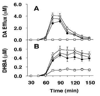
Figure 1: Effect of para-nonylphenol and bisphenolA on formation of •OH in the presence of MPP+ in the striatum of rats. After a 60-min washout with Ringer's solution (pH 7.4), stratum was infused with MPP+ (5 mM) for 15 min (solid bat; total dose, 75 nmol). Then, at 60 min after probe implantation, sodium salicylate (hatched bar; 0.5 nmol/µl/min) was infused through the microdialysis probe for 90 min to trap •OH radicals. The effects of MPP+alone (closed circle), MPP+ then para-nonylphenol (open circle), MPP+ then bisphenol A (triangle), and no MPP+ (square) on •OH generation to trapped as DHBA were compared. Brain dialysates were collected every 15 min in 0.1 N HClO4 and immediately assayed by an HPLC-EC. Values are means ± S.E.M. for five animals.
Para-nonylphenol (5 µM) significantly increased MPP+ - induced •OH formation trapped as DHBA in the dialysate by 137.1 ±11.5% (n = 6, P < 0.05), compared with MPP+ -onlytreated animals (100%). However, when corresponding experiments with bisphenolA (5 µM), the DHBA formation by 120.2 µ 10.4%, but this change was not significant. However, in the presence of tamoxifen (10 µM), a synthetic nonsteroidal antiestrogen, these compounds failed to increase the level of DA (Figure 2).
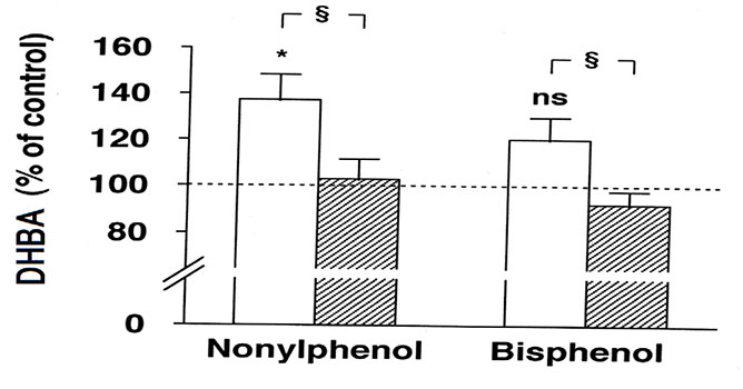
Figure 2: Effect of tamoxifen on para-nonylphenol or bisphenol A enhances MPP+-induced •OH formation. When tamoxifen (100 µM; hatched column) was administered to MPP+ then para nonylphenol or MPP+ then bisphenol A treated animals without tamoxifen (open column). The ordinate indicates that the level of DHBA, each measured 30-45 min after application of para nonylphenol or bisphenol A, and are shown as a percentage of the levels of DHBA, compared to that observed in MPP+-only treatment (100%). Values are means ± S.E.M. for six animals; *P < 0.05 versus MPP+ -only group (ANOVA and Fisher’s test).
These results indicate that tamoxifen attenuate the environmental substances enhanced DA efflux by the action of MPP+ in rat striatum. The blockage of DA release by tamoxifen may lead to prevent MPP+induced •OH generation. Moreover, to confirm the environmental substances evoked the •OH generation in the MPP+-rats by Fenton-type reaction, the •OH formation trapped as DHBA was measured in para-nonylphenol and bisphenol A-treated MPP+-rats. When iron (II) (2, 5 and 10 µM) was administered to para-nonylphenol (5 µM)-pretreated animals, iron (II) clearly produced a dose-dependent increase in the levels of DHBA, that showed a positive linear correlation between iron (II) and •OH formation trapped as DHBA (R2 = 0.985) in the dialysate. On the contrary, when corresponding experiments were performed with the bisphenol A (5 µM)-treated animals, same results were observed (Figure3).
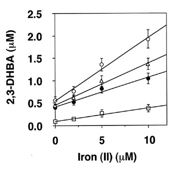
Figure 3: Cumulative dose-response relationship between iron (II) and the formation of •OH products of salicylic acid with without MPP+ treatment. Iron (II) and sodium salicylate (0.5 nmol/µl/min) were infused through the dialysis probe. Brain dialysates were collected every 15 min in 0.1 N HClO4 and immediately assayed by an HPLC-EC. Values are means ±S.E.M. for six animals. The ordinate shows the cumulative level of DHBA output over 90.
However, the levels of DHBA with bisphenol A-treated groups were lower than that of para-nonylphenol-treated group. Although there are many reports about MPP+ , the mechanism of •OH generation by MPP+ is obscure. If indeed the effect of environmental substances on •OH formation is due to DA release, environmental substances may increase •OH formation due to MPP+. It is known that estrogen significantly reduced MPP+-evoked DA release in both the corpus striatum and nucleus accumbens [5]. The sustained autoxidation of DA could lead to excessive accumulation of toxic quinones and potentially cytotoxic oxygen free radicals. These could be able to lead to the formation of •OH radicals and the radicals may induce lipid peroxidation, protein cross linking and DNA damage, mediated by base pair mutation [13]. Free radical formation enhanced by MPP+ in the striatum (but not in the cortex) appeared to be dose-dependent and positively correlated with amounts of sustained DA overflow [10]. Induction of both para-nonylphenol (5 µM) and bisphenolA (5 µM) enhanced the levels of DA due to MPP+ . However, tamoxifen significantly suppressed these compounds enhances MPP+ -released the level of DA. Therefore, environmental substances might be enhanced MPP+ -released the level of DA, via its estrogen action. On the other hand, nitric oxide (NO) can form peroxynitrite (ONOO- ) by interaction with oxygen free radicals and it can act as a neurotoxin by its interaction with thiol groups as well as through its decomposition to •OH radicals [14]. Tamoxifen was reported to inhibit NO synthase, which reduces NADPH and forms nitric oxide/superoxide ion various tissues [3,15]. Inhibitors of neuronal NO synthase such as 7-nitioindazole (7- NI) were found to prevent MPTP-induced striata DA depletion and nigral cell death [16-18]. Tamoxifen may inhibit •OH generation induced by MPP+ by suppressing NO synthase. However, further investigation is necessary to confirm the relationship between NO and environmental substances. In future, the present results may be used in answering some of the fundamental questions concerning the relevance of oxidant damage in the site effects of environmental substances.
ACKNOWLEDGEMENT
We are thankful to Prof. Lars Oreland (Department of Neuroscience, Section of Pharmacology, Box 593 Biomedical Centre, Uppsala University, 751-24, Uppsala, Sweden) for valuable discussions.
REFERENCES
- Nowak P, Kostrzewa RA, Skaba D and Kostrzewa RM. (2010). Acute L. DOPA effect on hydroxyl radical- and DOPAC-levels in striatal microdialysates of parkinsonian rats. Neurotox Res. 17(3): 299-304.
- Smith Y, Raju D, Nanda B, Pare JF, et al. (2009). The thalamostriatal systems: anatomical and functional organization in normal and parkinsonian states. Brain Res Bull. 78(2-3): 60-68.
- Obata T. (2006). Nitric oxide and MPP+ -induced hydroxyl radical generation. J Neural Transm. 113(9): 1131-1144.
- Sian-Hulsmann J, Mandel S, Youdim MB and Riederer P. (2011). The relevance of iron in the pathogenesis of Parkinson's disease. J Neurochem. 118(6): 939-957.
- Marin R, Casanas V, Perez JA, Fabelo N, et al. (2013). Oestrogens as modulators of neuronal signalosomes and brain lipid homeostasis related to protection against neurodegeneration. J Neuroendocrinol. 25(11): 1104-115.
- Samantaray S, Sribnick EA, Das A, Thakore NP, et al. (2010). Neuroprotective efficacy of estrogen in experimental spinal cord injury in rats. Ann NY Acad Sci. 1199: 90-94.
- Obata T and Kubota S. (2000). Formation of hydroxy radicals by environmental estrogen-like chemicals in rat striatum. NeurosciLett. 296(1): 41-44.
- Obata T and Yamanaka Y. (2002). Iron (III) attenuates hydroxyl radical generation accompanying nonenzymatic oxidation of noradrenaline in the rat heart. NaunynSchmiedebergs Arch Pharmacol. 365(2): 158-163.
- Hara S, Mizukami H, Kuriiwa F and Endo T. (2009). Hydroxyl radical generation dependent on extracellular ascorbate in rat striatum, as determined by microdialysis. Toxicology. 258(1): 10-16.
- Obata T, Takahashi S, Kashiwagi Y and Kubota S. (2008). Protective effect of captopril and enalaprilat, angiotensinconverting enzyme inhibitors, on para-nonylphenol-induced •OH generation and dopamine efflux in rat striatum. Toxicology. 250(2-3): 96-99.
- Navneet AK, Appukuttan TA, Pandey M and Mohanakumar KP. (2008). Taurine fails to protect against 1-methyl-4- phenyl-1,2,3,6- tetrahydropyridine-induced striatal dopamine depletion in mice. Amino Acids. 35(2): 457-461.
- Paxinos G and Watson C. (1982). The Rat Brain Stereotaxic Coordinates, 7th Edition 472.
- Dedon PC, Plastaras JP, Rouzer CA and Marnett LJ. (1998). Indirect Mutagenesis by oxidative DNA damage: formation of the pyrimidopurinone adduct of deoxyguanosine by base propenal. ProcNatlAcadSci USA. 95(19): 11113-11116.
- Samuni A and Goldstein S. (2011). One-electron oxidation of acetohydroxamic acid: the intermediacy of nitroxyl and peroxynitrite. J PhysChem A. 115(14): 3022-3028.
- Renodon A, Boucher JL, Sari MA, Delaforge M, et al. (1997). Strong inhibition of neuronal nitric oxide synthase by the calmodulin antagonist and anti-estrogen drug tamoxifen. BiochemPharmacol. 54(10): 1109-14.
- Hantraye P, Brouillet E, Ferrante R, Palfi S, et al. (1996). Inhibition of neuronal nitric oxide synthase prevents MPTPinduced parkinsonism in baboons. Nat Med. 2(9): 1017-1021.
- Serra PA, Pluchino S, Marchetti B, Desole MS, et al. (2008). The MPTP mouse model: cues on DA release and neural stem cell restorative role. Parkinsonism RelatDisord. 14(2): 189- 193.
- Kwon KJ, Kim HJ, Shin CY and Han SH. (2010). Melatonin Potentiates the Neuroprotective Properties of Resveratrol Against Beta-Amyloid-Induced Neurodegeneration by Modulating AMP-Activated Protein Kinase Pathways. J Clin Neurol. 6(3): 127-137.