Information Links
Related Conferences
Previous Issues Volume 8, Issue 2 - 2023
Enteroscrotal Fistula Following Incarcerated Inguinal Hernia that Presented as NEC in a Preterm Baby
Dilek Kurnaz*, Burcu Cebeci, Derya Buyukkayhan
Division of Neonatology, Haseki Training and Research Hospital, Health Sciences University, Sultangazi, 34265 Istanbul, Turkey
*Corresponding author: Dilek Kurnaz, Division of Neonatology, Haseki Training and Research Hospital, Health Sciences University, Sultangazi, 34265 Istanbul, Turkey, Tel: 090 212 453 20 00, ORCID: 0000-0001-9545-8820; Email: [email protected].
Received Date: October 11, 2023
Published Date: November 29, 2023
Citation: Kurnaz D, et al. (2023). Enteroscrotal Fistula Following Incarcerated Inguinal Hernia that Presented as NEC in a Preterm Baby. Mathews J Pediatr. 8(2):31.
Copyrights: Kurnaz D, et al. © (2023).
ABSTRACT
Inguinal hernia is a common surgical problem in neonates. It may be difficult to differentiate its complications and distinguish between strangulation and simple obstruction. The lack of clarification of the etiology and difficulties in diagnosis and treatment result in confusion of Necrotizing enterocolitis (NEC) with some other clinical entities. Enteroscrotal fistula following incarcerated inguinal hernia is an extremely rare entity, which requires emergency surgical intervention.We report a 24 weeker preterm infant who presented with enteroscrotal fistula following incarcerated inguinal hernia that has similar clinical presentation with NEC. Spontaneous enteroscrotal fistula is extremely rare complication in preterm infants. Inguinal hernia, which is especially common in preterm infants, should be closely monitored for complications. Non-NEC surgical pathologies should also be included in the differential diagnosis of NEC.
Keywords: Enteroscrotal Fistula, Inguinal Hernia, Preterm.
INTRODUCTION
NEC is one of the serious gastrointestinal emergencies in preterm infants. It is a gastrointestinal pathology that develops as a result of ischemic necrosis of the intestinal mucosa associated with inflammation. Prevalance is 3-15% and >90% of affected infants are preterm infants below 32 weeks of gestational age [1]. The diagnosis is based onclinical, laboratory and radiologic findings that are not specific for NEC. The lack of clarification of the etiology and difficulties in diagnosis and treatment result in confusion of NEC with some other clinical entities. The clinical findings of NEC include rectal bleeding, abdominal distension, gastric retention and moreover intestinal perforation.
Inguinal hernia is a common surgical problem in neonatal intensive care units. The most commonly affected group is preterm infants and the frequency increases inversely with gestational week. Ultrasound examination is adequate and easy to diagnose, but it may be difficult to differentiate its complications and distinguish between strangulation and simple obstruction. Frequently, surgical repair in preterm patients is postponed until after discharge under elective conditions due to accompanying morbidities [2].
Best of our knowledge, enteroscrotal fistula following incarcerated inguinal hernia is an extremely rare entity, which requires emergency surgical intervention. Incidence of incarceration is around 12% in childhood, increases to 30% in children less than one year of age and 60% in the first six months [3]. So far only one preterm infant and thirteen term infants were reported in the literature [4].
Herein, we report an extremely low birth weight (ELBW) preterm infant who presented with enteroscrotal fistula following incarcerated inguinal hernia that has similar clinical presentation with NEC.
CASE REPORT
A male preterm infant born at 24 weeks of gestational age, weighing 600 gr. He was initially supported with nasal intermittent positive airway pressure (n-CPAP) respiratory ventilation, 2 doses of surfactant were administered. He was intubated on the postnatal day 3 as a result of respiratory failure. After subsequent sepsis, reopening of the Patent Ductus Arteriosus (PDA) was detected on postnatal day 43 and ibuprofen was administered as medical treatment.
Dexamethasone treatment (DART protocol) was started because of extubation failure on the 45nd postnatal day. He was diagnosed with late-onset NEC with abdominal tenderness, sluggish bowel sounds, increased C-reactive protein level (CRP:37 mg/L), thrombocytopenia (PLT: 10000 uL) and dilatation in the intestinal loops on x-ray, the patient's enteral feeding was discontinued, broad-spectrum antibiotherapy was started, and steroid treatment was stopped. He was evaluated as suspected NEC (Bell’s Stage I) on day of life 49. We consulted the surgeon about our patient, whom we follow up with daily x-rays, with suspicion of NEC on day of life 52. Reducible inguinal hernia was dedected on the right side but elective surgical repair and continuation of medical treatment for NEC was recommended by pediatric surgeon. An increase in scrotum skin edema accompanying the existing abdominal wall edema was observed in the patient whose inguinal hernia became irreducible upon examination on the 55th postnatal day. Bowel loops herniated into the right inguinal canal, both testes were homogeneous with normal size, and blood flow was normal on ultrasonographic examination.
Whitin 22 hours while awaiting for erythrocyte transfusion of the patient who was scheduled for surgery due to incarcerated inguinal hernia, erosion and fecal leakage were observed in the erythematous area of the right scrotum skin. There was mild dilatation of the bowel loops on x-ray. He was diagnosed enteroscrotal fistula and a laparotomy was performed; there were no signs of NEC in the intestinal segments, there was strangulated inguinal hernia with ileal perforation leading to enteroscrotal fistula. Three cm intestinal segment was resected, anastomosis was performed and right inguinal canal defect was repaired. Postoperative follow-up and treatment was continued and the patient was switched to full enteral nutrition again.
Written informed consent was obtained from the parents of the patient.
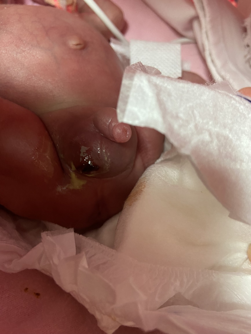
Figure 1. Clinical photograph of neonate showing faeces discharging from right scrotum.
DISCUSSION AND CONCLUSION
The incidence of inguinal hernia increases inversely with gestational age. In preterm male infants born at 24 weeks of gestation, the reported incidence reaches 40%. The higher incidence in male infants is explained by the fact that the inguinal canal remains open longer for the completion of testicular descent. The anatomy of the inguinal region of preterm infants, ventilator monitoring, conditions that increase intra-abdominal pressure such as constipation and bronchopulmonary dysplasia are predisposing factors for inguinal hernia [5]. In one study, exposure to dexamethasone, high-frequency ventilation and duration of ventilator stay were found to be associated with inguinal hernia development [6]. The incidence of strangulation in inguinal hernia is lower in preterm infants compared to term infants and has been reported to be 14-31%.
Edema and increased pressure that develop as a result of obstruction of venous return by internal or external fascial tissue in the inguinal hernia region disrupts perfusion in the tissue within the hernia and causes infarction in the tissue within the sac [2].
Emergency surgical intervention is indicated when an incarcerated inguinal hernia or strangulation that cannot be reduced is detected. However, although there is no clear timing for inguinal hernia operation in preterm infants under elective conditions, a large meta-analysis and review of systematic articles in this field found that early surgical repair performed before discharge was associated with increased recurrence. Late repair after discharge was not associated with an increased risk of incarceration compared with early repair [7].
Our patient was a preterm male infant and was in the high risk group for inguinal hernia and had many predisposing factors. Our patient had a diagnosed reducible inguinal hernia, but surgical operation was planned for after discharge due to reducibility and associated morbidities.
Although the pathogenesis of NEC is not fully understood, various mechanisms have been proposed. Although the fact that 90% of cases with NEC are premature suggests that the basic etiology is based on intestinal immaturity, it is known that multiple factors are effective in the development of the disease. Intestinal immaturity, microbial dysbiosis, hypoxia, ischemia and inflammation are the main factors involved in etiopathogenesis. Formula feeding, hypoxia, anemia, chorioamnionitis, birth asphyxia, low birth weight and presence of PDA are the main risk factors [8].
Ibuprofen is a nonsteroidal anti-inflammatory drug (NSAID) used in the treatment of PDA, but it is associated with some complications due to its potent vasoconstrictor effect on the renal and gastrointestinal (GI) system [9]. However, decreased GI blood flow due to systemic ductal steal in the presence of PDA and decreased prostaglandin-E synthesis in the GI mucosa are important risk factors for spontane intestinal perforation and NEC [10].
Our patient had a PDA that could not be closed with medical treatment and had recently been treated with NSAIDs and steroids, followed by feeding intolerance, vomiting and abdominal distension.
The earliest findings specific to the GI tract in NEC are feeding intolerance, vomiting and abdominal distension. Vomiting in infants with NEC is mostly bilious. Abdominal distension is another early sign and may be accompanied by rectal bleeding. Abdominal tenderness, abdominal skin erythema, palpable mass are more advanced GI findings. In 20-30% of cases, NEC is associated with bacteremia and clinical findings of bacteremia accompany the picture [10].
The development of NEC in preterm infants has been found to be associated with birth weight and birth week, and it has been reported that NEC can be observed up to 32 days postnatal in infants born under 1000 grams [11]. Similarly, another study reported that NEC development peaks at postmenstrual 34 weeks [12].
In our patient, NEC-like symptoms developed at postmenstrual 32 weeks and the clinical picture developed in our patient was evaluated as late-onset NEC. In preterm infants, inguinal hernia is common, incarceration is the most common known complication but strangulation following incarceration is very low [3]. However, spontaneous enteroscrotal fistula is extremely rare and only one preterm infant has been reported in the literature to our knowledge. Spontaneous enteroscrotal fistula has been reported as a complication of obstructed hernia that has not been treated for a while [4]. However, in our patient it developed within 22 hours, which may be explained by the fact that our baby was ELBW preterm.
Inguinal hernia, which is especially common in preterm infants, should be closely monitored for complications. Non-NEC surgical pathologies should also be included in the differential diagnosis of NEC.
REFERENCES
- Neu J, Walker WA. (2011). Necrotizing enterocolitis. N Engl J Med. 364(3):255-264.
- Ramachandran V, Edwards CF, Bichianu DC. (2020). Inguinal Hernia in Premature Infants. Neoreviews. 21(6):e392-e403.
- Bronsther B, Abrams MW, Elboim C. (1972). Inguinal hernias in children--a study of 1,000 cases and a review of the literature. J Am Med Womens Assoc (1972). 27(10):522-525.
- Houegban ASCR, Assan BR, Guedenon MA, Noukpozounkou SB, Gogan MVLSB, Yassegoungbe MG, et al. (2022). Spontaneous enteroscrotal fistula following an incarcerated inguinal hernia in a neonate: Case report and literature review. Int J Surg Case Rep. 90:106656.
- Unal S, Isik DU, Bas AY, Arslan Z, Demirel N. (2017). Inguinal Hernia Development in Very Low-Birth-Weight Infants: A Case-Control Study. Eur J Pediatr Surg. 27(4):341-345.
- Kumar VH, Clive J, Rosenkrantz TS, Bourque MD, Hussain N. (2002). Inguinal hernia in preterm infants (< or =32 week gestation). Pediatr Surg Int. 18(2-3):147-152.
- Ferrantella A, Sola JE, Parreco J, Quiroz HJ, Willobee BA, Reyes C, et al. (2021). Complications while awaiting elective inguinal hernia repair in infants: Not as common as you thought. Surgery. 169(6):1480-1485.
- Wertheimer F, Arcinue R, Niklas V. (2019). Necrotizing Enterocolitis: Enhancing Awareness for the General Practitioner. Pediatr Rev. 40(10):517-527.
- Groves AM, Kuschel CA, Knight DB, Skinner JR. (2008). Does retrograde diastolic flow in the descending aorta signify impaired systemic perfusion in preterm infants? Pediatr Res. 63(1):89-94.
- Hällström M, Koivisto AM, Janas M, Tammela O. (2006). Laboratory parameters predictive of developing necrotizing enterocolitis in infants born before 33 weeks of gestation. J Pediatr Surg. 41(4):792-798.
- Stoll BJ, Hansen NI, Bell EF, Walsh MC, Carlo WA, Shankaran S; Eunice Kennedy Shriver National Institute of Child Health and Human Development Neonatal Research Network. (2015). Trends in Care Practices, Morbidity, and Mortality of Extremely Preterm Neonates, 1993-2012. JAMA. 314(10):1039-1051.
- Sharma R, Hudak ML. (2013). A clinical perspective of necrotizing enterocolitis: past, present, and future. Clin Perinatol. 40(1):27-51.