Previous Issues Volume 3, Issue 1 - 2019
Effects of Trigonella Foenum Greacum in Combination with Ginger Zingiber Officinale on Nephrotoxicity in an Alloxan-Induced Diabetic Rats model
Alalwani AD*
Department of Biology, Science College, University of Jeddah, Saudi Arabia
Corresponding Author: Aisha D Alalwani, Department of Biology, Science College, University of Jeddah, Saudi Arabia.
Received Date: Sep 18, 2019 Published Date: Sep 30, 2019 Copyright © 2019 Alalwani AD Citation: Alalwani AD. (2019). Effects of Trigonella Foenum Greacum in Combination with Ginger Zingiber Officinale on Nephrotoxicity in an Alloxan-Induced Diabetic Rats model. Mathews J Cytol Histol 3(1): 10.
ABSTRACT
This study highlights the nephron-protective effects of Trigonella foenumgreacum (TFG) in combination with ginger, Zingiber officinale (GZO) in alloxaninduced diabetic rats. The alloxan-induced diabetic rat is a commonly used experimental animal model of diabetes and induces acute renal tubule interstitial nephritis, hence leading to nephrotoxicity. Forty rats were divided into four groups; normal control rats, diabetic control rats, diabetic rats treated TFG in combination with GZO, and diabetic rats treated with glibenclamide as a positive control. TFG in combination with GZO was administered orally for 6 weeks to alloxan-treated rats, and they were compared with the control and diabetic groups, respectively. The results of this study showed that diabetes mellitus induced by alloxan produced pronounced nephrotoxic effects, with a significant increase in the concentration of serum glucose and creatinine concentration, with a significant decrease in total proteins. Histopathologically, a fragmented and congested appearance of the glomerulus and degenerating and necrotic tubule epithelia were observed in the renal cortex. Administration of TFG in combination with GZO significantly decreased glucose and creatinine concentrations, while significantly increasing total protein concentrations in alloxan-induced diabetic rats. In addition, the medicinal plant treatment resulted in an improvement of histopathological changes.
KEYWORDS
Alloxan; Trigonella; Ginger; Creatinine; Total Protein; Nephrotoxicity; Diabetic Rats.
INTRODUCTION
The Diabetes mellitus is a common disease related to an increased death rate. Diabetes mellitus is a disorder of metabolic illness described by hyperglycemia coming about because of defects in insulin production, insulin activity, or both, altered metabolism of carbohydrates, lipids, and proteins, and extends to vascular disease complications [1-3]. In addition to hyperglycemia, dyslipidemia is likewise a significant risk factor for atherosclerotic patients with type-2 diabetes mellitus. Dyslipidemia in a diabetic patient is described by moderate hypertriglyceridemia and reduced high-density lipoprotein (HDL) cholesterol levels [4]. Diabetes induced by alloxan leads to beta cell-specific necrosis, insulin reduction, increase reactive oxygen species (ROS) in the status of alloxan mediated the toxic activity of these glucose analogs. Thus, expression of this glucose transporter GLUT2 in different organs like in renal cells and hepatocytes clarifies why the poisons can damage the liver and kidney [5].
The valuable impact of oral anti-diabetic medications on glycemic levels are widely reported, yet these medications cannot block the effects resulting from the complexities of diabetes-related dyslipidemia [6], Presently, herbs and plants are utilized to treat various as these have generally fewer sideeffects [7]. More than 400 kinds of plants have been noted to produce hypoglycemic effects; however, only a few of them have been tested and confirmed [8]. The anti-hyperlipidemic properties of TFG seed powder have been reported in humans and different animal species [9]. This finding emphasizes the importance of more detailed investigations using good experimental models to clarify the effects of oral treatment with TFG and GZO.
TFG seeds possess useful medicinal components, such as steroids, alkaloids, and sapogenin compounds, and have been utilized for numerous traditional treatments in Saudi Arabia. This plant, commonly applied as an antidiabetic herbal treatment, is a woody Asiatic species, which has adjusted well to Saudi Arabia, achieving heights up to 12m. There are few reports regarding the effectiveness of this plant in the literature, and some reports suggest conflicting or ineffective outcomes. The seed of the plant has been using for a long time, as a protein source for humans and animals. The fascinating point about TFG is the expansive scope of its effectiveness, including antidiabetic, anti-inflammatory, antiatherosclerotic, anticancer, triglyceride-lowering, digestive, and general metabolism improvement are some of the many properties ascribed to this plant [10]. Trigonelline is a considered as the most significant metabolite of fenugreek; this metabolite is synthesized from niacin, which is one therapeutic vitamin used for decreasing diabetes and blood cholesterol[11].
Ginger belongs to the family Zingiberaceae, and is an underground rhizome of the plant Zingiber Officinale and now, and is commonly consumed around the world [12]. Ginger is also used by traditional herbalists in south Asia for treating hypertension and cardiopathy. And its use was documented in ancient Arabic, Greek, Roman, and Chinese medical traditions [13-14].
Moreover, ginger is well-known globally for its utility in treating gastrointestinal tract disturbances such as dyspepsia, constipation, vomiting, and nausea [15]. Furthermore, it has been confirmed that ginger has medicinal properties against diabetes and rheumatism [16]. Ginger extracts have antioxidant properties that scavenge hydroxyl radicals and superoxide anions [17]. As well as ginger, prevented the initiated hyperglycemia and hyperinsulinemia [18].Sharma et al., confirm the hypolipidemic due from ginger effect, besides, Ajith et al., studied the success protective role of ginger extract against the prompted renal failure and nephrotoxicity [19-20]. This study aimed investigates the effects of the combination of Trigonella foenum greacum with Ginger Zingiber officinale and compare these effects with those of glibenclamide in a type-2 diabetes mellitus rat model.
MATERIALS AND METHODS
Plants
A single 250 mg dose used in the experimental groups contained the combination of the crushed seeds of Trigonella foenum-graecum and fresh rhizomes of Zingiber officinale (125 mg each). Authentic seeds of Trigonella foenum-graecum and fresh rhizomes of Zingiber officinale were purchased from a local market, specified and confirmed by the staff of the Botany Department from the University of Jeddah. After cleaning and drying, the seeds and roots were ground in a mechanical grinder, and 250 g of the powdered material was boiled in 2,500 ml distilled water for 30 min and cooled for 30 min at room temperature, filtered, and separated through a sieve twice [21].
Animals
Adult male Wister rats (200-250g) were purchased from King Fahd Medical Center Research (KFMCR) Laboratories in Jeddah, and housed in an animal room, with standard environmental conditions including a relative humidity of 55 ± 10%, a temperature of 23 ± 2°C, and 12-h light/dark cycles with free access to a standard commercial diet and water at least 10 times/h. All rats were sacrificed at 6 weeks after treatment. Experiments were performed according to the Guide for the Care and Use of Laboratory Animals.
Forty rats were divided into four groups; each group contained ten animals: the control group G1 (without treatment), the diabetic group G2 (injected with 150 mg/kg b. w. of alloxan), the treatment group G3 (alloxan-treated rats administered TFG in combination with GZO). The plant combination dose was administered daily in a single dose of 4ml/kg b.w. for 6 weeks. The G4 group (diabetic rats treated with a daily 4ml/ kg b. w. dose of glibenclamide for 6 weeks).
Drug treatments
The animals were fasted for 24 h prior to the induction of diabetes. Diabetes was induced in fasted rats, by a single freshly prepared dose in a normal saline intraperitoneal of 150 mg/kg body weight of alloxan monohydrate through the tail vein to induce diabetes. After injection, rats had free access to food and water; the rats were given a 5% glucose solution 6 hrs after the alloxan injection to counter hypoglycemic shock. This dose of alloxan was previously tested and proven to increase blood glucose levels above 200 mg/dl [2]. Alloxan and glibenclamide were obtained from Sigma Chemical Co. All orally administered drugs were dissolved in distilled water. Kits for glucose, creatinine, urea, and total protein measurement were purchased from Spinreact, S.A. Ctra. Santa Coloma, Spain. All other chemicals used were of analytical reagent grade.
Collection of blood samples
After 72 h, blood samples were collected from tail veins for evaluation of glucose levels using a glucometer. Animals with glucose levels above 196 mg/dL (11.1 mmol/L) were used for this study, i.e., those presenting glucose levels below 196 mg/dL were excluded from this study [22]. At 6 weeks posttreatment, blood samples were collected by sacrificing the animals, and the blood was collected in clean EDTA tubes, then plasma was separated by centrifugation and stored at -20°C for biochemical analysis. Glucose determination was performed according to the method of Trinder, while creatinine and urea were determined using an enzymatic method according to a previously described method. [23-24]
Histological studies Small portions of kidney tissues were fixed in 10% phosphatebuffered formalin, dehydrated, and then embedded in paraffin. Sections (4-μm-thick) were stained with hematoxylin [25]. Additionally, samples of kidney were fixed in 2.5% glutaraldehyde and 0.25 M sodium cacodylate, post-fixed in 1% osmium tetroxide and embedded in Spurr’s epoxy. Ultrathin sections were mounted on nickel grids and stained with uranyl acetate and lead citrate [26]. The sections were examined in a Philips TEM 100 in KFCMR and photographed.
Statistical analyses
Data were statistically analyzed using one-way analysis of variance followed by Duncan’s test (PC-stat computer program). Results were considered statistically significant when P < 0.05.
RESULTS
Blood glucose and body & kidney weights The administration of TFG in combination with GZO to diabetic rats (the G3 group) significantly reduced blood glucose levels when compared with diabetic rats (the G2 group). This reduction was not enough to affect normal rats, but it was still significantly higher when compared with the control group G1; these results are shown in (Table 1).
On the other hand, body and relative kidney weights were decreased in alloxan-induced diabetic rats and then treated with TFG in combination with GZO when compared with the control group, but it was still significantly higher than the G2 diabetic group and the G4 glibenclamide-treated group.
Table 1: Effect of Trigonella in Combination with Ginger on fasting blood glucose level and body weight.
|
Groups |
Blood glucose mg/dl |
Body weight |
Kidney weight |
|
G1 |
97.6 ± 4.6 |
237.5 ± 3.5 |
0.92 ± 0.2 |
|
G2 |
328 ± 10.83* |
206.0 ± 3.8 |
0.65 ± 2.1 |
|
G3 |
90.4 ± 8.7 |
226.0 ± 2.9 |
0.83 ± 0.5 |
|
G4 |
199 ± 3.0 |
223.0 ± 2.9 |
0.76 ± 0.3 |
|
P values; * >0.01 |
|||
Kidney functions
Indicates (Table 2) Alloxan produced a significant increase in the plasma levels of creatinine, urea, and uric acid in the G2 diabetic rat group when compared with the G1 control group. Conversely, treatment of TFG in combination with GZO in the G3 diabetic rat group significantly decreased the levels of plasma creatinine, urea, and uric acid when compared with the G2 diabetic group, but no significant changes were observed when compared with G1. Furthermore, G4 rats treated with glibenclamide exhibited plasma levels of measured markers significantly elevated relative to the G1 control rats group. In contrast, when compared with the control group, total proteins did not deviate from the normal range in the G3-treated group, as shown in (Table 2), while decreased total protein was noted in G2 diabetic animals.
Table 2: Effect of Trigonella in Combination with Ginger on plasma creatinine, urea and uric acid and total protein in alloxan diabetes.
|
Groups |
creatinine |
Urea |
Uric acid |
Total protein |
|
G1 |
0.98 ± 0.04 |
32.8 ±1.7 |
1.6 ± 0.1 |
6.32 ± 0.59 |
|
G2 |
2.86 ± 0.4* |
86.5 ± 3.2 |
4.8 ± 0.3* |
4.16 ± 0.35 |
|
G3 |
1.03 ± 0.01 |
35.3 ± 1.9 |
1.7 ± 0.1 |
6.51 ± 0.13 |
|
G4 |
1.68 ± 0.02 |
49.6 ± 2.0 |
2.8 ± 0.18 |
5.2 ± 038 |
|
P values; * >0.01 |
||||
Histological studies
The functional unit of the kidney is the nephron, formed by the glomerulus and the uriniferous tubule. The renal cortex is easily identified in the control group even at low magnification (Figure 1a) by the presence of renal corpuscles, which are dense rounded structures, the glomeruli, surrounded by Bowman’s spaces, normally filled with plasma ultra-filtrate and only just visible at this magnification. The tubules fill the bulk of the parenchyma between the corpuscles. However, the cortex consists mainly of proximal convoluted tubules lined by more eosinophilic epithelial cells, with smaller numbers of distal convoluted tubules (Figure 1b).
Electron microscopic examination of the G1 group showed glomerular cells and capillaries (Figure 1c, d). Some of the capillaries contain erythrocytes and are defined by the prominent glomerular basement membranes (GBM), where capillary endothelial cell nuclei are seen bulging into the capillary lumen. The mesangium consists of material similar to the basement membrane and contains mesangial cells, which have an essential role in the control of capillary flow in the glomerulus. They also secrete the mesangial matrix and have a phagocytic function. The mesangium is separated from the capillary lumen only by a thin layer of fenestrated endothelial cell cytoplasm. Podocytes also enclose the capillary loops and are adjacent to Bowman’s space; podocytes have large flattened cell bodies and bulging nuclei, and several long primary processes that surround one or more capillaries. The glomerular filtrate must cross the endothelial cell, GBM, and the foot processes of podocytes. The podocyte is united to the GBM and forms the interdigitating foot processes and the filtration slits, whose structure is essential in the filtration of proteins and other molecules.
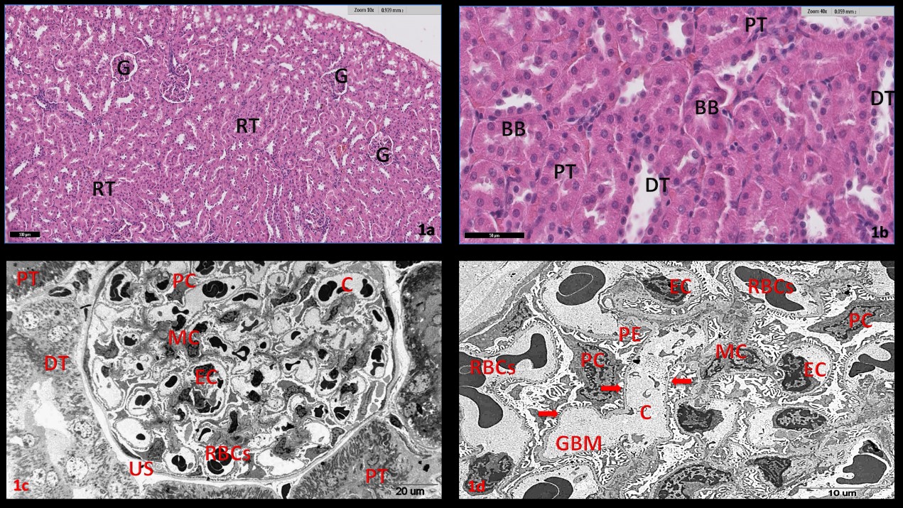
Figure 1: Plate (1a-d): sections of kidney cortex of male Wister rats; (a-b): Light microscope (L.M.) of kidney section of control rats (G1) H&E. :( a): showing the normal histological structure of the glomerulus (G) and renal tubules (RT) in the cortical portion. (b): High power from the previous section showing proximal convoluted tubules (PT), brush border (BB) and distal convoluted tubules (DT) in the cortical portion. (c-d): Electron micrographs (E.M.) of kidney cortex of control rat (G1): (c): apart from glomerulus showing podocytes (PC), mesangail cell (MC), endothelial cell (EN) and capillary (C) loops filled with red blood cells (RBCs). Also, note proximal convoluted tubules (PT) and distal convoluted tubules (DT). (d): showing podocytes (PC) with nearly normal nucleus, pedicles (PE), normalappearing glomerular basement membrane (GBM) and filtration slits with the diaphragm (arrows), also note mesangial cell (MC) with mesangial matrix and endothelial cell (EN) with a large irregular nucleus.
Both kidneys from rats in the G2 group showed similar lesions consisting of many fragmented, congested glomeruli; basal membranes were disrupted, and the urinary space observed in the cortex appeared obliterated (Figure 2a-e). Degeneration and necrosis of tubular epithelial cells were seen along with tubular obstruction due to cell debris (Figure 2a, b). Dilated regenerated tubules were lined with flattened and attenuated epithelia in the basophilic cytoplasm. These tubular epithelial cells often piled up, formed small cell clusters, and protruded into the lumen. In addition, we noted multinuclear foreign-body giant cells, and macrophages often infiltrated. Multinuclear foreign body giant cells contained minerals in their cytoplasm. Many degenerated tubules did not have a brush border, but some dilated and degenerated tubules did. Also, hyaline arteriolosclerosis affecting afferent and afferent arterioles of the glomeruli was present, as shown in (Figure 2c).
Electron micrographs (Figure 2d, e) confirmed that the observed effects in the diabetic state also occurred in the renal corpuscles and tubules in kidney cortex of G2 male rats. Many of the glomeruli showed congested capillaries filled with deformed red blood cells, focal glomerular basement membrane thickening (GBM), glomerular capillary endothelial abnormalities, and mesangial proliferation and were filled with mesangial matrix, and focal fused foot processes of podocytes (Figure 2d). The renal tubule cells, proximal tubules, increase of lysosomes and swelling of mitochondria become oval or rounded and increased volume. Note a part of distal tubular lumen filled with secretion and lipid droplet, destruction of cytoplasmic organelles, short and damage basal in folding with sever increased number of condense mitochondria and irregular of the basement membrane (Figure 2e).
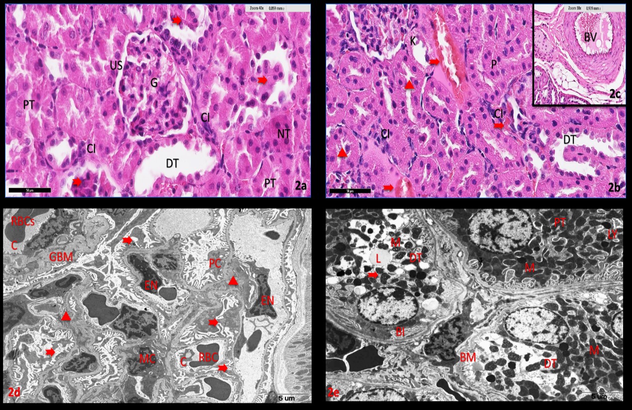
Figure 2: Plate (2a-g): sections of kidney cortex of diabetic rats (G2). (2ac): light micrographs of renal corpuscle and tubules in kidney cortex of male rats: H&E. (a): showing a fragmented and congested of glomerulus, basal membrane was disrupted (G), obliterate urinary space (US), many degenerations and necrosis tubules epithelium (NT) did not have brush border, tubular epithelial cells in some proximal tubules often piled up (PT), formed small cell clusters and protruded into the lumen (arrows), dilated of distal tubules (DT) and cellular infiltration(CI). (b): showing brush border fragmented (head arrows), dilated intercellular spaces (arrows), dilated and occluded distal tubules were segmentally (DT), Pyknosis (P), karyolysis (K) of tubular cells nuclei and cellular infiltration (CI). (c): showing dilation of the cortical blood vessel (BV) with red blood cells stasis. (2d-e): Electron micrographs (E.M.) of renal corpuscle and tubules in kidney cortex of male rats (G2): (d): apart from glomerulus showing congested capillaries (C), filled with deformed red blood cells (RBCs), focal glomerular basement membrane thickening (GBM), glomerular capillary endothelial abnormalities (EN), mesangial proliferation (MC) and filled with mesangial matrix (head arrows) also focal fused foot processes(arrows)of podocytes (PC). (e): a part of renal tubule cells, proximal tubules (PT), increase of lysosomes (LY) and swelling of mitochondria (M) become oval or rounded and increased volume. Note a part of distal tubular (DT) lumen filled with secretion and lipid droplet (L), destruction of cytoplasmic organelles(arrows), short and damage basal infolding (BI) with sever an increased number of condense mitochondria(M) and irregular of the basement membrane (BM).
Examination of sections of kidney cortex G3 of diabetic rats then given Trigonella in combination with ginger (Figure 3a, d), showing the normal histological structure of the glomerulus and renal tubules in the cortical portion, proximal convoluted tubules, good brush border and distal convoluted tubules in the cortical portion (Figure 3a). Electron micrographs of kidney cortex of rat (G3) showing podocytes, mesangial cells, endothelial cells, and capillary loops filled with red blood cells (Figure 3b). Podocytes with nearly normal nuclei, pedicles, and normal-appearing glomerular basement membranes (GBM) with clear filtration slits in the diaphragm (Figure 3c). Centrally located nuclei with normal chromatin distribution, numerous well-organized elongated mitochondria, brush border, and pinocytotic vesicles were noted in proximal convoluted tubules, and distal convoluted tubules showed apical spheroid nuclei, and well-developed basal in-folding surrounding filamentous elongated mitochondria (Figure 3d). renal cortical of rats a in the G4 diabetic rats treated with glibenclamide resembled healthy kidney tissue, and the regeneration of kidney parenchyma and glomerular apparatus appeared to be intact, but there was degeneration of the renal tubules limited to very small loci.
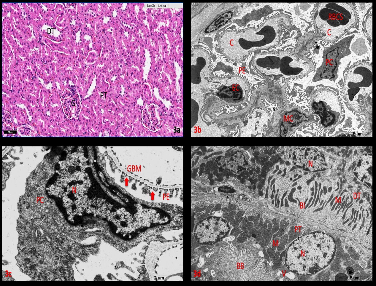
Figure 3: Plate (3a-d): sections of kidney cortex of diabetic rats then given Trigonella in Combination with Ginger (G3). (3a): light micrographs of renal corpuscle and tubules in kidney cortex of male rats: H&E. :( a): showing the normal histological structure of the glomerulus (G) and renal tubules in the cortical portion, proximal convoluted tubules (PT), brush border and distal convoluted tubules (DT) in the cortical portion. (3b-d): Electron micrographs (E.M.) of kidney cortex of rat (G3): (b): apart from glomerulus showing podocytes (PC), mesangail cell (MC), endothelial cell (EN) and capillary (C) loops filled with red blood cells (RBCs). (c): showing podocytes with nearly normal nucleus (N), pedicles (PE) also normalappearing glomerular basement membrane (GBM) and filtration slits with the diaphragm (arrows). (d): showing a central located nucleus (N) with normal chromatin distribution, numerous well-organized elongated mitochondria (M), brush border (BB) and pinocytotic vesicles (V) note in proximal convoluted tubules (PT), and a part of distal convoluted tubules (DT) apical spheroid nuclei (N) of DCT, well-developed basal infolding (BI) surrounding filamentous elongated mitochondria (M).
DISCUSSION
The structure of alloxan resembles glucose, and binds the GLUT2 glucose transporter, and can enter cells via the GLUT2 glucose transporter [5]. This entry produces hydroxyl radicals and superoxide; since beta cells typically have little protection against oxidative injury, they are sensitive to free radicaldamage by alloxan, and cellular necrosis ensues [27]. The GLUT2 glucose transporter is present in hepatocytes, beta cells, and epithelial cells of renal tubules [27]. The GLUT2 glucose transporter is located in the basal membrane of the epithelia of the proximal tubules and can cause damage to renal cells when exposed to alloxan.
Recently, interest in complementary medicine has expanded and utilizes plants and herbs because these often produce fewer side-effects when used to treat different illnesses [6,7]. Trigonella Foenum Greacum and ginger, Zingiber officinale are two plants utilized in the treatment of diabetes [28]. The therapeutic properties of these plants have been documented in human and animals [9]. Trigonella foenum-graecum increases insulin effects and defers carbohydrates absorption through the action of its sapogenins, which increases the secretion of biliary cholesterol [11].
In our study, we found that treatment with Trigonella in combination with ginger, in the G3 diabetic rats caused a decrease of plasma glucose levels when compared with the G2 diabetic rats. These results are in agreement with the findings of Akbari et al., who confirmed that post-treatment and pretreatment of diabetic rats with ginger extract increased insulin levels and significantly decreased blood glucose levels [29]. Kar et al., reported that the inorganic part of a therapeutic plant contains the most significant portion of mineral elements, which lead to hypoglycemic activity [30]. In support of this view, a large number of essential minerals (Ca, Zn, K, Mn, and Cr), are known to be associated with the mechanisms of insulin activity and its release in humans and various animals [31]. In addition, G4 diabetic rats treated with glibenclamide showed reduced plasma glucose levels when compared with the G2 diabetic group, but that reduction was not sufficient to reach levels seen in normal rats, though it was still higher than those observed in the G1 control group.
Alloxan-induced diabetes is described as a severe loss in body and kidney weight, and the same was seen in the present study (Table 1) [32]. The rats experienced a gradual reduction in body weight due to an accompanied reduction in food intake and urine volume following alloxan injection. Diabetes mellitus is due to insulin deficiency in the circulating blood and decreased responsiveness to insulin, or insulin resistance. These abnormalities lead to changes in the metabolism of proteins, lipids, and carbohydrates; the main feature of diabetes mellitus is hyperglycemia, muscle weakness, and weight loss. In our study G3 administration of TFG in combination with GZO could control weight loss compared with G4 rats treated with glibenclamide.
The results of this study suggest that the effects of TFG in combination with GZO in G3 diabetic rats reduced the levels of plasma creatinine, urea, and uric acid while increased total protein to normal levels and result improve the histological changes in the renal cortical. Whilst increased plasma creatinine levels and urea elevations in group G2 is evidence of acute kidney dysfunction [33]. The elevation of plasma creatinine, urea, and uric acid in diabetic rats are considered a significant marker of renal dysfunction.
Furthermore, Ajith et al., explained that ginger extracts protected against induced nephrotoxicity and elevation of serum creatinine and urea levels, which were apparent from the decreased serum urea, and creatinine levels in the animals that were treated with ginger extract [20].
Moreover, the treatment of ginger could significantly inhibit the depletion of antioxidant enzymes and increase antioxidant concentrations in the kidneys. In addition, Ajith et al., pointed out that the presence of flavonoids and polyphenols in ginger are responsible for the reduction of serum urea and creatinine levels and antioxidant nephroprotective activities [20].
The first description of the high capacity of alloxan to specifically injure pancreatic beta-cells was documented by Dunn et al. These researchers studied the nephrotoxicity of uric acid and found that alloxan caused the demolition of most of the pancreatic beta cells in the rabbit [34]. The nephrotoxic effect of alloxan had been known long before its diabetogenic activity was identified [35-36]. Renal changes induced by alloxan include extensive vacuolar, swelling, degeneration, and necrosis of tubular cells, cellular infiltration in the interstitium and tubular dilation. Renal damage may produce uremia and death.
Consequently, alloxan administration has been found to lead to diabetes in numerous animal species [37]. In this report, the histopathological changes observed in an alloxan-induced diabetic rat were similar to those described in previous reports and they were likely due to the nephrotoxicity of alloxan [38]. Unlike previous reports, serious mineralization was not found in rats with alloxan-induced nephrotoxicity. Diabetic hyperglycemia is a common finding in diabetes and is responsible for histopathological changes and vascular complications [39].
CONCLUSIONS
From our finding that both of TFG in Combination with GZO treatment use to controlling the blood glucose level and role antioxidant potential to prevention kidneys against damage caused due to diabetic. In addition, capable of improving the impaired kidney functions in alloxan-induced diabetic rats.
REFERENCES
- Zimmet P, Magliano D, Matsuzawa Y and Alberti G, et al. (2005). The metabolic syndrome: A global public health problem and a new definition. J Atheroscler Thromb. 12(6): 295-300.
- Zhang J, Huang Y, Hou T and Wang Y (2006). Hypoglycemic effect of Artemisia sphaerocephala Krasch seed polysaccharide in alloxan-induced diabetic rats. Swiss Med Wkly. 136 (33-34): 529-532.
- Yamagishi SI, Nakamura K, Matsui T and Ueda SI, et al. (2007). Role of postprandial hyperglycaemia in cardiovascular disease in diabetes. Int J Clin Pract. 61(1): 83-87.
- Chahil TJ and Ginsberg, HN (2006). Diabetic dyslipidemia. Endocrinol Metab Clin North Am. 35(3): 491-510.
- Elsner M, Tiedge M and Lenzen S (2003). Mechanism underlying resistance of human pancreatic beta cells against toxicity of streptozotocin and alloxan. Diabetologia. 46: 1713–1714.
- Kim J, Kang S, Seo B and Choi H, et al. (2006). Antidiabetic efficacy of SMK 001, a Poly Herbal Formula in Streptozotocin Induced Diabetic Rats: Therapeutic Study. Bio Pharma Bull. 29(3): 477-482.
- Gilani, AH and Rahman, AU (2005) Trends Ethnopharmacol J Ethanopharmacol. 100(1-2): 43-49.
- Silva KL, Biavatti MW, Leite SN, Yunes RA, Delle MF, Cechinel VJ (2000). Phytochemical and Pharmacognostic Investigation of Bauhinia forficata Link (Leguminosae). J Biosciences. 55: 478-480.
- Badary OA, Abdel-naim AB, Abdel Wahab MH and Hamada FM. (2000). The influence of thymoquinone on doxorubicin-induced hyperlipidemic nephropathy in rats. Toxicology. 143(3): 219-226.
- Salehi Surmaghi MH. (2003). Medicinal Plants and Herbal Therapy. [Vol 1]. Tehran University Press, Tehran, Iran, 253-254.
- Basch E, Ulbricht C, Kuo G, Szapary P and Smith, M. (2003). Therapeutic applications of fenugreek. Altern Med Rev. 8(1): 20-27.
- Sertie JA, Basile AC and Panizza S. (1991). Pharmacological assay of Cordia verbenacea. III: oral and topical anti-inflammatory activity and gastrotoxicity of a crude leaf extract, J Ethnopharmacol. 31(2): 239-247.
- Duke JA (2002). Handbook of Medicinal Herbs. CRC Press, Boca Raton, FL, USA, 327-329.
- Bhandari U, Sharma JN, and Zafar R. (2001). The protective action of ethanolic ginger (Zingiber officinale) extract in cholesterol-fed rabbits. J Ethnopharmacol. 61(2): 167-171.
- Tyler VE. (1993). The Honest Herbal [3rd Ed.] Pharmaceutical Products Press, NY, USA, 147-148.
- Afzal M, Al Hadidi D, Menon M, Pesek J, et al. (2001). Ginger: an ethnomedical, chemical and pharmacological review. Drug Metab Drug Interact. 18(3-4): 159-90.
- Krishnakantha TP and Lokesh BR. (1993). Scavenging of superoxide anions by spice principles. Indian J Biochem Biophys. 30(2): 133-134.
- Akhani SP, Vishwakarma SL, and Goyal RK. (2004). Anti-diabetic activity of Zingiber officinale in Streptozotocin-induced type I diabetic rats. J Pharm Pharmacol. 56: 101-105.
- Sharma I, Gusain D and Dixit VP (1996). Hypolipidemic and anti-atherosclerotic effects of Zingiber officinale in cholesterol-fed rabbits. Phyto Res. 10: 517-518.
- Ajith TA, Nivitha V and Usha S. (2007). Zingiber officinale Roscoe alone and in combination with alpha-tocopherol protect the kidney against cisplatin induced acute renal failure. Food Chem Toxicol. 45(6): 921–927.
- Xue WI, Li XS, Zhang JI, Lue YI, et al. (2007). Asia Pac J Clin Nur 16: 422-426.
- Onyije FM and Avwioro OG. (2012). Effect of ethanolic extract of Bauhinia monandra leaf on the liver of alloxan-induced diabetic rats. J Phys Pharm Adv. 2(1): 59-63.
- Trinder P. (1969). Enzymatic determination of glucose. Ann Clin Biochem. 6: 24.
- Tyler VE. (1993). The Honest Herbal, [3rd Ed.] Pharmaceutical Products Press, NY, USA, 147-148.
- Patton, CJ and Crouch SR. (1977). Spectrophotometric and kinetics investigation of the Berthelot reaction for the determination of ammonia Anal Chem. 49(3): 464-469.
- Bancroft JD and Gamble M. (2002). Theory and Practice of Histological Techniques. [5th Ed.], Churchill Livingstone, Edinburgh, UK.
- Woods AE and Stirling JW. (2002). Electron microscopy: The preparative teqhniques. In: Theory and practice of histological techniques. [5th Ed.] Bancroft JD and Gamble M. [Eds], Harcourt Publishers, USA.
- Lenzen S. (2008). The mechanisms of alloxan- and streptozotocininduced diabetes. Diabetologia. 51(2): 216-226.
- Abdel-barry JA, Abdel-hassan IA, Jawad AM and Al-hakiem MH. (2000). Hypoglycaemic effect of aqueous extract of the leaves of Trigonella foenum graecum in healthy volunteers. East Mediterr Health J. 6(1): 83-88.
- Akbari F, Ansari-Samani R, Karimi A, Mortazaei S, et al. (2013). Effect of turnip on glucose and lipid profiles of alloxan-induced diabetic rats. Iran J Endocrinol Metab. 14(5): 1-7.
- Kar A, Choudhary BK and Bandyopadhyay NG. (1999). Preliminary studies on the inorganic constituents of some indigenous hypoglycaemic herbs on oral glucose tolerance test. J Ethnopharmacol. 64 (2): 179-184.
- Castro VR. (1998). Chromium in series of Portuguese plants used in the herbal treatment of diabetes. Biol Trace Elem Res. 62(1-2): 101-106.
- Fernandes NP, Lagishetty Panda VS and Naik SR. (2007). BMC Complement Altern Med. 7: 1-8.
- Saumya RP, Satyaranjan M, Sabuj S and Prasana KP. (2011). Nephroprotective effect of Bauhinia variegata (Linn.) whole stem extract against cisplatin-induced nephropathy in rats. Indian J Pharmacol. 43(2): 200– 202.
- Memon A, Memon A, Shah S, Khand F, et al. (2010). Effect of combination of Nigella sativa and Trigonella foenum-graecum seeds with Glibenclamide on blood sugar levels in Type-2 Diabetes Mellitus patients. Isra Medical Journal. 2(2): 46-51.
- Evan AP, Mong SA, Connors BA, Aronoff GR, et al. (1984). The effect of alloxan, and alloxan-induced diabetes on the kidney. Anat Rec. 208(1): 33–47.
- Bruckmann G and Wertheimer E. (1947). Alloxan studies; the action of alloxan homologues and related compounds. J Biol Chem. 168(1): 241–256.
- Luz MMS, Santos CAM, Sato MEO and Arruda AMS. (1996). In Livro de Resumos. XIV Simpósio de Plantas Medicinais do Brasil, Florianopolis, 84.
- Vargas L, Friederici HH, and Maibenco HC. (1970). Cortical sponge kidneys induced in rats by alloxan. Diabetes. 19(1): 33–44.
- Kudchodkar BJ, Lee JC, Lee SM, DiMarco NM, et al. (1988). Effect of cholesterol homeostasis in diabetic rats. J Lipid Res 29: 1272-1287.