Information Links
Related Conferences
Previous Issues Volume 5, Issue 1 - 2021
Diagnostic Accuracy of Digital Radiography and Novel Diagnostic Tools versus Visual ICDAS Criteria: A Systematic Review
Shereen Hafez Ibrahim1*, Reham Nabil2, Peter Edward2
1Assistant professor of Conservative Dentistry, Faculty of Dentistry, Cairo University, Cairo, Egypt
2Assistant lecturer of Conservative Dentistry, Faculty of Dentistry, Cairo University, Cairo, Egypt
*Corresponding author: Shereen Hafez Ibrahim, Associate Professor of Conservative Dentistry, Department of Conservative Dentistry, Faculty of Dentistry, Cairo University, 11 EL-Saraya St. Manial, Cairo, Postal Code: 11553, Egypt, Tel:+201099296591; E-mail: [email protected]
Received Date: March 10, 2021
Publication Date: April 13, 2021
Copyrights: Ibrahim SH, et al. © (2021).
Citation: Ibrahim SH, Nabil R, Edward P. (2021 Diagnostic Accuracy of Digital Radiography and Novel Diagnostic Tools versus Visual ICDAS Criteria: A Systematic Review. Mathews J Dentistry. 5(1):27.
ABSTRACT
Purpose: To evaluate the diagnostic accuracy of digital radiography and other novel diagnostic tools compared to visual ICDAS criteria.
Materials and Methods: Two reviewers performed a literature search up to May 2020 in three databases: PubMed, Google Scholar, and Cochrane Library. Only in-vivo studies that evaluated the diagnostic accuracy of digital radiography, Diagnodent, Vistaproof, Soprolife, and ICDAS criteria were included.
Results: A total of 27 in vivo studies were included in the systematic review. Studies included suggested that digital dental radiography proved to be a reliable tool in detecting initial carious lesions/ especially proximal lesions. The Diagnodent device was proved to be an efficient adjunct to other detection methods, especially in the case of non-experienced clinicians. For VistaCam/VistaProof assessment, it was found that in some articles, VistaCam proved high accuracy in detecting early carious lesions with moderate to strong correlation in comparison to results obtained by visual and tactile methods ICDAS criteria. ICDAS criteria were considered to be sufficient alone in detecting occlusal caries.
Conclusions: The in vivo evidence suggested that None of the diagnostic methods alone are sufficient for diagnosis of dental caries.
Keywords: diagnostic accuracy, digital radiography, novel diagnostic tools, ICDAS visual criteria, systematic review.
INTRODUCTION
The diagnosis of dental caries is always a challenge since its patterns and prevalence have significantly changed in the last few years. The difficulty in diagnosing dental caries depends not only on the morphological changes of lesions and their rate of progression but also on the lack of a precise methodology to efficiently diagnose both the disease and the integrity of the dental structure (specificity) [1].
Conventional examination for caries detection is primarily done using visual inspection, tactile sensation, and radiographs. While these methods give satisfactory results in the detection of cavitated lesions, they are usually inadequate for the detection of initial lesions. Because of these deficiencies, new detection methods have been developed to aid better diagnosis [2].
To date, there are two main techniques aimed at helping clinicians in detecting caries represented by visual examination and by light-based caries diagnostic tools as fiber-optic transillumination (FOTI), DIAGNODent tool (KaVo), and SOPROLIFE. Visual diagnosing of caries has progressed by establishing the international caries detection and assessment system (ICDAS). ICDAS-II, the second version, was improved and provided a standardized system to enable clinicians to diagnose and detect the first visual change in enamel, leading to better information for clinical management [3].
There are various fluorescence-based methods, of which the most commonly studied are KAVO® DIAGNOdent and VistaProof by Durr Dental®, which may be used as complementary diagnostic tools in order to avoid the occurrence of false-positive and false-negative findings. VistaProof an intraoral camera which emits a 405 nm light wavelength and takes images that are computer-processed; the resulting mapping of the lesion is produced according to its depth [4,5].
The aim of this systematic review is to evaluate the diagnostic accuracy of digital radiography and other novel diagnostic tools in comparison to visual ICDAS criteria.
MATERIALS AND METHODS
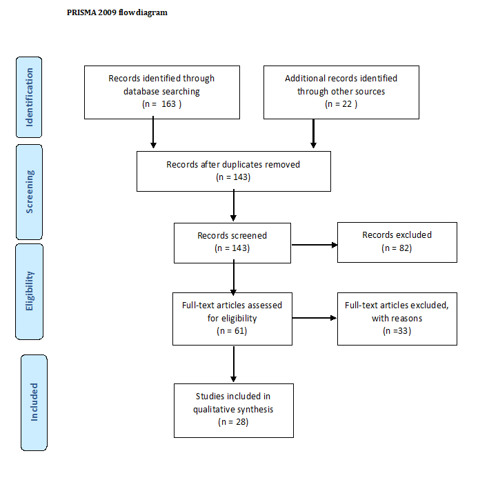
This systematic review was reported according to the guidelines of the PRISMA statement. The protocol was registered in the PROSPERO international database for systematic reviews (CRD-183388). The research question was: What is the diagnostic accuracy of digital radiography and other novel diagnostic tools in comparison to visual ICDAS criteria?
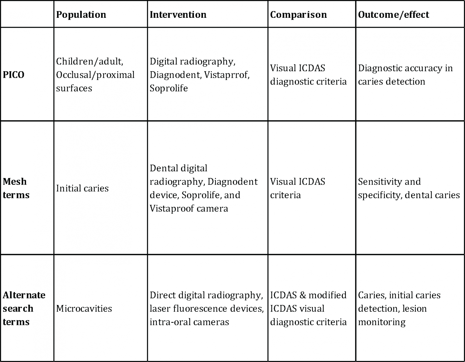
A detailed database search was conducted using Medical Subject Heading terms [Table 1] in PubMed, Cochrane Library, and google scholar. The search words included digital radiograph, dental digital radiograph, Diagnodent, laser fluorescence devices, Vistaproof/Vistacam, Soprolife, initial caries, and microcavities. The search was restricted to articles published in the English language with a time period limit from 2000 to 2020.
INCLUSION CRITERIA
• In vivo studies on caries detection methods
• Studies on diagnostic methods reporting diagnostic accuracy, i.e., sensitivity, specificity, and receiver operating characteristic curves
• Studies on primary/permanent teeth validated with reference/ gold standard
• Studies published in the English language only from 2000 to April 2020.
EXCLUSION CRITERIA
• In vitro studies
• Studies with an incomplete description of the sample size or outcome
• Studies reporting accuracy in laboratory work exclusively
• Animal studies, review articles, letters to the editor and conference abstracts.
Table 1: Medical subject heading terms and alternate terms enclosed in the search strategy.
TYPE OF PARTICIPANTS
All studies on human beings were included, regardless of age.
TYPES OF STUDY
All clinical trials on human beings and Occlusal/proximal surfaces were included. The English language was chosen.
TYPE OF OUTCOME
The main outcome was the absence or presence of dental caries using the following methods:
“Histological examination, the opening of cavities, or clinical examination based on ICDAS scores utilized in studies”.
REVIEW METHODS
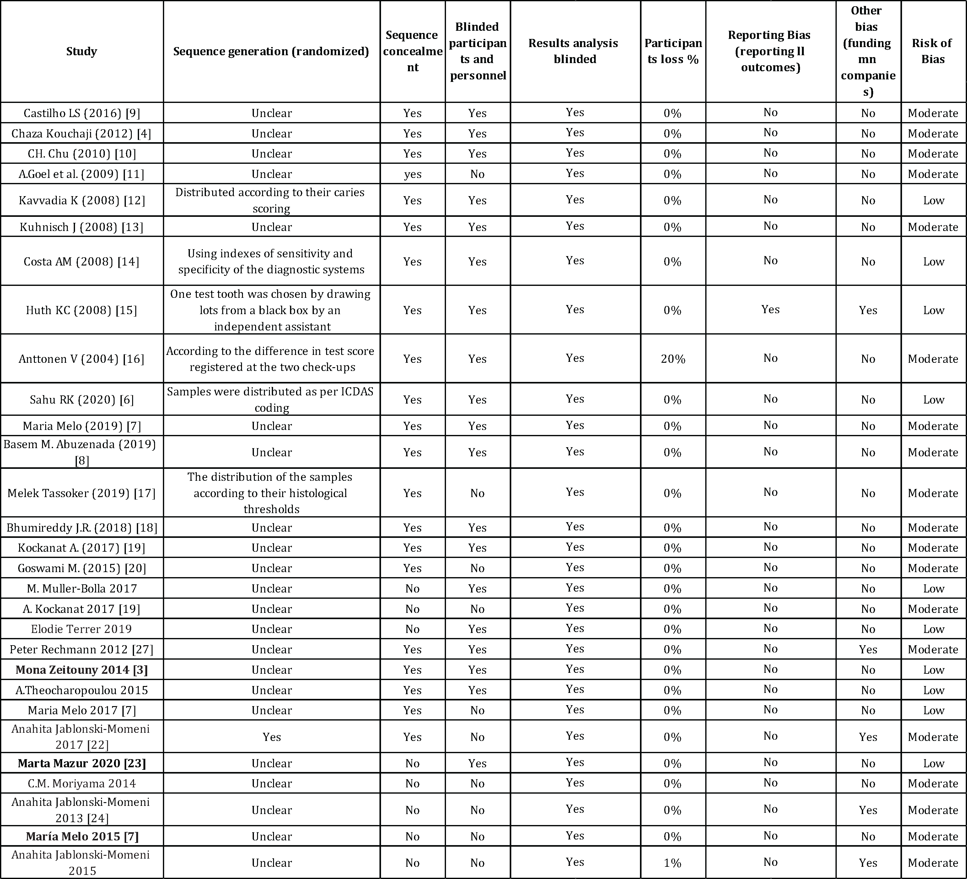
All reports yielded by the search were printed out and analyzed by two reviewers on the basis of title, keywords, and abstract to check if the study was likely relevant. A full report of all relevant papers was obtained, and also if a paper could not be classified.
The reviewers were not blinded to authors, journals, date of publication, financial support, or results. The inclusion criteria were applied, and the data assessed and extracted by two reviewers.
RESULTS
For Digital Radiography
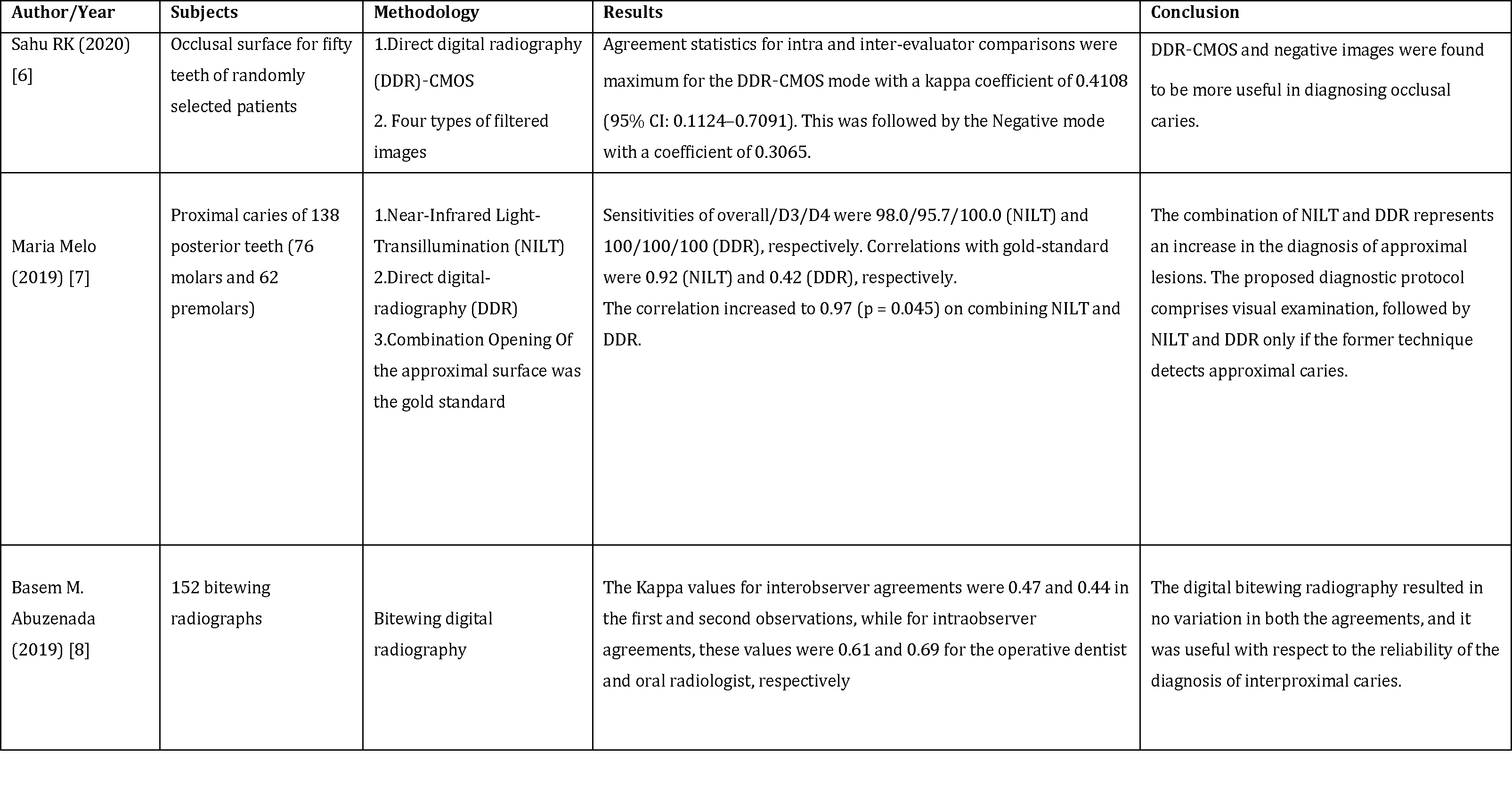
The search strategies yielded eleven reports from the search database. All were published in the English language between 2008 and 2020. Three of these met the selection criteria after reading the full articles. Of the eight reports excluded, three were literature reviews, and the remaining, one systematic review and four were in vitro investigations.
Therefore, three studies were selected for analysis of the methodology and data reliability (Table 2).
For Diagnodent
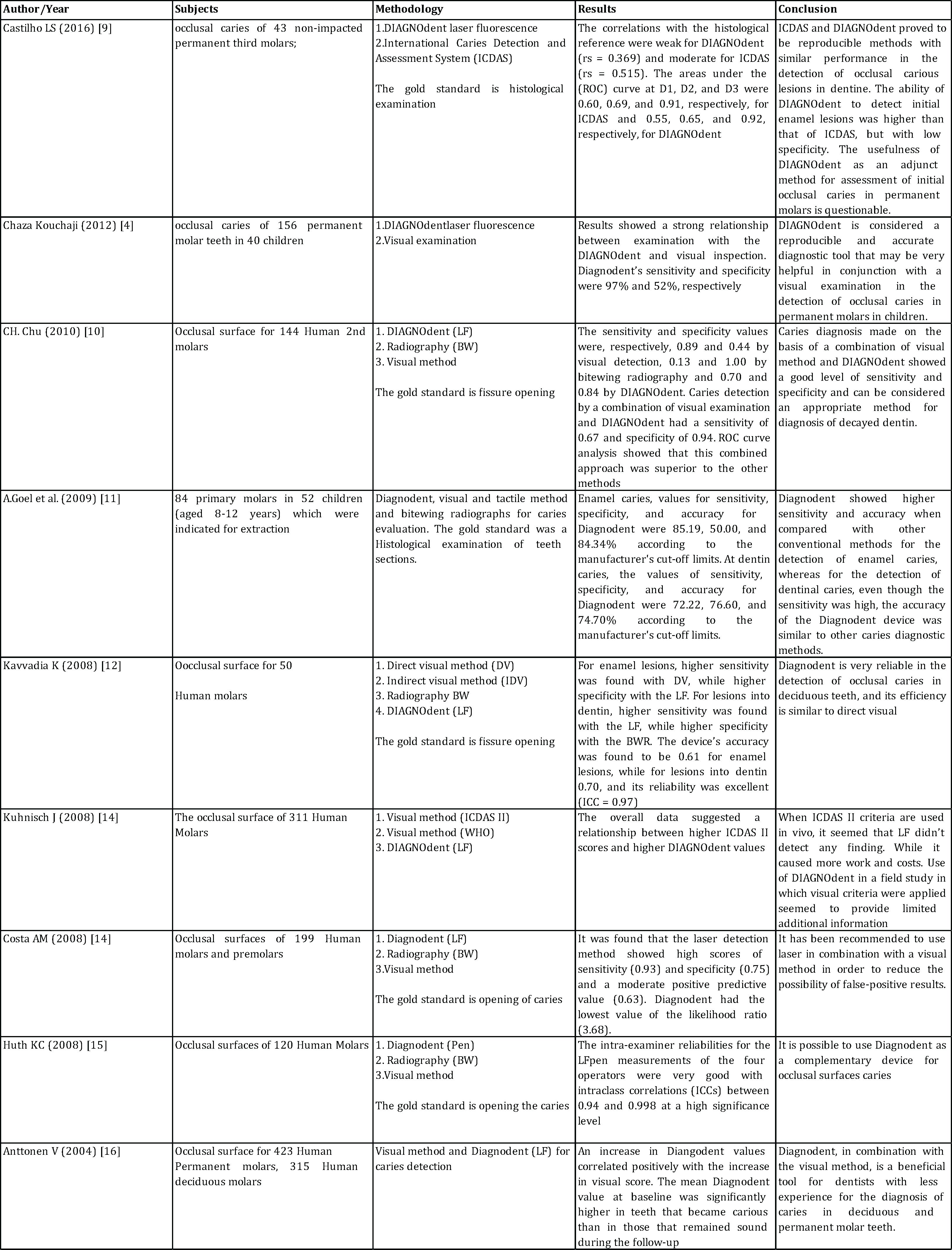
The search strategies yielded fifteen reports from the search database. All were published in the English language between 2004 and 2020. Eight of these met the selection criteria after reading the full articles. Of the seven reports excluded, three were systematic reviews, and the remaining, and five were laboratory investigations.
Therefore, eight studies were selected for analysis of the methodology and data reliability (Table 3).
For ICDAS Criteria
The search strategies yielded eight reports from the search database. All were published in the English language between 2010 and 2020. Four of these met the inclusion criteria after reading the full articles. Of the four reports excluded, one was a systematic review, one literature review, and two were in vitro investigations.
Therefore, three studies were selected for analysis of the methodology and data reliability (Table 4).
For Vista Cam/Vista Proof
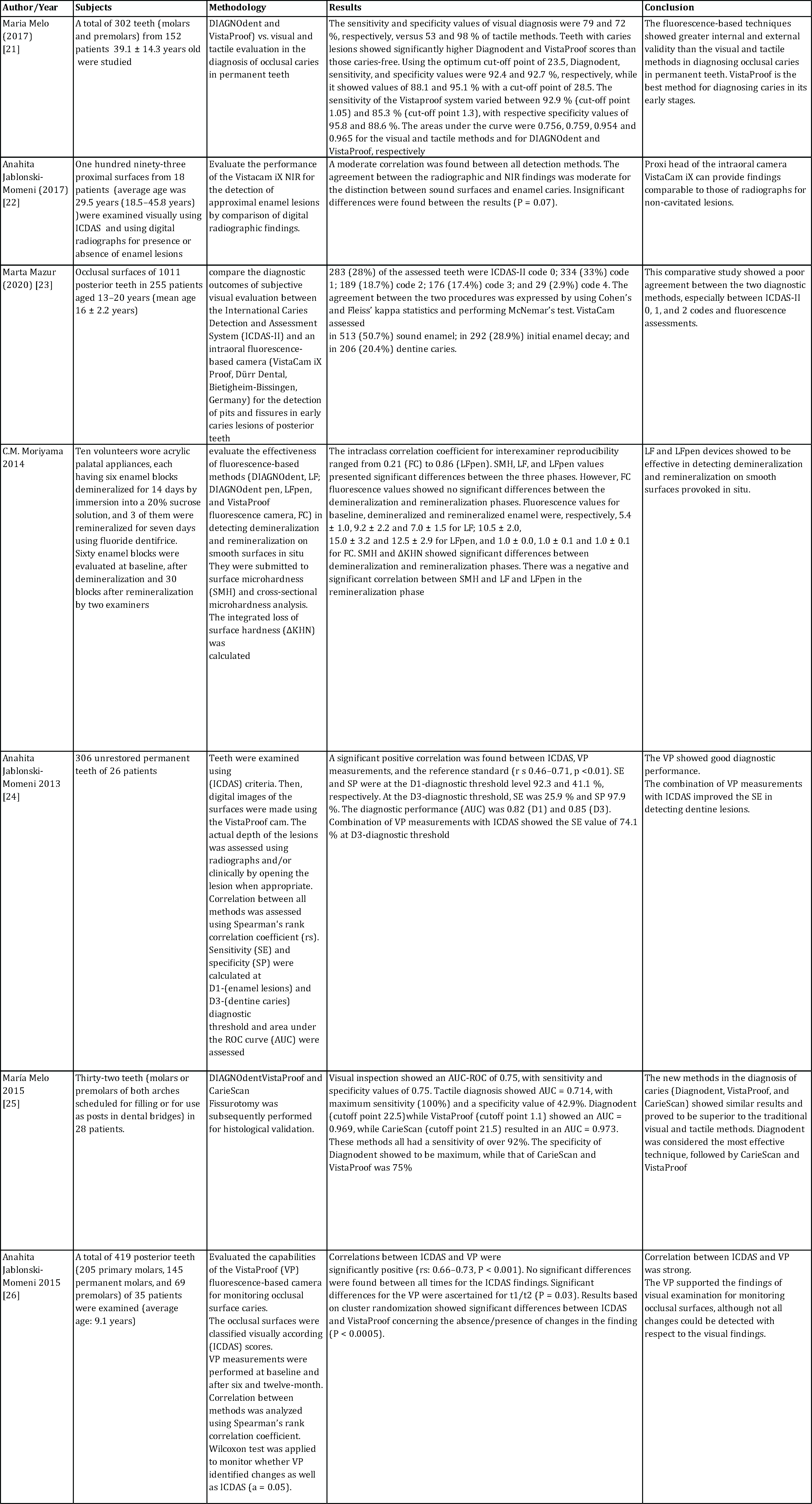
Following the search strategy, 17 articles were found in the search database. They were published between 2011 and 2020. Seven articles met the inclusion criteria after reading the full text, and the excluded ten articles included two literature reviews, 2 case reports, and five in-vitro studies, and one published in the Spanish language (Table 5).
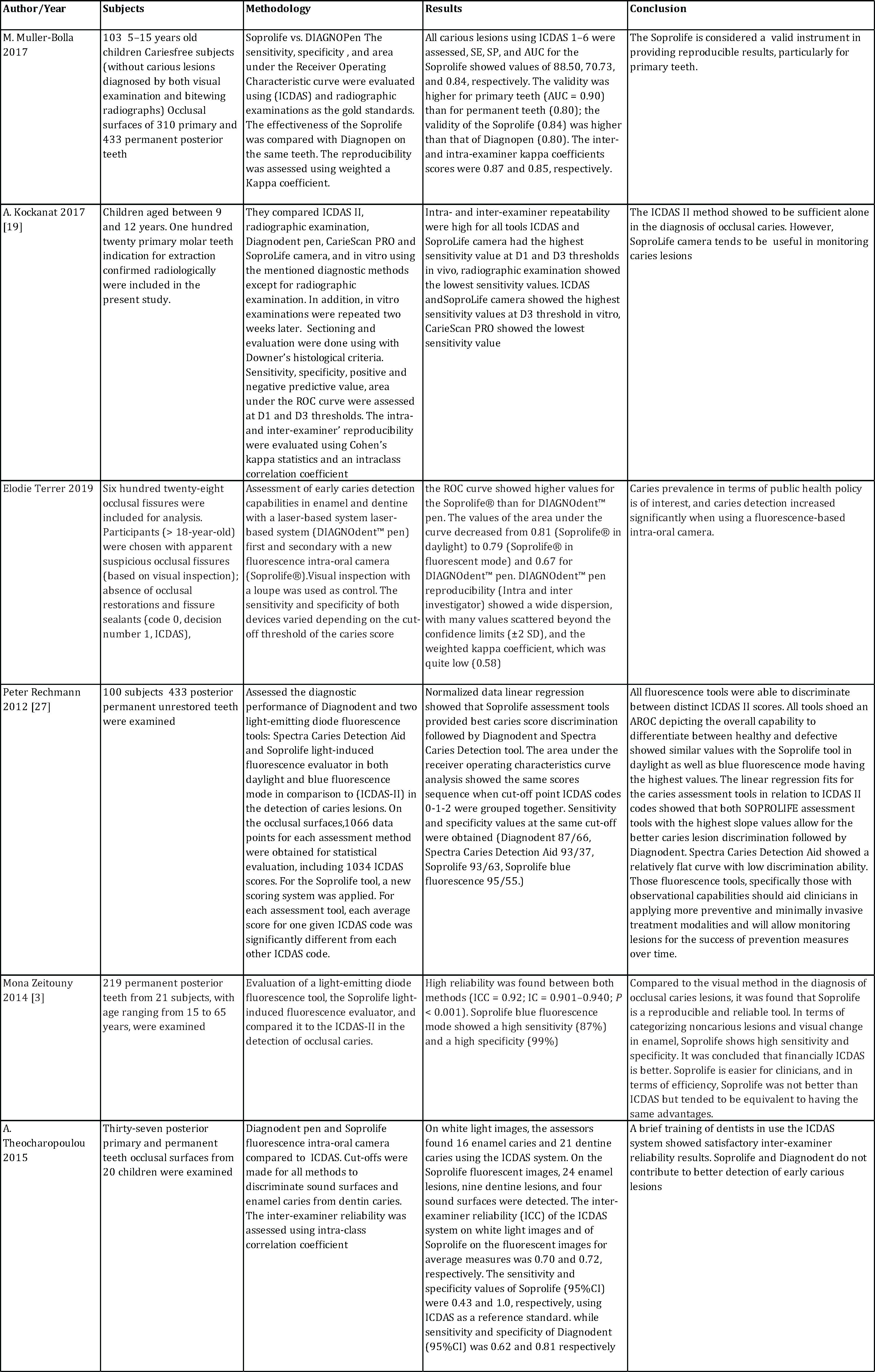
For Soprolife Camera
Searching the databases according to the search strategy yielded ten articles that were found published between 2014 and 2019. Six articles met the selection criteria after reading the full articles. The excluded four articles included two systematic reviews, one case report, and one in-vitro study (Table 6).
Table 2: Selected studies about digital radio
.png)
DISCUSSION
The current review was conducted to evaluate the in-vivo performance of digital dental radiography and some of the novel diagnostic tools in detecting initial carious lesions and microcavities on occlusal and proximal surfaces of both primary and permanent teeth in comparison to the visual and tactile inspection using the ICDAS diagnostic criteria.
Visual inspection has been the most frequently validated diagnostic technique for caries detection and using ICDAS diagnostic criteria proved good performance and accuracy in various in-vivo and in-vitro studies [28-33]. Accordingly, in our review visual inspection using ICDAS criteria was considered the comparator and gold standard protocol for detecting initial carious lesions.
Various studies assessed the digital radiography and recent diagnostic tools in detecting early carious lesions in-vivo and in-vitro, our review included recent in-vivo clinical trials as they are more relevant to the clinical diagnostic situations where the included studies assessed the diagnostic accuracy according to sensitivity, specificity, and receiver operating characteristic curves.
Previous systematic reviews related to our research’s scope were identified a review by [34] was conducted to evaluate various diagnostic tools, the article included some old diagnostic technologies which became rarely available and missed some of the recent tools which proved promising performance, Also in 2016 [35] published a systematic review but it was directed to evaluate the diagnostic capabilities of Soprolife camera only. A more recent review [36] was evaluating various detection technologies but in relation to caries activity of cavitated and non-cavitated lesions, Also [37] carried out a systematic review and meta-analysis, but it was limited to the use of Diagnodent and Vistaproof camera for pre-cavitated lesions.
According to previous reviews, none of them compared the widely available digital radiography and recent easily available and easy to use diagnostic tools side by side. Also, our review included clinical trial studies comparing other tools together and in comparison to the more commonly used visual and tactile methods following the ICDAS criteria on both occlusal and proximal surfaces of permanent or primary teeth.
The criteria of success and reliability of any caries detection tool depend on various factors; the tool should show high intrinsic and extrinsic validity with a high sensitivity values to limit as much as possible any false-negative test results to the test which means failing to detect active lesions; also it should have high specificity to avoid false-positive results referring to the presence of active lesion leading to unnecessary intervention [36]. The diagnostic accuracy of the discussed tools was also evaluated in relation to the Receiver Operating Characteristic (ROC) curves, which is a graphic presentation of the relation between true positive test results (sensitivity) and false positives ones (1-specificity). The area under the curve (AUC), allows proper assessment of the diagnostic performance of the tested tool and also allows determining the optimal cut-off point, which will discriminate between presence or absence of carious lesions [38].
Digital dental radiography proved to be a reliable tool in detecting initial carious lesions/ especially proximal lesions [7], also using filters as negative images allowed easier and more efficient detection of occlusal lesions [6]. The digital bitewing radiography was proposed to be high value and reliability in detecting proximal caries [8].
The studies published evaluating the Diagnodent device suggested that the device has high accuracy and reliability similar to or superior to visual and tactile methods [9]. The device was proved to be an efficient adjunct to other detection methods, especially in the case of non-experienced clinicians [16], but the limitation of its use was due to high cost and not providing valuable additional information [10].
ICDAS criteria for the diagnosis of dental caries were considered to be sufficient alone in detecting occlusal caries [19]. It was found that ICDAS II showed better performance accuracy than bitewing radiographs when the lesion is confined within enamel [18] but the use in conjunction with a recent diagnostic tool enhanced the outcomes [17,20].
Regarding the included articles evaluating the Soprolife camera, there was a consistent agreement upon the accuracy of the device with sensitivity values ranging from 43% up to 95% while specificity ranged from 55% to 99%; generally, it was found that the Intra- and inter-examiner repeatability was high and the device is a valid and reliable tool which can increase the clinicians' capabilities to detect early carious lesions. These observations were correlated to the ability of Soprolife to accurately transmit blue fluorescent light through the enamel into dentin cores and reflected image shows red areas indicating bacteria and their byproducts [26] and its recommended to be used in conjunction to the ICDAS criteria which is considered more financially efficient and to enhance visual examination [3].
Studies related to VistaCam/VistaProof assessment it was found that in some articles VistaCam proved high accuracy in detecting early carious lesions [21,24,25] with moderate to strong correlation in comparison to results obtained by visual and tactile methods ICDAS criteria [22,38]. Other studies found a low performance of VistaCam when compared to visual and tactile methods, and this result was justified by the need to modify the cut-off values of the device [39] moreover, poor correlation to visual and tactile diagnostic results were obtained in cases of ICDAS scores (0-2), and this result was due to the presence of saliva and blood which should be removed because they enhance the fluorescence and increases the scores obtained; also, removal of plaque from occlusal surfaces was required [23].
In agreement with previous reviews, the main aim of a recent diagnostic tool is to detect as early as possible the initial carious lesions to be able to apply a medical model rather than intervention. Generally, most of the recent diagnostic tools were found to be a very useful aid in diagnosing carious lesions and assessing their progress, the use of recent tools must be an addition to the standard visual and tactile protocol and not a replacement solely used tool to rely on completely.
CONCLUSION
The caries detection tools target the early detection of caries and prevent the progression of caries from demineralization to cavitation. None of the mentioned techniques were sufficient alone for the diagnosis of dental caries. In the future, with the advancements of the diagnostic tools, minimal changes in the tooth structure will be easily detected, and the dental structures will be protected by applying preventive treatments.
AUTHOR CONTRIBUTIONS
Shereen Hafez Ibrahim (associate professor of conservative dentistry): Corresponding author, project administration, supervision, conceptualization, validation, data curation, review.
Peter Edward: data extraction, methodology, resources, writing original draft
Reham Nabil: data extraction, methodology, resources, writing original draft
All authors have read and agreed to the published version.
FUNDING
Self-funded by the authors.
CONFLICTS OF INTEREST
The authors declare no conflict of interest.
DATA AVAILABILITY STATEMENT
The data presented in this study are available on request from the corresponding author.
DISCLOSURE OF STATEMENT
The authors declare that they have no conflicts of interest. This systematic review is not funded by any source.
REFERENCES
- Pinheiro IV, Medeiros MC, Ferreira MÂ, Lima KC. (2004). Use of laser fluorescence (diagnodent™) for In vivo diagnosis of occlusal caries: A systematic review. J Appl Oral Sci. 12(3):177-81.
- Yılmaz H, Keles Y. (2018). Recent Methods for Diagnosis of Dental Caries in Dentistry. Meandros Med Dent J. 19:1-8
- Zeitouny M, Feghali M, Nasr A, Abou-Samra P, Saleh N, et al. (2014). SOPROLIFE system: an accurate diagnostic enhancer. Scientific World Journal. 2014:924741.
- Chaza Kouchaji. (2012). Comparison between a laser fluorescence device and visual examination in the detection of occlusal caries in children. The Saudi Dental Journal. 24(3-4):169–174. https://doi.org/10.1016/j.sdentj.2012.07.002.
- Marmaneu-Menero A, Iranzo-Cortés JE, Almerich-Torres T, Ortolá-Síscar JC, Montiel-Company JM, et al. (2020). Diagnostic Validity of Digital Imaging Fiber-Optic Transillumination (DIFOTI) and Near-Infrared Light Transillumination (NILT) for Caries in Dentine. J. Clin. Med. 9:420. https://doi.org/10.3390/jcm9020420.
- Sahu RK. (2015). Assessment of diagnostic accuracy of a direct digital radiographic‑CMOS image with four types of filtered images for the detection of occlusal caries. Journal of Indian Society of Pedodontics and Preventive Dentistry. 33(1).
- Melo M. (2019). Combined Near-Infrared Light Transillumination and Direct Digital Radiography Increases Diagnostic In Approximal Caries. Scientific Reports. 9:14224.
- Abuzenada BM. (2019). Detection of proximal caries with digital intraoral bitewing radiography: An interobserver analysis, Saudi Journal for Health Sciences. 8(1):38-41. https://www.saudijhealthsci.org/text.asp?2019/8/1/38/256781
- Castilho LS, Cotta FV, Bueno AC, Moreira AN, Ferreira EF, et al. (2016). Validation of DIAGNOdent laser fluorescence and the International Caries Detection and Assessment System (ICDAS) in diagnosis of occlusal caries in permanent teeth: an in vivo study. Eur J Oral Sci; 124(2):188–194.
- Chu CH, Lo ECM, You DSH. (2010). Clinical diagnosis of fissure caries with conventional and laser-induced fluorescence techniques. Lasers Med Sci. 25:355–362. https://doi.org/10.1007/s10103-009-0655-6
- Goel A, Chawla HS, Gauba K, Goyal A. (2009). Comparison of validity of DIAGNOdent with conventional methods for detection of occlusal caries in primary molars using the histological gold standard: An in vivo study. J Indian Soc Pedod Prev Dent. 27(4):227-34.
- Kavvadia K, Lagouvardos P. (2008). Clinical performance of a diode laser fluorescence device for the detection of occlusal caries in primary teeth. Int J Paediatr Dent. 18(3):197-204.
- Kühnisch J, Berger S, Goddon I, Senkel H, Pitts N, et al. (2008). Occlusal caries detection in permanent molars according to WHO basic methods, ICDAS II and laser fluorescence measurements. Community Dent Oral Epidemiol. 36(6):475–484.
- Costa AM. (2008). Use of Diagnodent for diagnosis of non-cavitated occlusal dentin caries. J Appl Oral Sci. 16(1):18-23.
- Huth KC. (2008). Clinical performance of a new laser fluorescence device for detection of occlusal caries lesions in permanent molars. Journal of dentistry. 36:1033–1040.
- Anttonen V, Seppa L, Hausen H. (2004). A follow-up study of the use of DIAGNOdent for monitoring fissure caries in children. Community Dent Oral Epidemiol; 32(4):312–8. https://doi.org/10.1111/j.1600-0528.2004.00168.x.
- Tassoker M, Ozcan S, Karabekiroglu S. (2020). Occlusal Caries Detection and Diagnosis Using Visual ICDAS Criteria, Laser Fluorescence Measurements, and Near-Infrared Light Transillumination Images. Med Princ Pract. 29:25-31. doi: 10.1159/000501257.
- Bhumireddy JR, Rmasubbareddy C, Srikanthkumar M, Sivakumar N. (2018). Comparison of International Caries Detection and Assessment System and Digital Radiographs for Detecting Occlusal Dental Caries: An In vivo Study. European Journal of General Dentistry. 7(3):61-5. https://www.ejgd.org/text.asp?2018/7/3/61/241552
- Kockanat A, Unal M. (2017). In vivo and in vitro comparison of ICDASII, DIAGNOdent pen, CarieScan PRO and SoproLife camera for occlusal caries detection in primary molar teeth. European Journal of Paediatric Dentistry. 18(2):99- 104.
- Goswami M, Rajwar AS. (2015). Evaluation of cavitated and non-cavitated carious lesions using the WHO basic methods, ICDAS-II and laser fluorescence measurements. Journal of Indian Society of Pedodontics and Preventive Dentistry. 33(1):10-14. https://www.jisppd.com/text.asp?2015/33/1/10/148961
- Melo M, Pascual A, Camps I, Del Campo A, Ata-Ali J. (2017). Caries diagnosis using light fluorescence devices in comparison with traditional visual and tactile evaluation: a prospective study in 152 patients. Odontology. 105(3):283-290.
- Jablonski-Momeni A, Jablonski B, Lippe N. (2017). Clinical performance of the near-infrared imaging system VistaCam iX Proxi for detection of approximal enamel lesions. BDJ Open. 3:17012.
- Mazur M, Jedlinski M, Ndokaj A, Corridore D, Maruotti A, et al. (2020). Diagnostic Drama. Use of ICDAS II and Fluorescence-Based Intraoral Camera in Early Occlusal Caries Detection: A Clinical Study. Int J Environ Res Public Health. 17(8): 2937.
- Jablonski-Momeni A, Liebegall F, Stoll R, Heinzel-Gutenbrunner M, Pieper K. (2013). Performance of a new fluorescence camera for detection of occlusal caries in vitro. Lasers in Medical Science. 28(1):101-109.
- Melo M, Pascual A, Camps I, Del Campo A. (2015). In vivo study of different methods for diagnosing pit and fissure caries. J Clin Exp Dent. 7(3):e387-391.
- Jablonski-Momeni A, Stachniss V, Ricketts DN, Heinzel-Gutenbrunner M, Pieper K. (2015). Reproducibility and accuracy of the ICDAS-II for detection of occlusal caries in vitro. Caries Res. 42(2):79-87.
- Rechmann P, Rechmann B, Featherstone J, Charland D. (2012). Performance of laser fluorescence devices and visual examination for the detection of occlusal caries in permanent molars. Journal of Biomedical Optics. 17(3):036006.
- Diniz MB, Rodrigues JA, Hug I, Cordeiro Rde C, Lussi A. (2009). Reproducibility and accuracy of the ICDAS-II for occlusal caries detection. Community Dent Oral Epidemiol. 37(5):399-404.
- Lussi A, Megert B, Longbottom C, Reich E, Francescut P. (2001). Clinical performance of a laser fluorescence device for detection of occlusal caries lesions. European Journal of Oral Sciences. 109(1):14-19.
- Lussi A, Megert B, Longbottom C, Reich E, Francescut P. (2001). Laser fluorescence may increase diagnostic sensitivity in detecting class I caries. Journal of Evidence-Based Dental Practice. 1(2):95-96.
- Lussi A, Francescut P. (2003). Performance of conventional and new methods for the detection of occlusal caries in deciduous teeth. Caries Research. 37(1):2-7.
- Lussi A, Hibst R, Paulus R. (2004). DIAGNOdent: an optical method for caries detection. J Dent Res. 83 Spec No C:C80-3.
- Lussi A. Hellwig E. (2006). Performance of a new laser fluorescence device for the detection of occlusal caries in vitro. Journal of Dentistry. 34(7):467-471.
- Pretty IA, Ekstrand KR. (2016). Detection and monitoring of early caries lesions: a review. Eur Arch Paediatr Dent. 17(1):13-25.
- Domejean S, Rongier J, Muller-Bolla M. (2016). Detection of Occlusal Carious Lesion using the SoproLife((R)) Camera: A Systematic Review. J Contemp Dent Pract. 17(9):774-779.
- Drancourt N, Roger-Leroi V, Martignon S, Jablonski-Momeni A, Pitts N, et al. (2019). Carious lesion activity assessment in clinical practice: a systematic review. Clinical Oral Investigations. 23(4):1513-1524.
- Iranzo-Cortes JE, Montiel-Company JM, Almerich-Torres T, Bellot-Arcis C, Almerich-Silla JM. (2019). Use of DIAGNOdent and VistaProof in diagnostic of Pre-Cavitated Caries Lesions-A Systematic Review and Meta-Analysis. J Clin Med. 9(1):p.20. https://doi.org/10.3390/jcm9010020.
- Marczuk-Kolada G, Luczaj-Cepowicz E, Obidzinska M, Rozycki J. (2020). Performance of ICDAS II and fluorescence methods on detection of occlusal caries-An ex vivo study. Photodiagnosis Photodyn Ther. 29:101609.
- Jablonski-Momeni A, Heinzel-Gutenbrunner M, Vill G. (2016). Use of a fluorescence-based camera for monitoring occlusal surfaces of primary and permanent teeth. Int J Paediatr Dent. 26(6):448-456.