Information Links
Related Conferences
Current Issue Volume 5, Issue 1 - 2026
Bilateral Middle Ear Cholesteatoma: Epidemiological, Clinical, and Therapeutic Challenges in a Moroccan Cohort
H Boudhar*, ON Laraqui, M lahjaouj, M loudghiri, W Bijou, Y Oukessou, RL Abada, S Rouadi, M Roubal, M Mahtar
ENT Head and Neck Surgery Department, 20 August Hospital, Ibn Rochd University Hospital, Faculty of Medicine and Pharmacy, Hassan II University Casablanca, Morocco
*Corresponding author: Hajar Boudhar, ENT Head and Neck Surgery Department, 20 August Hospital, Ibn Rochd University Hospital, Faculty of Medicine and Pharmacy, Hassan II University Casablanca, Morocco, Phone: 0678328423; Email: [email protected]
Received Date: March 10, 2026
Published Date: April 23, 2026
Citation: Boudhar H, et al. (2026). Bilateral Middle Ear Cholesteatoma: Epidemiological, Clinical, and Therapeutic Challenges in a Moroccan Cohort. Mathews J Otolaryngol. 5(1):16.
Copyrights: Boudhar H, et al. © (2026).
ABSTRACT
Introduction: Bilateral middle ear cholesteatoma is a rare but severe form of chronic otitis media, characterized by keratinizing squamous epithelium in the middle ear, leading to complications such as hearing loss, facial paralysis, and intracranial involvement. Despite extensive research on unilateral cases, bilateral cholesteatoma remains understudied, particularly in resource-limited settings. This study aims to analyze its epidemiological, clinical, and therapeutic particularities. Materials and Methods: A retrospective study was conducted in our department from 2021 to 2025 involving 54 patients with surgically confirmed bilateral cholesteatoma. Data included demographic characteristics, clinical presentation, imaging (CT scans), surgical techniques (canal-wall-up vs. canal-wall-down), and postoperative outcomes. Statistical analysis was performed using SPSS. Results: The cohort showed a male predominance (55.5%) and a mean age of 17 years, with 33.3% aged 10–20. Diagnostic delay averaged 10 years, and complications included meningitis (3.7%) and facial paralysis (1.85%). CT scans revealed ossicular erosion (81.5%) and tegmen tympani erosion (44.4%). Closed techniques were preferred (77.5%), yielding a 7.4% recurrence rate vs. 1.85% for open techniques. Hearing improvement averaged 5–32 dB, with 27.3% achieving a Rinne gap <20 dB. Discussion: Bilateral cholesteatoma poses unique challenges due to its aggressive nature and need for bilateral hearing preservation. The high diagnostic delay underscores gap in early detection, while conservative surgical approaches balanced recurrence risk with functional outcomes. CT imaging proved critical for preoperative planning. Limitations included lost-to-follow-up (61.1%) and short follow-up duration (22 months). Conclusion: Bilateral cholesteatoma requires tailored surgical strategies and rigorous follow-up to mitigate recurrence. Future efforts should prioritize early screening, advanced imaging (e.g., MRI), and standardized protocols in similar settings.
Keywords: Bilateral Cholesteatoma, Chronic Otitis Media, Ossicular Erosion, Canal-Wall-Up, Hearing Preservation, Diagnostic Delay, Surgical Outcomes.
INTRODUCTION
Bilateral middle ear cholesteatoma is a rare but severe form of chronic otitis media, characterized by the presence of keratinizing squamous epithelium in the middle ear cavities, leading to potentially serious complications such as hearing loss, vestibular dysfunction, facial paralysis, and intracranial involvement [1]. Although unilateral cholesteatoma is well-documented, bilateral cholesteatoma remains less studied, with distinct epidemiological, clinical, and therapeutic features that warrant special attention.
Epidemiological data indicate that bilateral cholesteatoma accounts for 8% to 17% of cholesteatoma cases, with a male predominance and a higher incidence among children and young adults [2,3]. Its pathogenesis is multifactorial, involving theories such as epithelial migration, metaplasia, papillary proliferation, and retraction pocket formation due to Eustachian tube dysfunction [4]. Diagnosis primarily relies on clinical examination and imaging, particularly computed tomography (CT), which helps assess lesion extent and guide therapeutic decisions [5].
Treatment is primarily surgical and aims to achieve complete cholesteatoma eradication, prevent recurrence, and preserve or improve hearing. Surgical techniques include closed (canal-wall-up) and open (canal-wall-down) approaches, each with its own advantages and disadvantages [6,7]. However, managing bilateral cholesteatoma is particularly challenging due to the need to preserve hearing in both ears and the high risk of recurrence, estimated between 7.4% and 9.25% in reported series [3].
this study aimed to analyze the epidemiological, clinical, and therapeutic particularities of this condition while highlighting the challenges associated with its management in a setting where long-term patient follow-up is often limited. The findings underscore the importance of early diagnosis, tailored surgical strategies, and rigorous surveillance to improve functional outcomes and reduce complications.
MATERIALS AND METHODS
This retrospective study was conducted in the ENT, Head and Neck Surgery Department over a 5-year period (2021–2025). We included 54 patients who underwent surgery for confirmed bilateral cholesteatoma, representing 10.3% of the 524 cases of cholesteatomatous chronic otitis media recorded during this period.
Inclusion criteria consisted of a diagnosis confirmed by clinical examination and computed tomography (CT), while incomplete records or unilateral cholesteatomas were excluded.
Data were systematically collected from medical records using a standardized form, including epidemiological parameters (age, sex, medical history), clinical signs (otorrhea, hearing loss, complications), and results of complementary tests. Otoscopic evaluation under a microscope allowed for the characterization of tympanic lesions (retraction pockets, marginal perforations), while pure-tone audiometry determined the type and degree of hearing loss. Temporal bone CT scans, performed for all patients, provided essential information on lesion extension, ossicular erosion, and bone destruction.
Regarding treatment, two main surgical approaches were used: the closed technique (canal-wall-up, CWU) with anatomical preservation and cartilage reconstruction, and the open technique (canal-wall-down, CWD) via mastoidectomy. The choice of technique depended on preoperative lesion extent, with reported recurrence rates of 9–70% for CWU and 4–15% for CWD [6,7]. Ossiculoplasty was performed in 25% of patients during the first surgical intervention, primarily using cartilage grafts or titanium prosthesis.
Postoperative follow-up combined clinical evaluations (assessing otorrhea or recurrence), audiometric tests (measuring hearing improvement), and radiological examinations (follow-up CT scans). Statistical analysis was performed using SPSS software, with appropriate descriptive and comparative tests. This rigorous methodology allowed us to comprehensively evaluate the specific characteristics of bilateral cholesteatoma in our clinical setting.
RESULTS
The analysis of 54 cases of bilateral cholesteatoma revealed a slight male predominance, with 30 males (55.5%) compared to 24 females (44.4%), yielding a sex ratio of 1.25. Age distribution showed a clear predominance in pediatric and young adult populations: 18 patients (33.3%) were between 10 and 20 years old, with an overall mean age of 17 years (range: 10–66 years). The pediatric population (<15 years) accounted for 14.8% of cases. Otologic history was particularly frequent: 20 patients (37%) reported recurrent otitis media, and 19 (35.2%) had previously undergone tympanoplasty. A history of retraction pocket reinforcement was noted in 8 patients (14.8%).
The study revealed a characteristic clinical presentation marked by chronic otologic symptoms. Patients sought consultation after an average disease duration of 10 years (range: 4 months to 30 years), with nearly 40% having a disease course exceeding 10 years. The primary reason for consultation was fetid otorrhea, present in 96.3% of cases, often bilateral (55.5%). Hearing loss, the second most frequent symptom (72.2%), was also frequently bilateral (37%). Clinical examination revealed objective signs: retraction pockets in 77.8% of cases (predominantly attic retractions at 62%), marginal perforations (11.1%), and external auditory canal polyps (20.4%). Severe complications, though rare, included peripheral facial paralysis (1.85%) and meningitis (3.7%).
Systematic complementary evaluation combined audiometry and imaging. Pure-tone audiometry (performed in 50 patients) demonstrated conductive hearing loss in 70.4% of cases (bilateral in 55.5%), mixed hearing loss (22.2%), and, less frequently, sensorineural hearing loss (3.7%). Temporal bone CT scans, performed in all patients, provided crucial information on lesion extent: ossicular erosion (81.5% of cases), erosion of the scutum (75.9%), attic filling (46.3%), and more severe findings such as tegmen tympani erosion (44.4%) or facial canal involvement (3.7%). These precise radiological findings guided therapeutic strategy.
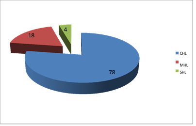
CHL: Conductive hearing loss; MHL: Mixed hearing loss; SHL: Sensorineural hearing loss.
Figure 1. Audiogram Results.
Table 1. Audiogram data
|
Audiogram Data Rinne |
Number of Affected Ears |
Number of Contralateral Ears |
|
< 20 dB |
2 |
3 |
|
20-30 dB |
18 |
11 |
|
30-40 dB |
15 |
17 |
|
40-50 dB |
7 |
14 |
|
50-60 dB |
6 |
8 |
|
60-70 dB |
3 |
1 |
|
80-90 dB |
0 |
0 |
|
Anacusis |
3 |
0 |
The surgical approach was tailored to each case based on lesion extent. Initial unilateral surgery was preferred (74.1% of cases), with a clear predominance of closed techniques (77.5%). For the 14 patients who underwent bilateral surgery (25.9%), the average interval between interventions was 24 months. Frequent intraoperative findings included inflammatory mucosa (78.6%), primarily attic involvement (71.4%), often requiring antroatticotomy (55%). Ossiculoplasty, mainly type II, was performed in 42.9% of bilateral cases, using cartilage grafts or titanium prosthesis.
Table 2. Techniques Used in Bilaterally Operated Cases
|
Case No |
First stage on the right |
First stage on the left |
Second stage |
Interval between the two stages |
|
1 |
CT |
CT |
OT Right |
4 years |
|
2 |
CT |
CT |
Ossiculoplasty |
1 year and half |
|
3 |
CT |
CT |
CT Left |
2 years |
|
4 |
CT |
CT |
OT G |
1 year |
|
2 years |
||||
|
5 |
CT |
CT |
CT L |
|
|
Ossiculoplasty |
Right Ossiculoplasty |
|||
|
6 |
CT |
2 years |
||
|
OT |
||||
|
7 |
CT |
CT |
Ossiculoplasty L\ OT R |
2 years |
|
8 |
CT |
CT |
Right Ossiculoplasty |
3 years |
|
9 |
CT |
CT |
- |
3 years |
|
10 |
CT |
CT |
- |
4 years |
|
11 |
CT |
OT |
- |
2 years |
|
12 |
CT |
CT |
- |
1 year |
|
2 years |
||||
|
13 |
CT |
Ossiculoplasty |
- |
|
|
Ossiculoplasty CT |
Ossiculoplasty OT |
|||
|
14 |
- |
2 years |
Postoperative follow-up, available for 21 patients (38.9%), with complete evaluations for 11 of them, showed after a mean follow-up period of 22 months: satisfactory anatomical outcomes with an intact neotympanum in 54.5% of cases, but recurrences in 36.4% of cases, more frequent after closed techniques. Functionally, an average hearing improvement of 5–32 dB was observed, with 27.3% of patients achieving a Rinne gap below 20 dB. These findings highlight the importance of prolonged follow-up for early detection of recurrences.
DISCUSSION
The results of our study provide significant insights into the epidemiological, clinical, and therapeutic particularities of bilateral cholesteatoma. Our series of 54 cases confirms and refines several essential aspects of this complex ENT pathology [8-10].
From an epidemiological perspective, the observed male predominance (55.5%) aligns with data from the literature [2,3]. This characteristic may be explained by several pathophysiological mechanisms. On one hand, anatomical differences in the Eustachian tube typically narrower and more horizontal in males could contribute to tubal dysfunction [4]. On the other hand, hormonal factors might influence epithelial proliferation and keratinization processes. The mean age of 17 years, with a peak incidence in the 10–20 age group (33.3% of cases), confirms the early onset of this pathology, likely linked to local immune system immaturity and the high frequency of ENT infections in this age group.
The clinical presentation is particularly revealing. The mean consultation delay of 10 years significantly longer than European data [11,12] highlights the challenges in accessing specialized care in our case. This diagnostic delay likely explains the high frequency of complications observed (5.55%), including potentially severe cases such as meningitis (3.7%). The predominance of attic retraction pockets (62%) supports the pathogenic theory of tympanic invagination as the primary mechanism in bilateral forms [4,13]. These clinical findings underscore the need to strengthen early screening efforts, particularly in children with recurrent otitis media.
Table 3. Reported Cases of Bilateral CC [14]
|
Author |
Year |
Age |
Sex |
Presentation |
Comment |
|
Peron et Schuknecht |
1975 |
20 years |
Male |
Mixed hearing loss |
Lesions detected postmortem during temporal bone study |
|
Wingert |
1976 |
2 years |
Male |
Recurrent otitis media |
Lesions detected simultaneously |
|
Curtis |
1979 |
23 years |
Male |
Conductive hearing loss |
Lesions detected 2.5 months apart |
|
Wang |
1984 |
4 years |
Male |
Abnormal finding on examination |
Lesions detected 2.5 months apart |
|
Fedok |
1990 |
4 years |
Male |
Abnormal finding on examination |
Lesions detected 7 months apart |
|
Braganza et Kearns |
1993 |
4 years |
Male |
Abnormal finding on examination |
Second lesion detected by CT scan |
|
Nishizaki |
1996 |
6 years |
Male |
Conductive hearing loss |
Second lesion detected by CT scan |
|
Litman |
1996 |
2 years |
Male |
Abnormal finding on examination |
Lesions detected 18 months apart |
|
Litman |
1996 |
4 years |
Male |
Abnormal finding on examination |
Lesions detected simultaneously |
|
El Bitar |
2002 |
4 years |
Male |
Abnormal finding on examination |
Lesions detected simultaneously |
The analysis of paraclinical data provides major diagnostic insights. Audiometry revealed more severe (70.4%) and more frequently bilateral (55.5%) conductive hearing losses compared to unilateral forms, reflecting the often symmetrical involvement of the ossicular chain. CT scans, with a sensitivity of 85–93% for detecting bone lesions [5], proved indispensable for assessing lesion extent. Findings such as ossicular erosion (81.5%), scutum erosion (75.9%), and tegmen tympani erosion (44.4%) directly influenced our therapeutic choices.
From a therapeutic standpoint, our conservative approach (77.5% closed techniques) differs from some authors' recommendations [15,16] but appears justified by several arguments. First, it allows better preservation of residual hearing particularly crucial in bilateral cases. Second, it reduces immediate surgical morbidity. However, this strategy has limitations, including a higher recurrence rate (7.4% vs. 1.85% for open techniques) and requires rigorous follow-up, often difficult to maintain in our context.
Functional outcomes, though modest (mean hearing improvement of 5–32 dB), remain encouraging. They confirm that despite the aggressiveness of bilateral forms, satisfactory auditory rehabilitation is achievable. The proportion of patients achieving a Rinne gap <20 dB (27.3%) demonstrates the efficacy of modern ossiculoplasty techniques [10].
Several methodological limitations must be acknowledged. The high rate of lost-to-follow-up cases (61.1%) and the limited mean follow-up period (22 months) restrict the generalizability of our long-term conclusions. The absence of a unilateral control group prevents certain statistically significant comparisons.
Future improvements are multifaceted. The development of postoperative MRI could enhance recurrence detection [5,17]. Less invasive endoscopic techniques warrant evaluation for this indication. Finally, optimizing follow-up protocols emerges as a priority to reduce the lost-to-follow-up rate.
Despite these limitations, our study provides valuable insight into the clinical behavior and surgical management of bilateral cholesteatoma in a resource-limited setting.
CONCLUSION
Bilateral middle ear cholesteatoma represents a challenging clinical entity due to its aggressive behavior and the need to preserve hearing in both ears. Our findings highlight the predominance of young patients, the long diagnostic delay, and the high frequency of ossicular and bony erosion at presentation. Computed tomography remains essential for accurate assessment and surgical planning. Although canal-wall-up techniques allowed better anatomical preservation, they were associated with a higher recurrence risk, emphasizing the importance of careful case selection and strict postoperative surveillance. Strengthening early detection and improving long-term follow-up strategies are crucial to optimize functional outcomes and reduce complications in bilateral disease.
DECLARATIONS
Funding
This research received no specific grant from any funding agency in the public, commercial, or not-for-profit sectors.
Ethics approval and consent to participate
The study was conducted in accordance with the principles of the Declaration of Helsinki. Ethical approval was obtained from the Ethics Committee of [Institution Name]. Written informed consent was obtained from all participants.
Consent for publication
Written informed consent for publication was obtained from the participants.
Competing interests
The authors declare that they have no competing interests.
REFERENCES
- Olszewska E, Wagner M, Bernal-Sprekelsen M, Ebmeyer J, Dazert S, Hildmann H, et al. (2004). Etiopathogenesis of cholesteatoma. Eur Arch Otorhinolaryngol. 261(1):6-24.
- Tsuji K, Sone M, Kakibuchi M, Sakagami M. (2002). Bilateral cholesteatoma and habitual sniffing. Auris Nasus Larynx. 29(2):111-114.
- Vartiainen E. (1993). Fate of patients with bilateral cholesteatoma. Am J Otolaryngol. 14(1):49-52.
- Beltaief N, Sellami M, Tababi S, Zainine R, Charfeddine A, Sahtout S, et al. (2012). Le choléstéatome de l’oreille moyenne. J. tun ORL — No. 28, June–December 2012.
- Marciano S, Thomassin JM, Braccini F, Moulin G. (1999). Imagerie et pathologies traumatique, inflammatoire et tumorale de l'oreille externe et moyenne [Imaging and traumatic, inflammatory and neoplastic pathologies of the external and middle ear]. Rev Laryngol Otol Rhinol (Bord). 120(3):185-193.
- Göçmen H, Kiliç R, Ozdek A, Kizilkaya Z, Safak MA, Samim E. (2003). Surgical treatment of cholesteatoma in children. Int J Pediatr Otorhinolaryngol. 67(8):867-872.
- Lesinskas E, Vainutiene V. (2004). Closed tympanoplasty in middle ear cholesteatoma surgery. Medicina (Kaunas). 40(9):856-859.
- Deguine C. (1990). L'oreille contro-latérale du cholestéatome [The contralateral ear in cholesteatoma]. Rev Laryngol Otol Rhinol (Bord). 111(5):463-468.
- Samari H. (2001). Otite moyenne chronique cholestéatomateuse: Apport de l’imagerie. Thèse Med Casa. N ° 278.
- Khalil HS, Windle-Taylor PC. (2003). Canal wall down mastoidectomy: A long term commitment to the outpatients? BMC Ear Nose Throat Disord. 3(1):1.
- Deguine C. (1990). L'oreille contro-latérale du cholestéatome [The contralateral ear in cholesteatoma]. Rev Laryngol Otol Rhinol (Bord). 111(5):463-468.
- Kemppainen HO, Puhakka HJ, Laippala PJ, Sipilä MM, Manninen MP, Karma PH. (1999). Epidemiology and aetiology of middle ear cholesteatoma. Acta Otolaryngol. 119(5):568-572.
- Sudhoff H, Tos M. (2000). Pathogenesis of attic cholesteatoma: clinical and immunohistochemical support for combination of retraction theory and proliferation theory. Am J Otol. 21(6):786-792.
- El Bitar MA, Sukgi Chol S. (2002). Bilateral occurrence of congenital middle ear cholesteatoma Otolaryngology Head and Neck Surgery. 127(5):480-482.
- Artuso A, di Nardo W, De Corso E, Marchese MR, Quaranta N. (2004). Canal wall down tympanoplasty surgery with or without ossiculoplasty in cholesteatoma: hearing results. Acta Otorhinolaryngol Ital. 24(1):2-7.
- Rachidi-Alaoui F, Benchekroun L, Jazouli N, Lazrak A, Kzadri M. (1996). Le cholestéatome de l'oreille moyenne chez l'enfant. A propos de 160 cas [Cholesteatoma of the middle ear in children. Apropos of 160 cases]. Rev Laryngol Otol Rhinol (Bord). 117(1):47-50.
- Thomassin JM, Braccini F. (1999). Place de l'imagerie et de l'endoscopie dans la surveillance et la prise en charge des cholestéatomes opérés en technique fermée [Role of imaging and endoscopy in the follow up and management of cholesteatomas operated by closed technique]. Rev Laryngol Otol Rhinol (Bord). 120(2):75-81.
