Information Links
Related Conferences
Previous Issues Volume 6, Issue 1 - 2022
Anti-Bacterial Mechanism for Metallic Ag+, Cu2+, Zn2+ Ions-Induced Bactertiolysis on Disruptive OM Lpp and PGN Inhibitive Elongations Against S. aureus and E. coli
Ishida Tsuneo*
2-3-6, Saido, Midori-Ku, Saitama-Shi, Saitama-Ken, Japan
*Corresponding author: Dr. Sci. Tsuneo Ishida, 2-3-6, Saido, Midori-Ku, Saitama-Shi, Saitama-Ken, 〒336-0907, Japan, Phone: 048-881-3970; Email: [email protected]
Received Date: October 04, 2022
Published Date: November 04, 2022
Citation: Ishida T. (2022). Anti-Bacterial Mechanism for Metallic Ag+, Cu2+, Zn2+ Ions-Induced Bactertiolysis on Disruptive OM Lpp and PGN Inhibitive Elongations Against S. aureus and E. coli. Mathews J Cytol Histol. 6(1):18.
Copyrights: Ishida T. © (2022).
ABSTRACT
Anti-bacterial mechanism for complete-ionized Ag+, Cu2+, Zn2+ ion solutions has been established against S. aureus and E. coli. Anti-bacterial mechanism against S. aureus is involved that bacterolysis and destruction of S. aureus cell wall occur by inhibition of PGN elongation through metallic Ag+, Cu2+, Zn2+ ions-induced PGN inhibitive transglycosylase (TG) and transpeptidase (TP) syntheses (TG for Zn2+) and PGN activated major autolysin of amidase. The other, anti-bacterial mechanism against E. coli has been clarified that bacteriolysis and destruction of E. coli cell wall occur by disruption of E. coli outer membrane (OM) structure with OM lipoprotein-endopeptidase activation, and by inhibition of PGN elongation through inhibitive TG and TP syntheses (TG for Zn2+) and PGN activated major autolysins. Ag+, Cu2+, Zn2+ ions-induced ROS generation of O2- and H2O2 and ROS-mediated oxidative stress in bacterial cell lead to killing by stress damage for silver ions, cell membrane damages due to high reactive •OH and OH- are formed by Haber-Weiss and Fenton reactions for Cu2+ ions, and DNA molecular damage for Zn2+ ions.
Keywords: Ag+, Cu2+, Zn2+ ions, Bacteriolysis, PGN synthesis and autolysin, PGN elongation, Autolysin amidase, ROS-mediated oxidative stress
ABBREVIATIONS
BLP: Braun’s lipoprotein; CTD: C-terminal domain; E. coli: Escherichin coli; IMP: integral membrane protein; LdtF: l,d- transpeptidase factor; Lpp: lipoprotein; LPS: lipopolysaccharide; MBP: maltose-binding protein; NAG: N-acetylglucosamine; NAM: N-acetylmuramic acid; NTD: N-terminal domain; OM: outer membrane; OMP: outer membrane protein; Omp: outer membrane porin; Pal: Proteinasso-ciated lipoprotein; PGN: peptidoglycan; PGRPs: peptidoglycan recognition proteins, ROS: reactive oxygen species; S. aureus: Staphylococcus aureus; SNF: silver nanoformulation form; TG: transglycosylase; Tol: Tol proteins; TP: transpeptidase; ZnPT: zinc pyrithione.
INTRODUCTION
Silver, copper, and zinc of transition metals have highly antibacterial activities and are utilized as chemotherapy agents. The high antibacterial activities for these Ag+, Cu2+, Zn2+ ion solutions have the processes of bacteriolyses and destructions of bacterial cell walls against Staphylococcus aureus (S. aureus) peptidoglycan (PGN) and Escherichia coli (E. coli) outer membrane cell walls.
Anti-bacterial activity of silver (I) ions depend on bacteriolysis and destruction of bacterial cell walls that silver ions inhibit PGN elongation and PGN biosynthesis, and enhance PGN autolysin activation [1]. Especially, the interaction of silver ions with Escherichia coli (E. coli) used as a model microorganism is characterized by energy-filtering transmission electron microscopy (EFTEM) that the outer membrane and the interior cell membrane with cytoplasmic protein were destructed by silver ions [2], in which bacterial killing of silver ions is shown to have a strong highest function for the destructions of E. coli outer membrane lipoprotein and inner membrane protein.
Copper ions destroy the bacterial cell wall, which becomes thick and coarse, the cytoplasm is then degraded and disappears, leading finally to cell death. The antibacterial mechanism is attributed mainly to the strong adsorption of copper ions to bacterial cells, which imparts antibacterial efficacy in a concentration-dependent manner [3]. The bacteriolytic mechanisms by copper (II) ions had been revealed that bacteriolysis of S. aureus PGN cell wall by Cu2+ ions is ascribed to the inhibition of PGN elongation due to the damages of PGN biosynthesis of transglycosylase (TG) and transpeptidase (TP), and the Cu2+ ions-induced activated PGN autolysins, whereas bacteriolysis of E. coli outer membrane cell wall by Cu2+ ions is attributed to the destruction of outer membrane structure and the inhibition of PGN elongation due to the damage of PGN biosynthesis TP and the activations of PGN autolysins [4].
Zn2+ ions can be internalised into the bacterial cell and disrupt the enzymatic system. ROS production (causing the destruction of cellular components such as DNA, proteins and lipids): O2− and HO2− do not penetrate the membrane, but direct contact causes damage and H2O2 is internalised. Internalisation within the bacteria cell and direct contact cause damage such as the loss of cellular integrity [5]. Zinc ions-induced anti-bacterial mechanism also may be clarified. It had been appeared that the anti-bacterial effects had the order of Zn2+ > Cu2+ > Ag+ > Al3+ in metallic ion concentration 100 mL of the sulfate solution under the halo inhibitory tests, in which Zn2+ ion indicated to be the highest effect in the sulfates [6].
As described above, Ag+, Cu2+. Zn2+ ion solutions having very high antibacterial abilities call attention to potential treatments such as preventions of serious diseases, restriction of viral infection, and regulation of cancer tumor cells. Furthermore, antibacterial Ag, Cu, Zn metallic ion solution materials are raised such as silver compound (silver chloride), silver nanoparticles for Ag+ ion solutions, copper sulfate, copper chelators, CuO nanoparticles for Cu2+ ion solutions, and zinc chloride, zinc sulfate, zinc pyrithione, zinc oxide for Zn2+ ion solutions.
In this semi-review article, silver (I)-, copper (II)-, zinc (II)-induced, respectively, bacteriolytic functions of inhibition or activation of E. coli outer-membrane lipoprotein, bacterial PGN synthesis and PGN major autolysins are investigated against S. aureus and E. coli, subsequently anti-bacterial mechanisms for silver, copper, and zinc ion solutions is elucidated from the bactericidal viewpoint that relates metallic ions-induced bacteriolytic denaturation of outer-membrane lipoprotein (Braun’s lipoprotein), bacterial PGN elongation, syntheses, and autolysins.
Molecular structures of bacterial cell walls, PGN synthesis and PGN autolysins in S. aureus and E. coli
The bacterial cell walls are a strong flexible meshwork of PGN that gives a bacterium structural integrity, in which to accommodate a growing cell, the walls are remodeled by PGN synthesis and PGN autolysin. PGN is the main constituent of bacterial cell walls and must be continuously synthesized and degraded to maintain the integrity and viability of the cells that bacterial cell wall hydrodases of amidase, gycosidase, and peptidase display a modular architecture combining multiple and different catalytic domains, including some lytic transglycosylases as well as cell wall binding domains [7]. Bacterial PGN structure of both Gram-positive and Gram-negative bacteria comprises repeating disaccharide backbones of N-acetylglucosamine (NAG) and β-(1-4)-N- acetylmuramic acid (NAM) that are cross linked by peptide stem chains attached to the NAM residues [8].
S. aureus surface layer consists of teichoic acids, lipoteichoic acids, and thick PGN cell wall, in which the molecular structure of S. aureus PGN cell wall and the action sites of synthesis TG/TP enzymes and PGN forth autolysins, as shown in Figure 1. For Staphylococcus aureus (S. aureus) PGN layer, there are biosynthesis TG/TP and forth autolysins of N-acetylmuramidase and N-acetylglucosamidase, N-acetylmuramidase-L-alanine amidase and PGN chain cross-linkage DD-endopeptidase.
The other, E. coli cell wall consists of lipid A, lipopolysaccharide, porin proteins, outer membrane of lipoprotein, and thinner 2-7 nm PGN layer in 30-70 nm periplasmic space [9]. E. coli cell wall is constituted of lipopolysaccharide (LPS), lipoproteins (LPT), and PGN, thinner layer within periplasmic space. The first permeability barrier of zinc ions in the E .coli cell wall is highly anionic LPS with hydrophobic lipid A, core polysaccha-ride, O-polysaccharide, in which zinc ions may be possible for the inhibition of LPS biosynthesis, owing to that promotes formation of metal-rich precipitates in a cell surface [10]. E. coli Braun’s lipoprotein (BLP) of outer-membrane (OM) lipoprotein that BLP is anchored in the OM via a lipidated N-terminus, whereas the C-terminus is covalently attached to the peptide chain of PGN and that BLP exists in PGN-bound and PGN-unbound states, the length of BLP has a direct influence on the distance between the peptidoglycan layer and the outer membrane of E. coli [11]. BLP facilitates interactions of OmpA monomer with PGN, in which The OmpA dimer readily binds to PGN, in which The E. coli outer membrane porin OmpA is a multidomain protein whose N-terminal domain (NTD) is made of a b-barrel and C-terminal domain (CTD) is a globular periplasmic unit that binds to PGN, connected by an unstructured 20-residue linker region and that BLP and OmpA CTD are able to form nonspecific electrostatic interactions in the periplasm [11]. Penicillin binding protein4 (PBP4) localizes specifically at midcell as part of the division machinery that PBP4 is a periplasmic endopeptidase with a C-terminal amphipathic alpha-helix that associates with membranes and has three domains [12]. Despite its conservation throughout evolution among pathogenic and non-pathogenic bacteria, OmpA interacts with specific receptors for initiating pathogenesis in some Gram-negative bacterial infections [13].
The gram‐negative bacterial cell envelope is made up of an outer membrane (OM), an inner membrane (IM) that surrounds the cytoplasm, and a periplasmic space between the two membranes containing peptidoglycan (PGN or murein). PGN is an elastic polymer that forms a mesh-like sacculus around the IM, protecting cells from turgor and environmental stress conditions. In several bacteria, including Escherichia coli, the OM is tethered to PGN by an abundant OM lipoprotein, Lpp (or Braun’s lipoprotein), that functions to maintain the structural and functional integrity of the cell envelope. Since its discovery, Lpp has been studied extensively, and although L,D-transpeptidases, the enzymes that catalyze the formation of Lpp-PGN linkages, have been earlier identified, it is not known how these linkages are modulated. Recently, LdtF is identified as an endopeptidase that cleaves the Lpp-PGN cross-links and as a glycine-specific carboxypeptidase [14]. For Escherichia coli (E. coli) cell wall, there are endopeptidase and aminopeptidase of degrading enzyme at lipoprotein of N- and C-terminals, and amidase, peptidase, and caboxypeptidase at thin PGN layer in periplasmic space [15].
Figure 2 shows the schematic structure of E. coli, in which the molecular bonding manner of E. coli cell wall and periplasmic PGN, and the action sites of the hydrolases and degradative enzymes of lipoproteins E. coli PGN synthetic enzymes TG/TP and the PGN autolysins such as Muramidase, Glucosamidase, Amidase, Peptidase, and Carboxypeptidase. Interactions of PGN molecular structure, PGN biosynthesis TG/TP and PGN autolysins influence in any event for the bacteriolysis of bacterial cell walls.
Bacterial PGN biosynthesis autolysins against S. aureus and E. coli are summarily represented in Table 1 that these PGN biosynthesis and autolysin sites are shown in Figure 1 and Figure 2. The S. aureus killing mechanism was more likely due to activation autolysins along with minimum membrane disruption [16]. In these autolysins, zinc-dependent PGN major autolysin of amidases chiefly may be enhanced induced anti-bacterial activities.
.png)
Figure 1. PGN molecular structure and the action sites of PGN synthesis TG/TP and PGN autolysins of N-acetylmuramidase, N-acetylglucosamidase, N-acetylmuramyl- L-alanine amidase, DD-endopeptidase against S. aureus thick PGN layer.
.png)
Figure 2. E. coli outer-membrane and periplasmic space PGN molecular structures, and the action sites of degrading endopetidase enzyme of outer-membrane lipoprotein at C-and N-terminals, PGN synthesis TG/TP enzymes, and PGN autolysins of N-acetylglucosaminidase, N-acetylmuramidase, amidase, peptidase, and carboxypeptidase against E. coli cell wall.
Table 1. Bacterial PGN synthesis and autolysins against S. aureus, and outer membrane lipoprotein degrading enzyme, PGN synthesis and autolysins against E. coli
|
PGN synthesis TG/TP and PGN autolysins against S. aureus |
Outer membrane lipoprotein degrading enzymes, and PGN synthesis TG/TP and PGN autolysins against E. coli |
|
・PGN synthesis N-acetylmuramidase, TG Transpeptidase, TP ・PGN autolysins N-acetylmuramidase and N-acetylglucosamidase N-acetylmuramidase-L- alanine amidase, PGN chain cross-linkage DD-endopeptidase. |
・Endopeptidase of degrading enzyme at lipoprotein of N-terminal and Endopeptidase or OM lipoprotein, Lpp(Braun’s lipoprotein), L,D-transpeptidase, LdtF of degrading enzyme at lipoprotein of C-terminal. ・PGN synthesis N-acetylmuramidase, TG Transpeptidase, TP ・PGN autolysins Muramidase, Glucosamidase, Amidase, Peptidase, and Carboxypeptidase. |
Anti-bacterial activity for silver ions against S. aureus and E. coli
1. Silver ions induced PGN inhibitive synthesis TG/TP against S. aureus cell wall
Silver (I) ions-induced PGN inhibitive synthesis TG/TP and PGN activated major autolysin against S. aureus and destructive outer membrain lipoprotein, PGN inhibitive synthesis TG/TP and PGN activated major autolysin against E. coli are described. In proteins, the coordination is limited by His, Cys, Glu, and sulfur donors from the side chains of a few amino acids. In silver nitrate solution, AgNO3 is dissociated into aqua silver ion [Ag (H2O)2]+ and nitrate ion (NO3)―, aqua silver ions are liable to be bound to ligand L having negative charge. The nitrate ion has bactericidal inactivity.
For silver nitrate in solution is
AgNO3 + 2H2O = [Ag(H2O)2]+ + (NO3) ―
[Ag(H2O)2]+ + L― = Ag(H2O)2L
Ag(H2O)2L = AgL + 2H2O
or 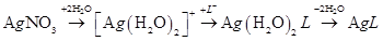
The released Ag+ ions from AgNO3 solution penetrate into bacterial cells, can inhibit the growth of Gram-positive B. subtilis bacterium which exerts toxicity by damaging cellular membrane, degrading chromosomal DNA, lowering reductase activity, and reducing protein expression. Wall teichoic acids are spatial regulators of PGN crosslinking biosynthesis of transpeptidase (TP), and silver ions could inhibit both transglycosylase (TG) and TP enzymes of the PGN that Ag+-induced bacteria may inactivate PGN biosynthesis TG and TP [17]. Lysostaphin-like PGN hydrolase and glycylglycine endopeptidase LytM may function as TP enzyme. Silver ions could inhibit both TG and TP enzymes of the PGN that Ag+-induced bacteria may inactivate PGN synthesis transglycosylase TG [17] and transpeptidase TP [18,19]. Thus, Ag+-induced S. aureus can inactivate PGN synthesis of TG and TP.
2. Silver ions induced PGN major autolysins against S. aureus cell wall
Silver ions enhance activation of PGN autolysins of amidases [20]. For the sake of growth of S. aureus thick PGN layer cell wall, there is necessarily required for the adequate balance between PGN synthesis and PGN autolysin. When the balance was broken to be imbalanced, bacteriolysis and destruction of the cell wall should occur. Hence, it became apparent that bacteriolysis of S. aureus PGN cell wall by Ag+ ions is caused by inhibition of PGN elongation due to inactivation of PGN TG or TP [17] and enhancement of activation of PGN autolysins of amidases [15].
Thus, Ag+ ions activate PGN major autolysins of Bacteriolysis of S. aureus PGN cell wall, in which wall teichoic acids control PGN synthesis cross-linking TP, is due to the inhibition of PGN elongation by enhancing the activities of PGN autolysins; amidase AmiA and AmiE, and PGN hydrolase Lysostaphin-like endopeptidase (Glycine-Glycine bond cleavage).
3. Silver ions induced disruption of E. coli outer membrane structure by hydrolases of lipoproteins at C- and N-terminals
E. coli outer-membrane lipoprotein structure had been observed to be destructed by silver ions [2], in which silver ion is shown to have interaction with protein Braun lipoprotein. Silver nitrate has interaction with protein Braun lipoprotein and is capable of making interaction with many proteins by that bioinformatic interaction of silver nitrate with Braun lipoprotein [21].
Tol protein (Tol)-protein-associated lipoprotein (Pal) system is composed of five proteins that TolA, TolQ, and TolR are inner membrane proteins, TolB is a periplasmic protein, and Pal, the peptidoglycan associated lipoprotein, is anchored to the outer membrane. Ag+ ions induced Tol-Pal complex is antimicrobial agents widely used, it has recently been demonstrated to be essential for bacterial survival and pathogenesis that outer membrane structure may be disrupted [22].
It is unclear whether both Aminopeptidase and Endopeptidase (or L,D-transpeptidase, LdtF) of lipoprotein at C- and N-terminals are simultaneously activated by Ag+ ions. However, outer membrane may be considered to be disrupted probably by predominant activation of lipoprotein-endopeptidase. There is no data about Ag-lipoprotein aminopeptidase, LdtF enzyme interactions, hence, whether Ag+ ion react with endopeptidase enzyme or not [14].
Both silver nanoparticles and ionic silver may interact with proteins associated to the bacterial cell wall and membrane disrupion and thereby form detrimental complexes that alter its physicochemical properties. Silver quickly reacts with the sulfhydryl groups on the bacterial cell membrane by exchanging the terminal hydrogen atom, generating a stable S–Ag bond and thereby fully blocking the respiratory chain, electron transfer, protein secretion and lipid biosynthesis [23].
Silver inhibits outer membrane proteome (OMP) that The molecular mechanism of the antibacterial activity of silver and molecular changes in bacterial cells strongly depend on the physical and chemical properties of the tested silver nanoformulation form (SNF) [24]. A silver-binding peptide, AgBP2, was identified from a combinatorial display library and fused to the C terminus of the E. coli maltose-binding protein (MBP) to yield a silver-binding protein exhibiting nanomolar affinity for the metal [25]. Silver ions may be accumulated and damaged in E. coli PGN synthetic enzyme of silver protein endopeptidase in periplasmic space, in which the silver ions are spent to the activation of bacteriolysis of the cell wall and efflux activity to extracellular cell. Then, Endopeptidase (L,D-transpeptidase, LdtF) of lipoprotein endopeptidase is degradative by Ag+ binding proteins.
4. Silver ions-induced PGN activated major autolysins of amidase, peptidase, and carboxypeptidase against E. coli
It is unclear that the silver-induced PGN syntheses TG/TP should be inhibited by the silver ions. However, silver ions inactivate TP of endopeptidase by because of destructive observation of bacterial cell walls. Silver ions could activate E. coli PGN autolysins of amidase, peptidase, Carboxypeptidase, such as silver depending PGN autolysin, AmiC, AmiD, Muramidase, Amino acid amidase, Carboxypeptidase A, Bacteriolysis and destruction for E. coli cell wall also are considered to be due to the damage of LPS synthesis, destructing of outer membrane structure by degrading of lipoprotein at C-, N-terminals, and to be owing to inhibition of PGN formations by inactivation of carboxypeptidase and TP-endopeptidase, and activities of PGN autolysins of amidase, peptidase and carboxypeptidase.
Thus, the anti-bacterial mechanism of Ag+ ion solution has been found that bacteriolysis and destruction of E. coli cell wall by silver ions are caused by the destruction of outer membrane structure owing to the activation of endopeptidase of lipoprotein at C-, and N-terminals, and inhibition of PGN elongation due to the damage of PGN synthetic TG/TP enzyme and PGN major activated autolysins of Amidase, Peptidase, and Carboxypeptidase in silver-protein amidases in periplasmic space. Specially, the inhibition of PGN elongation had occurred by silver ion induced activities of PGN hydrolases and autolysins.
5. Siver ions induced ROS generation in S. aueus and E. coli
For the penetration of Ag+ ions to S. aureus PGN cell wall, the ROS production such as superoxide anion radical O2-, hydroxyl radical ・OH, hydrogen peroxide H2O2 occurred from superoxide radical O2- molecular. O2- and H2O2 permeate into membrane and cytoplasm, and then, DNA molecular is damaged by oxidative stress [26]. Silver ions react with -SH, and H+ in E. coli that free radicals O2-, OH-,・OH and H2O2 are formed as follows:
O2 + e → O2-
2O2 + 2H+ → H2O2+ O2
O2- + H2O2 → OH- + ・OH + O2.
In cell wall, reacting with polyunsaturated fatty acids:
LH + OH・ → L・ + HOH L・ + O2 → LOO・ LH + LOO・ → L・ + LOOH
Thus, Ag+-containing peptidoglycan recognition proteins (PGRPs) induce ROS production of H2O2, O-, HO・, and then the ROS occur the oxidative stress, and killing by stress damage [27].
Accordingly, anti-bacterial mechanism for silver ion solution is found that bacteriolysis and destruction of S. aureus cell wall occur by inhibition of PGN elongation through Ag+ ions-induced damages PGN both synthetic TG/TP and PGN major activated autolysins of amidase, the other, bacteriolysis destruction of E. coli cell wall occur by the disruption of outer membrane structure owing to the activation of endopeptidase of lipoprotein at N-terminal, inhibition of PGN elongation due to Ag+ ions induced damage of PGN synthetic TG/TP enzyme and PGN major activated autolysins of Amidase, Peptidase, and Carboxypeptidase in silver-protein amidases in periplasmic space.
Anti-bacterial activity for copper (II) ions against S. aureus and E. coli
1. Copper (II) ions-induced S. aureus with coordinated limited ligand
Copper is redox-inert and has only one valence state of Cu (II). In proteins, the coordination is limited by His, Cys, Glu, and sulfur donors from the side chains of a few amino acids. In copper sulfate solution, CuSO4 is dissociated into aqua Cu ion [Cu (H2O)6]2+ and sulfuric ion (SO4)2- aqua Cu ions are liable to be bound to ligand L having negative charge. The sulfuric ion has bactericidal inactivity
Cu(NO3)2 + 6H2O = [Cu(H2O)6]2+ + 2(NO3)-
[Cu(H2O)6]2+ + 2L- = CuL2 + 6H2O
2. Inhibition of polymerization of glycan chains bonding and cross-linking of side peptide
Cu2+ ions may inhibit polymerization of glycan chains, forming copper complex in which is partial action sites of glycan saccharide chains [4]. L is coordinated molecular.
Cu2+ + LH → CuL+ + H+ CuL+ + LH → CuL2 + H+
Copper-complexes on saccharide chains may be,
Glycan chaine: ―NAG-(NAM-Cu-2O-2N-NAG)-NAM―.
The other, Cu2+ ions may inhibit cross-linked reaction by peptide copper complex formation bonding to sidepeptide chains.
Cu2+ + 2LH → CuL2 + H+
Peptide copper complex may be 3N-Cu-O, Cu (Gly-L-Ala) H2O. Specially, Cu2+ ions react with cross-molecular penta glycine (Gly)5, copper-glycine complex may be formed.
Amino acid: Cu2+ + Gly- → Cu (Gly)+, Cu (Gly)+ + Gly― → Cu(Gly)2,
Peptido: Cu2+ + GlyGly → Cu (GlyGly), Cu (GlyGly) + Gly―→ Cu(GlyGlyGly)―.
3. Cu2+ Ions induced Bacteriolysis of S. aureus PGN Cell Wall by inhibition of PGN elongation through inhibitive TG/TP enzymes and PGN activated major autolysins
Bacteriolysis by balance deletion between synthesis enzyme and decomposition enzyme (autolysin) in PGN cell wall: For the sake of growth of S. aureus PGN cell wall, there is necessarily required for the adequate balance between PGN synthesis and PGN autolysin. When the balance is broken by Cu2+ penetration, Cu2+ ions are self-catalytically treated as coenzyme, that this is indicated that activation of autolysin is preceded, in which bacteriolysis and killing may result.
Copper ions inhibit PGN synthesis TG/TP against S. aureus that damages PGN synthetic TG/TP [28]. Cu2+ ions could activate PGN autolysin, AmiA [29,30]. Hence, bacteriolysis of S. aureus PGN cell wall by Cu2+ ions is due to inhibition of PGN elongation owing to the damages of PGN synthetic TG/TP and the activation of PGN major autolysins of AmiA.
4. Bacteriolysis and Destruction of E. coli Outer Membrane Cell Wall by Cu2+ Ions
Inhibition of outer membrane cell wall: Cu2+ ions inactivate catalyst enzyme with forming Cu+ ions
Cu2+ + ―SH →―SCu(I) + H+
By the penetration of Cu2+ ions, the activations of amidase enzyme of N-terminal and endopeptidase enzyme of C-terminal are enhanced. Interaction of copper ion with E. coli Braun lipoprotein is considered that copper dramatically decreases the minimal inhibitory concentration of ampicillin in E. coli strain with a resistance mechanism relying on LD-transpeptidases (LDTs) and inhibits purified LDTs at submillimolar concentrations [31].
Accordingly, the activations of decomposition at N-, C-terminals of lipoproteins may occur with the disruption of outer membrane structure. Hence, bacteriolysis of E. coli cell wall by Cu2+ ions occurs by disruption of outer membrane structure due to degradation of lipoprotein at N-, C-terminals, damage of TP enzyme and activations of PGN autolysins. Furthermore, deletion of PGN autolysin also becomes bacteriolytic factor.
5. Cu2+ ions-induced ROS production in S. aureus and E. coli
Cu2+ ions-induced reactive oxygen species (ROS) O2- and H2O2 generated in the cell wall, and permeate into cell membrane and cytoplasm, in which in cell membrane high reactive •OH and OH- are formed by Haber-Weiss and Fenton reactions.
Haber-Weiss reaction: H2O2 + O2-→ •OH + OH- + O2
Fenton reaction: Cu+ + H2O2 → •OH + OH- + Cu2+
Furthermore, new ROS productions occur by Fenton-like type. L=Ligand
LCu(II) + H2O2 → LCu(I) + ・OOH + H+ LCu(I) + H2O2 → LCu(II) + •OH + OH-
Production of reactive oxygen species (ROS) against S. aureus. O2- and H2O2 permeate into membrane and cytoplasm, that DNA molecular is damaged by oxidative stress [32]. By the penetration of copper ions into bacterial cell wall, productions of O2-, H+, H2O2, ONOO- occurs. The other, in E. coli cell wall, the productions of O2-, H+ in outer membrane, and H2O2, OH-, ・OH in periplasmic space occur. These ROS and H2O2 damage the cell membrane and the DNA molecules by oxidase stress [33].
Anti-bacterial activity for Zinc (II) ions against S. aureus and E. coli
1. Zinc induced zinc-proteins complex formation against S. aureus
In Bacteriolysis of S. aureus PGN Cell Wall by Zn2+ Ions against S. aureus, zinc is redox-inert and has only one valence state of Zn(II). In proteins, the coordination is limited by His, Cys, Glu, and sulfur donors from the side chains of a few amino acids. In zinc sulfate solution, ZnSO4 is dissociated into aqua zinc ion [Zn (H2O)6]2+ and sulfuric ion (SO4)2- aqua zinc ions are liable to be bound to ligand L having negative charge. The sulfuric ion has bactericidal inactivity [34].
ZnSO4 + 6H2O → [Zn(H2O)6]2+ + (SO4)―
[Zn(H2O)6]2+ + 2L- → [Zn(H2O)L2] + 5H2O
Zn(H2O)L2 → ZnL2 + H2O
Structural Zn2+ ions are most commonly coordinated by cysteine, followed by histidine, aspartate, and glutamate that Zn-cysteine complex in bacteria and Zn2+ chelation represents a potential therapeutic approach for combating biofilm growth in a wide range of bacterial biofilm-related infections [35].
2. Zinc induced PGN inhibitive synthesis enzymes of transglycosylase TG and transpeptidase TP against S. aureus
Zinc disrupts PGN synthesis in bacterial cell wall [36] and wall teichoic acids are spatial regulators of PGN cross-linking biosynthesis TP, however, it is not explicit whether zinc ions could inhibit both TG and TP enzymes of the PGN, wherein is due to uncertain relation between wall teichoic acids biosynthesis and PGN biosynthesis [37].
Metallation of Per R with Zn(II) disrupts this coordination, resulting in depression of heme synthesis but continued repression of catalase that Zn(II) intoxication leads to intracellular heme accumulation from measurement of heme content of crude extract of cells treated with zinc concentration 50 μM Zn(II) [38]. Zinc intoxication also is observed to disrupt or inhibit PGN biosynthesis [39].
The bactericidal activity of Zn2+-dependent peptidoglycan recognition proteins (PGLYRPs) is salt insensitive and requires N-glycosylation of PGLYRPs that the LD99 of PGLYRPs for Gram-positive and Gram-negative bacteria is 0.3–1.7 M, and killing of bacteria by PGLYRPs does not involve permeabilization of cytoplasmic membrane, namely, zinc may be shown to inhibit PGN biosynthesis TG [40]. But, these limited PGLYRPs don't be applicable for Gram-negative bacteria. Thus, zinc ions could inhibit PGN synthesis TG against S. aureus.
3. Zinc induced PGN inhibitive elongation due to the activations of autolysins against S. aureus
Zn2+ binding Rv3717 showed no activity on polymerized PGN and however, it is induced to a potential role of N-Acetylmuramyl L-alanine Amidase [41], PGN murein hydrolase activity and generalized autolysis; Amidase MurA [42], Lytic Amidase LytA [43], enzymatically active domain of autolysin LytM [44], Zinc-dependent metalloenzyme AmiE [45] as prevention of the pathogen growth, and Lysostaphin-like PGN hydrolase and glycylglycine endopeptidase LytM [46].
Zn2+ ions-induced bacteriolysis and destruction of S. aureus PGN cell wall could be enhanced by the inhibitions of PGN elongation simultaneously with the activations of these PGN autolysins. Thus, zinc(II) ions can impair the activity of PGN biosynthesis TG and PGN elongation by bacteriolytic destruction of bacterial cell walls, causing bacterial lysis [47].
Accordingly, zinc induced PGN inhibitory biosynthesis corresponds to disruption of bacterial cell wall, but zinc ions may be possible to inhibit PGN synthesis TG and PGN elongation by PGN activated major autolysin of amidase against S. aureus.
4. Zinc induced disruption of outer membrane structure by hydrolases of lipoproteins at C-, N-terminals against E. coli
In zinc ion uptake across the outer membrane, the lipoproteins of Omp A, Omp C, Omp F porins have a role for at least some of these proteins in Zn2+ uptake, in which the lipoproteins have metallic cation selective and hydrophilic membrane crossing pore, to be effective for zinc transfer [48]. Zinc (II) ions react with -SH base, and then H2 generates. Zinc bivalent is unchangeable as
-SZn―S― bond 4-coordinated.
Zn2+ + 2(-SH) → -SZn(II) –S– + 2H+
ZnPT (zinc pyrithione) and Tol (Tol proteins)-Pal (Proteinassociated lipoprotein) complex are antimicrobial agents widely used, however, it has recently been demonstrated to be essential for bacterial survival and pathogenesis that outer membrane structure may be disrupted [49,50]. Interaction zinc ions with E. coli Braun lipoprotein may be considered that Lpp as a new target of antimicrobial peptides is Gram-negative bacterial cell surface receptor for cationic antimicrobial peptides [51].
5. Zinc induced PGN inhibitive elongation through the damage of PGN synthesis enzyme of zinc-protein in periplasmic space and the activation of PGN autolysins against E. coli
The zinc-induced decrease of protein biosynthesis led to a partial disappearance of connexin-43 of protein synthesis in neurons [52], but it is unknown whether PGN synthesis is inhibited. Further, it is also unclear whether the both TG/TP should be inhibited by the zinc ions [53-55]. The other, zinc ions were accumulated in E. coli periplasmic space, in which the zinc ions are spent to the activation of bacteriolysis of the cell wall. Zinc depending PGN autolysin, amidase PGRPs [56], zinc metallo enzymes AmiD [57], zinc-containing amidase; AmpD [58], zinc-present PGLYRPs [59] serve to be effective for the PGN autolysins. It is particularly worth noting that enhancement of the activities of autolysins is characterized on PGN carboxypeptidase-transpeptidase IIW [60] requiring divalent cations. Thus, the inhibition of PGN elongation had been occurred by zinc ion-induced activa tions of PGN hydrolases and autolysins.
Accordingly, bacteriolysis of E. coli cell wall by Zn2+ ions is due to disruption of outer membrane structure by degrading of lipoprotein at C-, N-terminals through PGN formation inhibition by PGN inhibitive synthesis TG and PGN activated autolysins of amidase and carboxypeptidase-transpeptidase
6. Zinc induced ROS generation against S. aureus and E. coli
Zinc induced production of reactive oxygen species (ROS) against S. aureus: O2- and H2O2 permeate into membrane and cytoplasm, that DNA molecular is damaged by oxidative stress [61]. For the penetration of zinc ions to PGN cell wall, the ROS production such as superoxide anion radical O2-, hydroxyl radical •OH, hydrogen peroxide H2O2 occurred from superoxide radical O2- molecular [62]. O2- and H2O2 permeate into membrane and cytoplasm, and then, DNA molecular is damaged by oxidative stress [61].
O2 + e- + H+ → • HO2
•HO2 → H+ + O2
H2O2 + e- → HO― + •OH
2H+ + • O2- + • O2- → H2O2 + O 2
H2O → •OH + •H + e- → H2O2
Zinc induced ROS production and oxidative stress against E. coli: Zinc ions react with -SH, and H+, ROS generate. In E. coli, free radicals O2-, OH-, •OH) and H2O2 are formed as follows [63]:
O2 + e → O2― 2 O2― + 2H+ → H2O2 +O2 O2―+ H2O2 → OH― + •OH + O2-
In the cell wall, reacting with polyunsaturated fatty acids:
LH + OH• → L• + HOH L• + O2 → LOO• LH + LOO• → L• + LOOH
Zinc-containing Peptidoglycan Recognition Proteins (PGRPs) induce ROS production of H2O2, O2-, HO•, the ROS occur the oxidative stress, and killing by stress damage [64].
Accordingly, as mentioned above, metallic Ag+, Cu2+, Zn2+ ions-induced PGN inhibitive synthesis TG/TP, distruptive OM Lpp, and PGN activated autolysin against S. aureus and E. coli cell walls are summarized in Table 2, including bactericidal mechanism for metallic Ag+, Cu2+, Zn2+ complete-ionized ion solutions.
Table 2. Metallic Ag+, Cu2+, Zn2+ ions-induced PGN inhibitive synthesis TG/TP, disruptive OM Lpp, and PGN activated autolysin against S. aureus and E. coli cell walls
|
Ag+,Cu2+, Zn2+ Ions |
S. aureus Cell Wall |
E. coli Cell Wall |
|||
|
|
PGN Synthesis TG/TP |
PGN Autolysins |
OM lipoprotein- endopeptidase |
PGN Synthesis TG/TP |
PGN Autolysins |
|
|
➡ Ag+, Cu2+, Zn2+, ROS, O2―, OH- H2O2, O― |
➡ Ag+, Cu2+, Zn2+, ROS, O―, OH-H2O2, O― |
➡Ag+, Cu2+, Zn2+, ROS |
➡Ag+, Cu2+, Zn2+, ROS, O2―, OH-, ・OH and H2O2 |
➡ Ag+, Cu2+, Zn2+, ROS O2―, OH-, ・OH and H2O2 |
|
Ag+ ➡ |
Both inhibitive TG and TP |
PGN activated major autolysins of AmiA, AmiE |
Disruptive OM Lpp by Endopeptidase (L,D-transpeptidase, LdtF) |
Inhibitive both TG and TP |
PGN activated major autolysins |
|
Cu2+ ➡ |
Both inhibitive TG and TP |
PGN activated major autolysin of AmiA |
Disruption of outer membrane structure |
Inhibitive both TG and TP |
PGN activated major autolysins |
|
Zn2+ ➡ |
Inhibitive TG |
PGN activated major autolysin of AmiD |
Interaction zinc ions with E. coli Braun lipoprotein occurs |
Inhibitive synthesis TG |
PGN activated autolysins of amidase, carboxypeptidase- transpeptidase |
|
|
Bactericidal mechanism: Bacteriolysis and destruction of S. aureus cell wall by metallic Ag+, Cu2+, Zn2+ ions occur by inhibition of PGN elon- gation through PGN inhibitive TG/TP (TG for Zn2+ ion) and PGN activated major autolysins. |
Batericidal mechanism: Metallic Ag+, Cu2+, Zn2+ ions can disrupt OM Lpp and inhibit PGN elongation through PGN inhibitive TG/TP (TG for Zn2+ ion) and PGN activated major autolysins against E. coli. |
|||
CONCLUSIONS
Anti-bacterial activity for Ag+ ion solution against S. aureus has been found that bacteriolysis and destruction of S. aureus PGN cell wall occurs by the inhibition of PGN elongation by Ag+-induced S. aureus inactivating PGN synthesis transglycosylase TG and transpeptidase TP and enhancing the activation of PGN autolysins of amidase AmiA and AmiE. The other, against E. coli, the anti- bacterial activity for Ag+ ion solution has been found that bacteriolysis and destruction of E. coli cell wall by silver ions are caused by the disruption of outer membrane structure owing to the activation of Endopeptidase, L,D-transpeptidase, LdtF of lipoprotein at C- and N-terminals, and inhibition of PGN elongation through the damage of PGN TG/TP synthetic enzyme of silver-protein Amidase in periplasmic space, and PGN activated autolysins of amidase, peptidase, and carboxypeptidase.
Thus, antibacterial mechanism for Ag+ ions solution is clarified that bacteriolysis and destruction of bacterial cell wall occur by the disruption of E. coli outer membrane structure owing to the activation of Endopeptidase (L,D-transpeptidase, LdtF) of lipoprotein at C- and N-terminals, and by inhibition of PGN elongation through the damage of PGN synthetic TG/TP enzymes and PGN activated autolysins of amidase against S. aureus and E. coli.
Bacteriolysis of S. aureus PGN cell wall by Cu2+ ions are thought to be due to inhibition of PGN elongation owing to the damages of PGN both synthetic TG/TP and the activations of PGN major autolysin of AmiA. The other, bacteriolysis of E. coli cell wall by Cu2+ ions occur by disruption of outer membrane structure due to degradation of lipoprotein at N-, C-terminals, damage of PGN syntheses TG and TP enzyme, and activations of PGN major autolysins. Furthermore, deletion of PGN autolysin also becomes bacteriolytic factor.
Anti-bacterial activity of Zn2+ ions against S. aureus has been found that Zn2+ ions-induced PGN autolysin activation could be enhanced the inhibitions of PGN elongation simultaneously, with bacteriolysis and destruction of S. aureus PGN cell wall. The activations of these PGN autolysins by Zn2+ ions could be enhanced the inhibitions of PGN elongation simultaneously, with bacteriolysis of S. aureus PGN cell wall. The other, antibacterial mechanism of Zn2+ ions against E. coli was found that Bacteriolysis and destruction of E. coli cell wall by Zn2+ ions are due to disruption of outer membrane structure by degrading of lipoprotein at C-, N-terminals, owing to PGN formation inhibition by damage of PGN synthesis TG and PGN autolysins of amidase and carboxypeptidase-transpeptidase.
Ag+, Cu2+, Zn2+ ions-induced ROS generation of O2― and H2O2 and ROS-mediated oxidative stress in bacterial cell lead to killing by stress damage for silver ions, cell membrane damages due to high reactive •OH and OH- are formed by Haber-Weiss and Fenton reactions for Cu2+ ions, and DNA molecular damage for Zn2+ ions.
Accordingly, bactericidal mechanism for complete-ionized metallic Ag+, Cu2+, Zn2+ ions solutions has been established that Ag+, Cu2+, Zn2+ ions, respectively, induced the bacteriolyses and destructions of bacterial cell walls occur by disruption of E. coli outer- membrane lipoprotein and by inhibition of PGN elongation through PGN both inhibitory syntheses TG/TP (TG for Zn 2+ ion) and PGN activated major autolysin of amidase. Ag+, Cu2+, Zn2+ ions-induced ROS generation of O2- and H2O2 and ROS-mediated oxidative stress in bacterial cell lead to killing by stress damage, cell membrane damages due to high reactive •OH and OH -, and DNA molecular damage.
REFERENCES
- Ishida T. (2018). Antibacterial mechanism of Ag+ ions for bacteriolyses of bacterial cell walls via peptidoglycan autolysins, and DNA damage. MOJ Toxicology. 4(5): 345-350.
- Yamanaka M, Hara K, Kudo J. (2005). Characterization of the Interaction between Silver Ions and Escherichia Coli by Energy-Filtering Transmission Electron Microscopy. Sharp Technical Reports. 91: 45-49.
- Raffi M, Mehrwan S, Bhatti TM, Akhter JI, Hameed A, Yawar W, et al. (2010). Investigations into the antibacterial behavior of copper nanoparticles against Escherichia coli. Ann Microbiol. 60: 75–80.
- Ishida T. (2017). Mechanism of Antibacterial Activities of Cu(II) Ions against Staphylococcus aureus and Escherichia coli on the Ground of Results Obtained from Dilution Medium Method. Virology & Immunology Journal. 1(3): 1-8.
- da Silva BL, Abuçafy MP, Manaia EB, Oshiro Junior JA, Chiari-Andréo BG, et al. (2019). Relationship between Structure and Antimicrobial Activity of Zinc Oxide Nanoparticles: An Overview. Int J Nanomedicine. 14: 9395–9410.
- Ishida T. (2017). Halo inhibitory zone tests and antibacterial activities for metallic salts aqueous solutions. Int J Prev Med. 11(3): 93-99.
- Vermassen A, Leroy S, Talon R, et al. (2019). Cell wall hydrolases in bacteria: Insight on the diversity of cell wall amidases, glycosidases and peptidases toward peptidoglycan. Front Microbiol. 331: 1-27.
- Ishida T. (2016). Bacteriolyses of Cu2+ solution on bacterial cell walls/cell membrane and DNA base pairing damages. Japanese Biomedical Research on Trace Elements. 27(4): 151-161.
- Silhavy TJ, Kahne D, Walker S. (2010). The Bacterial Cell Envelope. Cold Spring Harb Perspect Biol. 2(5): a000414.
- Langley S, Beveridge TJ. (1999). Effect of O-Side-Chain-LPS Chemistry on Metal Binding. Appl Environ Microbiol. 65(2): 489-498.
- Samsudin F, Boags A, Piggot TJ, Khalid S. (2017). Braun’s Lipoprotein Facilitates OmpA Interaction with the Escherichia coli Cell Wall. Biophys J. 113(7): 1496-1404.
- Verheul J, Lodge A, Yau HCL, Liu X, Boelter G, et al. (2022). Early midcell localization of Escherichia coli PBP4 supports the function of peptidoglycan amidases. PLoS Genet. 18(5): 1-36.
- Krishnan S, Prasadarao NV. (2012). Outer membrane protein A and OprF–Versatile roles in Gramnegative bacterial infections. FEBS J. 279(6): 919–931.
- Bahadura R, Chodisettia PK, Reddya M. (2021). Cleavage of Braun’s lipoprotein Lpp from the bacterial peptidoglycan by a paralog of L, d-transpeptidases, LdtF. Proc Natl Acad Sci U S A. 118(19): e2101989118.
- Ishida T. (2019). Comparative bacteriolytic mechanism for Ag+ and Zn2+ ions against S. aureus and E. coli: A review. Annals of Microbiology and Infectious Diseases. 2(1): 1-12.
- Yasir M, Dutta D, Willcox MDP. (2019). Mode of action of the antimicrobial peptide Mel4 is independent of Staphylococcus aureus cell membrane permeability. PLoS One. 14(7): e0215703.
- Baizman ER, Bransttrom SA, Longley CB, et al. (2000). Antibacterial activity of synthetic analogues based on the disaccharide structure of moenomycin,an inhibitor of bacterial transglycosylase. Microbiology (Reading). 146(Pt 12): 3129-3140.
- Oka T, Hashizumre K, Fujita H. (1980). Inhibition of peptidoglycan transpeptidase by beta-lactam anbiotics: structure-activity relationships. J Antibiot (Tokyo). 33(11): 1357-1362.
- Ortiz-Gila MA, Nunez-Anita RE, Srenas-Arrocena MC, et al. (2015). Silver nanoparticles for the inhibition of S. aureus. Entreciencias. 3(7): 133-142.
- Mellroth P, Sandalova T. (2014). Structural and Functional Insights into Peptidoglycan Access for the Lytic Amidase LytA of Streptococcus pneumoniae. M bio. 15(1): e01120-13.
- Golestannejad Z, Gavanji S, Doostmohammadi M. (2014). In silico analysis of interaction of silver nitrate with Braun lipoprotein in bacterial cell wall. J Chem Pharm Res. 6(12): 366-369.
- Slavin YN, Asnis J, Hafeli U, et al. (2017). Metal nanoparticles: understanding the mechanisms behind antibacterial activity. J Nanobiotechnology. 15(1): 15–65.
- Godoy-Gallardo M, Eckhard U, Delgado LM, de Roo Puente YJD, et al. (2021). Antibacterial approaches in tissue engineering using metal ions and nanoparticles: From mechanisms to applications.Bioactive Materials. 6(12): 4470-4490.
- Kedziora A, Speruda M, Wernecki M, Dudek B, Kapczynska K, et al (2021). How Bacteria Change after Exposure to Silver Nanoformulations: Analysis of the Genome and Outer Membrane Proteome. Pathogens. 10(7): 817.
- Sedlak RH, Hnilova M, Grosh C, Fong H, Baneyx F, Schwartz D, et al. (2012). Engineered Escherichia coli Silver-Binding Periplasmic Protein That Promotes Silver Tolerance. Appl Environ Microbiol. 78(7): 2289-2296.
- Gaupp R, Ledala N, Somerville GA. (2012). Staphylococal response to oxidative stress. Front Cell Infect Microbiol. 2: 33.
- Kashyap DR, Kuzma M, Kowalczyk DA, Gupta D, Dziarski R. (2017) Bactericidal peptidoglycan recognition protein induces oxidative stress in Escherichia coli through a block in respiratory chain and increase in central carbon catabolism. Mol Microbiol. 105(5): 755–776.
- Egan AJF, Biboy J, Veer IV, Breukink E, Vollmer W. (2015). Activities and regulation of peptidoglycan synthases. Philos Trans R Soc Lond B Biol Sci. 370(1679): 20150031.
- Zoll S, Patzold B, Schlag M, Gotz F, Kalbacher H, et al. (2010). Structural Basis of Cell Wall Cleavage by a Staphylococcal Autolysin. PLoS Pathog. 6(3): e1000807.
- Humann J, Lenz LL. (2009). Bacterial peptidoglycan degrading enzymes and their impact on host muropeptido detection. J innate Immun. 1(2): 88-97.
- Petersa K, Pazosa M, Edoob Z, Hugonnetb JE, Martoranac AM, et al. (2018). Copper inhibits peptidoglycan LD-transpeptidases suppressing β-lactam resistance due to bypass of penicillin-binding proteins. Proc Natl Acad Sci U S A. 115(42): 10786-10791.
- Gaupp R, Ledala N, Somerville GA. (2012). Staphylococal response to oxidative stress. Front Cell Infect Microbiol. 2: 33.
- Chautrand T, Souak D, Chevalier S, Duclairoir-Poc C. (2021). Gram-Negative Bacterial Envelope Homeostasis under Oxidative and Nitrosative Stress. Microorganisms. 10(5): 924.
- Faiz U, Butt T, Satti L, Hussain W, Hanif F. (2011). Efficacy zinc as an antibacterial agent against enteric bacterial pathogens. J Ayub Med Coll Abbottabad. 23(2): 8-21.
- Conrady DG, Brescia CC, Horii K, Weiss AA, Hassett DJ, Herr AB. (2008). A zinc-dependent adhesion module is responsible for intercellular adhesion in staphylococcal biofilms. Proc Natl Acad Sci U S A. 105(49): 19456-19461.
- Erin B. Brazel, Aimee Tan, Stephanie L. Neville, Amy R. Iverson, et al (2022); Dysregulation of Streptococcus pneumoniae zinc homeostasis breaks ampicillin resistance in a pneumonia infection model Cell Reports 38, Issue 2: 1-18.
- Atilano ML, Pereira PM, Yates J, Reed P, Veiga H, Pinho MG, et al. (2010). Teichoic acid are temporal and spatial regulators of peptidoglycan cross-linking in S. aureus. Proc Natl Acad Sci U S A. 107(44): 18991-18996.
- Chandrangsu P, Helmann JD. (2016). Intracellular Zn(II) Intoxication Leads to Dysregulation of the PerR Regulon Resulting in Heme Toxicity in Bacillus subtilis. PLoS Genet. 12(12): 1-18.
- Wang M, Liu LH, Wang S, Li X, et al. (2007). Human Peptidoglycan Recognition Proteins Require Zinc to Kill Both Gram-Positive and Gram-Negative Bacteria and Are Synergistic with Antibacterial Peptides. J Immunol. 178(5): 3116-3125.
- Wang M, Liu LH, Wang S, Li X, et al. (2007). Human Peptidoglycan Recognition Proteins Require Zinc to Kill Both Gram-Positive and Gram-Negative Bacteria and Are Synergistic with Antibacterial Peptides. J Immunol. 178(5): 3116-3125.
- Prigozhin DM, Mavrici D, Huizar JP, Vansell HJ, Alber T. (2013). Structural and Biochemical Analyses of Mycobacterium tuberculosis N-Acetylmuramyl-L-alanineAmidaseRv3717 Point to a Role in peptidoglycan Fragment Recycling. J Biol Chem. 288(44): 31549-31555.
- Carroll SA, Hain T, Technow U, Darji A, Pashalidis P, Joseph SW, et al. (2003). Identification and Characterization of a Peptidoglycan Hydrolase, MurA of Listeria monocytogenes, a Muramidase Needed for Cell Separation, J Bacteriology. 185(23): 6801-6808.
- Mellrotha P, Sandalovab T, Kikhneyc A, Vilaplanad F, Heseke D, Leee M, et al (2014). Structural and Functional Insights into Peptidoglycan Access for the Lytic Amidase LytA of Streptococcus pneumonia. m Bio.5(1): e01120-13.
- Jagielsksa E, Chojnacka O, Sabata I. (2016). LytM Fusion with SH3b-Like Domain Expands Its Activity to Physiological Conditions. Microb Drug Resist. 22(6): 461-469.
- Zoll S, Pätzold B, Schlag M, Götz F, Kalbacher H, Stehle T. (2010). Structural Basis of Cell Wall Cleavage by a Staphylococcal Autolysin. PLoS Pathog. 6(3): e1000807.
- Ramadurai L, Lockwood KJ, Nadakavukaren MJ, Jayaswal RK. (1999). Characterization of a chromosomally encoded glycyglycine endopeptidase of S. aureus. Microbiology (Reading). 145(4): 801-808.
- Ishida T. (2017). Antibacterial Mechanism of Bacteriolyses of Bacterial Cell Walls by Zinc(II) Ion Induced Activations of PGN Autolysins, and DNA damages. J Genes Proteins. 1(1): 1-7.
- Blindauer CA. (2015). Advances in the molecular understanding of biological zinc transport. Chem Commun. 51(22): 4544-4563.
- Dinning AJ, AL-Adham IS, Austin P, Charlton M, Collier PJ. (1998). Pyrithione biocide interactions with bacterial phospholipid head groups. J Appl Microbiol. 85(1): 132-140.
- Godlewska R, Wiśniewska K, Pietras Z, Jagusztyn-Krynicka EK. (2009). Peptidoglycan-associated lipoprotein (Pal) of Gram- negative bacteria: function, structure, role in pathogenesis and potential application in immunoprophylaxis. FEMS Microbiol Lett. 298(1): 1-11.
- Chang TW, Lin YM, Wang CF, Liao YD. (2012). Outer Membrane Lipoprotein Lpp Is Gram-negative Bacterial Cell Surface Receptor for Cationic Antimicrobial Peptides. J Biol Chem. 287(1): 418-428.
- Alirezaei M, Mordelet E, Rouach N, Nairn AC, Glowinski J, Prémont J. (2002). Zinc-induced inhibition of protein synthesis and reduction of connexin-43 expression and intercellular communication in mouse cortical astrocytes. Eur J Neurosci. 16(6): 1037-1044.
- Egan AJF, Biboy J, Veer IV, Breukink E, Vollmer W. (2015). Activities and regulation of peptidoglycan synthases. Philos Trans R Soc Lond B Biol Sci. 370(1679): 20150031.
- Singh SK, Sai Sree L, Amrutha RN, Reddy M. (2012). Three redundant murein endopeptidases catalyze an essential cleavage step in peptidoglycan synthesis of E. coli K12. Mol Microbiol. 86(5): 1036-1051.
- Ramachandran V, Chandrakala B, Kumar VP, Usha V, Solapure SM, de Sousa SM. (2006). Screen for Inhibitors of the Coupled TransglycosylaseTranspeptidase of Peptidoglycan Biosynthesis in E. coli. Antimicrob Agents Chemother. 50(4): 1425-1432.
- Rivera I, Molina R, Lee M, Mobashery S, Hermoso JA. (2016). Orthologous and Paralogous AmpD Peptidoglycan Amidases from Gram-Negative Bacteria. Microb Drug Resist. 22(6): 470-476.
- Pennartz A, Généreux C, Parquet C, Mengin-Lecreulx D, Joris B. (2009). Substrate-Induced Inactivation of the E. coli AmiD N- AcetylmuramoylL-AlanineAmidase Highlights a New Strategy To Inhibit This Class of Enzyme. Antimicrob Agents Chemother. 53(7): 2991-2997.
- Carrasco-López C, Rojas-Altuve A, Zhang W, Hesek D, Lee M, Barbe S, et al. (2011). Crystal Structures of Bacterial Peptidoglycan Amidase AmpD and an Unprecedented Activation Mechanism. J Biol Chem. 286(36): 31714-31722.
- Wang M, Liu LH, Wang S, Li X, Lu X, Gupta D, et al. (2007). Human Peptidoglycan Recognition Proteins Require Zinc to Kill Both Gram-Positive and Gram-negative Bacteria and Are Synergistic with Antibacterial Peptides. J Immunol. 178(5): 3116-3125.
- Gupta HD, Fan DP. (1979). Purification and Characterization of a Carboxypeptidase-Transpeptidase of Bacillus megaterium Acting on the Tetra peptide Moiety of the Peptidoglycan. J Biol Chem. 254(13): 5672-5682.
- Gaupp R, Ledala N, Somerville GA. (2013). Staphylococal response to oxidative stress. Front Cell Infect Microbiol. 2: 33.
- Morina F, Vidović M, Kukavica B, VeljovićJovanović S. (2015). Induction of peroxidase isoforms in the roots of two Verbascum Thapsus L. populations is involved in adaptive responses to excess Zn2+ and Cu2+. Botanica SERBICA. 39(2); 151-158.
- Kashmiri ZN, Mankar SA. (2014). Free radicals and oxidative stress in bacteria. Int J Curr Microbiol App Sci. 3(9): 34-40.
- Kashyap DR, Rompca A, Gaballa A, Helmann JD, Chan J, Chang CJ. (2014). Peptidoglycan Recognition Proteins Kill Bacteria by Inducing Oxidative,Thiol,and Metal Stress. PLoS Pathog. 10(7): e1004280.