Previous Issues Volume 1, Issue 1 - 2017
Canine Mammary Carcinoma: Comparative Study Between Cytopathological and Histopathological Assays
Isadora maria moreira*, isabela emy kamiguchi, fabrício de silva trindade, paula bilbau sant’ anna, rozany m. Dufloth, alessandre hataka, noeme sousa rocha
School of Medicine Veterinary and Animal Science (FMVZ) - Sao Paulo State University (UNESP) - Campus Botucatu, Sao Paulo, Brazil. Corresponding Author: Isadora Maria Moreira, School of Medicine Veterinary and Animal Science (FMVZ) - Sao Paulo State University (UNESP) - Campus Botucatu, Sao Paulo, Brazil, E-Mail: [email protected]
Received Date: 09 Jun 2017 Accepted Date: 14 Jun 2017 Published Date: 19 Jun 2017
Copyright © 2017 Moreira IM
Citation: Moreira IM, kamiguchi IE, Trindade FDS, Sant’ anna PB, et al. (2017). Canine Mammary Carcinoma: Comparative Study between Cytopathological and Histopathological Assays. Mathews J Cytol Histol. 1(1): 003.
ABSTRACT
Breast cancer is one of the most frequent injuries in female dogs and often constitutes considerable aggressiveness. Your definitive diagnosis is established by histopathological examination, and the Pap test is still poorly used to help diagnose this injury in Veterinary Medicine. On the other hand, in Human Medicine, the Pap test has been widely used as an important auxiliary tool in the diagnosis of breast cancer, contributing to the decision of the therapeutic management and prognosis. Studies have shown that breast carcinoma in bitches and women share many similarities in the pathogenesis and morphological characteristics, mainly. On this track, this study aimed to carry out a comparative study between cytopathology and histopathology examinations of bitches and to establish their malignancy criteria, in order to verify the agreement between the parameters found and the applicability of the Pap test in the diagnosis of this injury in dogs, since the study of this lesion in these animals can also serve as a study model for the disease in women. The study of the samples and establishment of the criteria proved to be satisfactory, enabling conclusive diagnosis of breast cancer in bitches.
INTRODUCTION
Mammary tumors are the most frequent neoplasm in dogs and the risks of developing the affliction increase after the animal is five years old. About 40% to 50% of mammary carcinomas are malignant and studies suggest that benignant tumors may evolve to malignant tumors. This neoplasm is very common in humans, but the incidence in dogs is three times higher. The primary cause of the affliction is unknown, but there are several studies outlining risk factors that influence its development, including hormones (either physiologic or iatrogenic in the case of progesterone intake to prevent estrus) and obesity, as well as nutritional and genetic aspects [1-7]. It is important to classify mammary tumors both in canines and in humans since the afflictions share similarities regarding their etiopathogenesis and morphological expression. The classification of mammary carcinomas also aid in determining the prognosis of each patient and in choosing a therapeutic approach. Mastectomy is still the main treatment for canine mammary carcinomas, with complementary treatments such as radiotherapy and chemotherapy employed in cases of inoperable tumors and inflammatory carcinomas. However, survival after the procedure and the risk of metastasis are the main clinical complications for the treatment [7-9]. In dogs, the diagnosis of mammary cancer is usually reached through an histopathological assay after the mastectomy, which means that it’s uncommon to perform diagnostic tests before the surgical procedure. On the other hand, in humans, the diagnosis is confirmed before any therapy is started [10-15]. Cytopathological assays have been widely used in humans for establishing the diagnosis, the prognosis and for determining the treatment due to the high compatibility between the histopathological assay and the cytopathological assay. In women, the cytopathological assay presented sensitivity of 65-96% and specificity of 83-100%, but there are few studies on this subject in the field of veterinary medicine [6, 16-20].
OBJECTIVE
In the light of this, this study aims at verifying the expression of parameters employed to reach the cytopathological and the histopathological diagnosis of malignant mammary tumors in dogs. The results of the assays were compared in order to verify a possible concordance between cytopathological and histopathological parameters for these neoplasms.
MATERIALS AND METHOD
The project was submitted to the Ethics Commission for Animal Use (CEUA, from Portuguese Comissão de Ética no Uso de Animais), receiving protocol number 76/2014 for evaluation and later approval. The commission was created and is maintained by FMVZ, UNESP, Botucatu campus The validity of using a cytopathological assay for diagnosing and classifying mammary tumors is controversial, but the activities performed for this study have evidenced patterns that repeat across several samples, paving the way for creating a table with the observed criteria. The materials employed in the study were obtained through the diagnosis archive of the Veterinary Pathology Service at the FMVZ Veterinary Hospital, Botucatu Campus from a timeframe between 2009 and 2012. The samples were selected and assessed for diagnosis and quality of the material through Giemsa staining. For the histopathological and cytopathological diagnosis, we applied the evaluation system established by the Brazilian National Cancer Institute (2002). The reading was taken in a light microscope (ZEISS 3 – I Model AXIO Imager A1). We analyzed 32 samples of cytopathological assays that had a corresponding histopathological assay. For each sample, we considered quality control to be either satisfactory or unsatisfactory. We also considered the nature of the wound as epithelial or myoepithelial. The criteria observed were grouped in four major categories: arrangement, cytoplasm, nucleus and microenvironment. For arrangement, we used the following key to indicate percentage bands: “++++” for 100%, “+++” for 75%, “++” for 50% and “+” for 25%. Aside from the aforementioned criteria, we also included criteria related to nuclear characteristics, cytoplasmic characteristics and microenvironmental characteristics in tumor cells. All these characteristics were organized in a table to classify malignancy.
STATISTICAL ANALYSIS AND RESULTS
Of the 32 samples, two were considered unsatisfactory due to poor staining quality preventing reading and later classification. Therefore, 30 samples were assessed and compared adequately both for cytopathological and histopathological assays. Most of the tumors were of epithelial origin. Cell arrangement in the cytopathological assays was classified as cribiform, papillary, lobular/alveolar, cohesive, isolated, filiform and naked cell. In the histopathological assay, it was classified as simple duct, papillary duct, apocrine duct, solid, cribiform, complex and mixed. Most wounds presented more than one arrangement, but the papillary arrangement was the most common in both assays, being present in 17 cytopathology samples and 15 histopathology samples, as shown in Charts 1 and 2. A cribiform arrangement was also present in 8 histopathology samples, as shown in Figure 5.

Chart 1: Cytopathological assay – carcinoma tissue architecture of 30 sample.
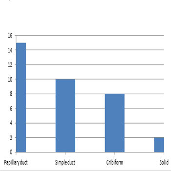
Chart 2: Hitopathological assay � tissue architecture.
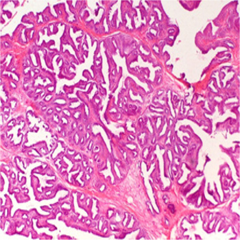
Figure 1: Papillary canine mammary carcinoma. Neoplastic proliferation is observed forming papillae projecting towards the center . H&E, 50x
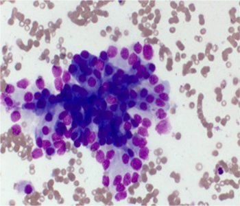
Figure 2: Canine mammary carcinoma. Notice a group of epithelial cells arranged in papillary form. Giemsa, 200x
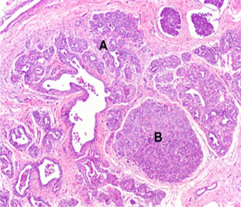
Figure 3: Canine mammary carcinoma. Neoplastic proliferation is observed forming ducts (A) and solid formations (B). Giemsa, 200x.
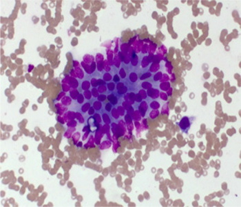
Figure 4: Canine mammary carcinoma. Notice a group of epithelial cells arranged in papillary form. Giemsa, 200x
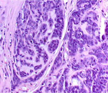
Figure 5: Canine mammary carcinoma. Neoplastic proliferation arranged in cribiform organization. Giemsa, 200x
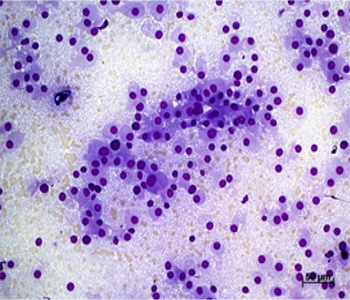
Figure 6: Canine mammary carcinoma. Notice a groups of epithelial cells in a cribform formation. Giemsa, 100x.
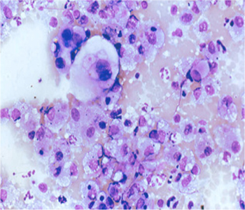
Figure 7: Canine mammary carcinoma. Notice epithelial dispersed cells, with vacuolated cytoplasm. Giemsa, 200x.
For the nucleus criteria, the most prevalent on the cytopathological assays were multinucleation (21 samples), evident nucleolus (14 samples) and grooves (9 samples), as shown in Chart 4. In the histopathological assay, we identified only single nucleolus as a characteristic (11 samples).
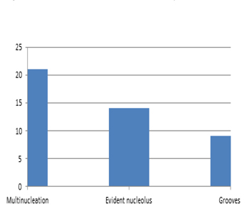
Chart 3: Nucleus – cytopathological assay
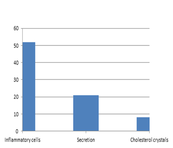
Chart 4: Microenvironment – cytopathological assay.
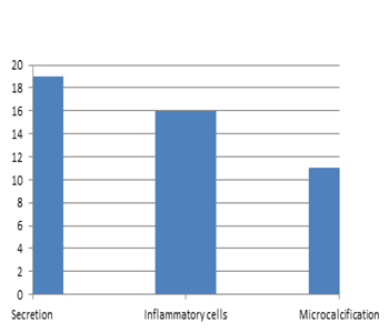
Chart 5: Microenvironment – histopathological assay
DISCUSSION
The assessment of the cytopathological samples allowed us to verify that neoplasms present varying degrees of aggressive- ness depending on the criteria observed and the frequency some criteria repeat themselves across different samples. For an assay to become a diagnostical method for mammary neoplasms, we need to understand not only the technique, but also the structures being analyzed and comparing their normal states to what is observed [21, 22]. Given these facts, we can also verify the lack of methods for assessing the malignancy of carcinomas, a characteristic that would be very important due to how often these neoplasms display varying degrees of aggressiveness, which may be observed both in cytopathological and histopathological assays: the papillary arrangement was prevalent in both assays. This, aside from showing a considerable degree of concordance between the results, also indicates a certain degree of aggressiveness for the wound since the formation of papillae differs from the morphology of a normal acinus Another important finding was the presence of a secretion in a considerable number of samples across both assays, implying hormonal activity and, therefore, a possible progression of the wound if not treated adequately It is important to stress that there are some cellular characteristics that are easily detectable in a cytopathological assay, but hard to detect in the histopathological assay, such as those regarding cytoplasm and nucleus. For example, cytopathology enabled the visualization of fibrillar cytoplasm and multinucleation, which indicates a considerable degree of aggressiveness if we consider the normal morphology of the cell. This highlights the fact that the assays share some characteristics, but there are also some characteristics from the cytopathological assay that cannot be observed in the histopathological assay [17]. In addition, the nomenclature and terminology created in cytopathology aim at grouping diseases in categories to reduce subjective variations and add value to the diagnosis and the care of the patient. Communication through a universal language is a field that has been widely studied and applied to medicine. Considering this, we observed the fact that organizing the malignancy criteria in a table is something that warrants further studies given the lack of such information in the literature [23].
CONCLUSION
Under the light of these results, the cytopathological assay may, in fact, be used as an auxiliary or alternative tool in the diagnosis of canine mammary cancer. In addition, the assay has its own characteristics that cannot be observed in the histopathological assay. This dichotomy also contributes towards the qualification of the assay as part of an array of diagnostic possibilities for the affliction.
ACKNOWLEDGEMENTS
This work was partly supported by National Institutes of Health grant 5 T32 HL 69764-9, granted to Dr. Kwon.
REFERENCES
- Sonnenschein EG, Glickman LT, Goldschmidt MH and Mckee LJ. (1991). Body conformation, diet, and risk of breast cancer in pet dogs: a case-control study. Am J Epidemiol. 133(7): 694-733.
- Alenza DP, Pena L, Del Castillo and N Nieto AI. (2000). Factors influencing the incidence and prognosis of canine mammary tumors. J Small Animal Practice. 41(7): 287-291.
- MISDORP W. (2002). Tumors of the mammary gland. Tumours in Domestic Animals, 4th Edit, DJ. Meuten Ed, Iowa State Press, Iowa. 575-606
- Davidson EB. (2003). Treatment of mammary tumors in dogs and cats. Proceedings of the 2003 North American Veterinary Conference, Orlando, Florida.10361038
- Johnston SD. (2003). Reproductive systems. In: Textbook of Small Animal Surgery. Slatter D (ed.). Philadelphia: W.B. Saunders Company. 2177-2192.
- Sontas BH, Öztürk GY, Toydemir TFS, Arun SS, et al. (2012). Fine-Needle Aspiration Biopsy of Canine Mammary Gland Tumours: A Comparison Between Cytology and Histopathology. Reprod Dom Anim. 47(1): 125-130.
- Petrov EA, llievska K, Trojacanec P, Celeska l, et al. (2014). Canine mammary tumours–Clinical Survey. Macedonian Veterinary Review. 37(2): 129-134.
- Baptista CS, Bastos E, Santos S, Ut IG, et al. (2010). Gene: First insights in Felis catus. Current Genomics, 11: 212-220.
- Rivera P and Von Euler H. (2011). Molecular biological aspects on canine and human mammary tumours. Veterinary Pathology, 48(1): 132-146.
- Rutten V, Misdorp W, Gauthier A, Estrada M, et al. (1990). Immunological aspects of mammary tumors in dogs and cats: A survey including own studies and pertinent literature. Veterinary Immunology and Immunopathology. 26(3): 211-225.
- Morris J, dobson J and Bostock D. (1993). Use of tamoxifen in the control of canine mammary neoplasia. Veterinary Record. 133(22): 539-542.
- Teske E, Rutteman GR, VD Ingh TS, Van Noort R, et al. (1998). Liposomeencapsulated muramyl tripeptide phosphatidyl-ethanolamine (L-MTP-PE): A randomized clinical trial in dogs with mammary carcinoma. Anticancer Research. 18(2A): 1015-1020.
- Novosad CA, Bergman PJ, O‘Brien MG, Mcknight JA, et al. (2006). Retrospective evaluation of adjunctive doxorubicin for the treatment of feline mammary gland adenocarcinoma: 67 cases. Journal of the American Animal Hospital Association. 42(2): 110-120.
- Simon D, Schoenrock D, Baumgartner W and Nolte I. (2006). Postoperative adjuvant treatment of invasive malignant mammary gland tumors in dogs with doxorubicin and docetaxel. Journal of Veterinary Internal Medicine. 20(5): 1184-1190.
- Matos AJF, Baptista CS, Gärtner MF and Rruteman GR. (2011). Prognostic studies of canine and feline mammary tumours: The need for standardized procedures. The veterinary journal. 193(1): 24-31.
- Rotten D, Levaillant JM, Leridon H, Letessier A, et al. (1993). Ultrasonographically guided fine needle aspiration cytology and core-biopsy in the diagnosis of breast tumors. Eur J Obstet Gynecol Reprod Biol. 49(3): 175-186.
- Zuccari Dapc, Santana AE and Rocha NS. (2001). Correlação entre a citologia aspirativa por agulha fina e a histologia no diagnóstico de tumores mamários de cadelas. Braz. J. Vet. Res. Anim. Sci. 38(1): 38-41.
- Simon D, Schoenrock D, Nolte I, Baumgartner W, et al. (2009). Cytologic examination of fine-needle aspirates from mammary gland tumors in the dog: diagnostic accuracy with comparison to histopathology and association withpostoperative outcome. Vet Clin Pathol. 38(4): 521-528
- haziroglu R, Yardimci B, Aslan S, Yildirim MZ, et al. (2010). Cytological evaluation of canine mammary tumours with fine needle aspiration biopsy technique. Rev Med Vet. 161(15): 212-218.
- Hodges J. (2013). Using cytology to increase small animal practice revenue. Veterinary Clinics of North America. Small Animal Practice. 43(6): 1385-1408.
- Dolka. (2014). The value of cytological examination of canine mammary tumours. Journal of Comparative Pathology. 152(65).
- BRAZIL. (2002). Ministry of health. National Cancer Institute. Coordination of Prevention and Surveillance - COMPREV. Histopathological and cytopathological diagnosis of breast lesions. Rio de Janeiro: COMPREV. 7-13.
- Collaço LM. (2014). The importance and usefulness of terminology and nomenclature for Cythopathology. Jornal Brasileiro de Patologia e Medicina Laboratorial. 394-395.
- CASSALI. (2011). Consensus for the Diagnosis, Prognosis and Treatment of Canine Mammary Tumors. Braz J Vet Pathol. 4(2): 153-180.
- Hellmen E and lindgren A. (1989). The accuracy of cytology in diagnosis and DNA analysis of canine mammary tumours. J Comp Pathol. 101(4): 443-450,
- Macewen EG, Harvey HJ, Patnaik AK, Mooney S, et al. (1985). Evaluation of effects of levamisole and surgery on canine mammary cancer. Journal of Biological Response Modifiers. 4(4): 418-426.