Information Links
Related Conferences
Previous Issues Volume 2, Issue 1 - 2021
Thyroid Cartilage Radiological Clues Following Exhumation in a Suspected Case of Strangulation
Elmas Shaqiri1, Bledar Xhemali1, Gentian Vyshka2*
1Forensic Pathologist, Department of Forensic Pathology, Institute of Legal Medicine, Tirana, Albania
2Professor of Human Physiology, Biomedical and Experimental Department, Faculty of Medicine, University of Medicine in Tirana, Albania
*Corresponding Author: Gentian Vyshka, Faculty of Medicine, University of Medicine in Tirana, Rr. Dibres 371, Tirana, Albania, Tel: +355-692-828-140; E-mail: [email protected].
Received Date: December 28, 2020
Published Date: January 27, 2021
Copyrights: Shaqiri E, et al. © 2021.
Citation: Shaqiri E, et al. (2021). Thyroid cartilage radiological clues following exhumation in a suspected case of strangulation. Mathews J Foren. (2)1:8.
IMAGES IN FORENSIC MEDICINE
Suspected cases of strangulation are as a rule clarified during investigations following the immediate after-death period. However, cases of suspected strangulation might be disguised as natural deaths, especially in areas where medical assistance is inexistent or superficial. These cases will come sometimes into the scope of the coroner, or legal medicine physician, even several years after burial.
The state attorney office in an Albanian district requested exhumation of the corpse three years after the death. The corpse was of a 74-years old Caucasian female, whose official cause of death prior to the raised suspicion of murder was that of natural death.
On autopsy the laryngeal structures were cautiously scrutinized. The hyoid bone was found intact (Figure 1); but the ossified thyroid cartilage was found comminuted into small fragments such as assembling its right lamina was impossible.
The left lamina (Figure 2) of the ossified thyroid cartilage was preserved up to some extent; but a radiological evaluation showed as well signs of osteitic changes that were compatible with external pressure applied on the laryngeal area during the presumed strangulation (Figure 3).
The age of the dead person (74 years) would explain the ossification of the thyroid cartilage that rendered on the other hand possible the radiological evaluation of the structure. Due to its particular character, the cartilage remains nonossified in a majority of cases and therefore, computed tomography (CT) might be the examination of choice [1]. However, plain radiographs are still of some value in post-mortem cases, when the state of the cadaver renders the CT evaluation uneasy, if not feasible at all.
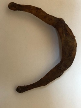
Figure 1: Intact hyoid bone (exhumation finding).
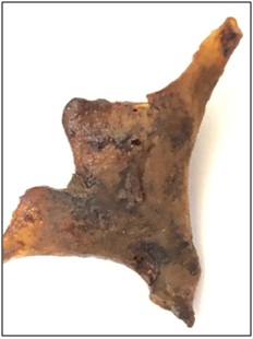
Figure 2: Left lamina of the thyroid cartilage (exhumation finding).
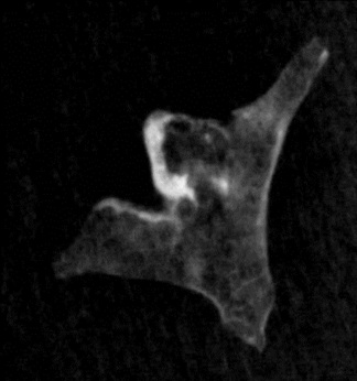
Figure 3: Osteitic changes, left lamina of thyroid cartilage (plain radiograph).
Laryngeal structures should be carefully examined every time there is a suspicion of violent death or blunt trauma [2,3]. Thyroid cartilage fracture might remain uncovered in cases of unsuspected violence, when in fact even minor blunt trauma related to neck hyperflexion, as well as other overlooked occurrences, will leave their imprint on the cartilage – be it ossified or not [4].
REFERENCES
- Dadfar N, Seyyedi M, Forghani R, Curtin HD. (2015). Computed tomography appearance of normal nonossified thyroid cartilage: implication for tumor invasion diagnosis. J Comput Assist Tomogr. 39(2):240-3.
- Chand S, Solanki R, Aggrawal A, Dikshit PC, Ranjan R. (2017). Study of Postmortem Findings of Neck Structures in Cases of Asphyxial Deaths. Int J Sci Stud. 5(4):248-56.
- Becker M, Leuchter I, Platon A, Becker CD, Dulguerov P, et al. (2014). Imaging of laryngeal trauma. Eur J Radiol. 83(1):142-54.
- Lucerna A, Espinosa J, Hertz R. (2017). Thyroid Cartilage Fracture in Context of Noncompetitive “Horseplay” Wrestling. Emergency Medicine. 49(9):422-5.