Previous Issues Volume 3, Issue 1 - 2018
The Accuracy of Clinical Ultrasonography in the Diagnosis of Shoulder Joint Dislocation
Behnam movahedi1*, Leila azizkhani1, Sarvenaz Salahi2, Arman mohammadi1, Enayatollah Noori3
1 Department of Emergency Medicine &Traumatology, Besat Hospital, Kurdistan University of Medical Sciences, Sanandaj, Iran. 2 Department of minimal invasive surgery research committee, School of Medicine, Iran University of Medical Sciences, Teran, Iran. 3 Department of research committee, School of Medicine, Qom University of Medical Sciences, Qom, Iran. Corresponding Author: Behnam Movahedi, Department of Emergency Medicine &Traumatology, Besat Hospital, Kurdistan University of Medical Sciences, Sanandaj, Iran, Tel: +989355155124; Email: [email protected] Received Date: 10 Oct 2018 Accepted Date: 06 Dec 2018 Published Date: 12 Dec 2018 Copyright © 2018 Movahedi B Citation: Movahedi B, Azizkhani L, Salahi S, Mohammadi M, et al. (2018). The Accuracy of Clinical Ultrasonography in the Diagnosis of Shoulder Joint Dislocation. M J E-Med. 3(1): 031.
ABSTRACT Introduction: When the head of the humerus is out of the shoulder joint, it is termed as shoulder joint dislocation. This type of dislocation can be easily diagnosed through taking precise medical history and accurate physical examination. In the case of dislocation with fracture, clinical diagnosis and confirmation of this pathologic condition are done by using radiography. After joint reduction, radiography of shoulder is requested which includes the confirmation of reduction and evaluation about any new fracture during the shoulder reduction. According to the common use of ultrasonography in the emergency, and the lack of studies about its precision in diagnosis of shoulder dislocation and its reduction, we designed this study with the aim of evaluating the accuracy of ultrasonography in these fields. Methodology: In this case-control study, patients who visited the Emergency Department of Besat Hospital with a clinical diagnosis of shoulder dislocation were examined by ultrasonography in terms of diagnostic criteria of the shoulder joint dislocation and fracture before radiography. In addition, patients’ status was also evaluated through clinical examination and then they were sent for radiography. Results: In this study, 100 patients diagnosed with shoulder dislocation were examined. The mean age of patients was 39.15. In terms of gender, 85% of patients were male, and 71% of them were complaining of a shoulder dislocation for the first time. All patients were examined by both ultrasonography and radiography. None of the patients were afflicted with a shoulder dislocation. Positive and negative predictive value, sensitivity, feature, and accuracy of ultrasonography were equal to 100%. Conclusion: Based on the study results, ultrasonography has an appropriate accuracy in diagnosing the shoulder dislocation and confirming the reduction. Therefore, it can be considered as a beneficial alternative to radiography in the diagnosis of shoulder dislocation and confirmation of its reduction. However, considering the shortage of patients with a shoulder dislocation, it recommended that more studies be conducted on patients with a possible shoulder dislocation and complications of dislocation to assess the diagnostic power of ultrasonography. KEYWORDS Shoulder Dislocation; Ultrasonography.
INTRODUCTION
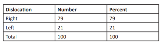
The shoulder dislocation is one of the most common types of dislocation among large joints of the body [1] which accounts for 50% of large joints dislocation [2]. The incidence rate of it was established about 23.9 in 100,000 persons annually in United States which has two main and independent risk factors of age and gender [3]. This dislocation is occurred when the head of humerus moving out of shoulder joint [3]. Because of its anatomical and biomechanical conditions, the shoulder is more vulnerable to be dislocated than any large joint of the body. Shoulder joint dislocation is divided into two types of acute and chronic [3]. The acute shoulder dislocation is divided into anterior and posterior forms [4]. About 95-97% of cases of acute shoulder dislocation are of anterior type and others are of posterior and lower form [5, 6]. The major clinical signs of shoulder dislocation include severe pain and limitation of the range of joint motions [7]. The patient’s hand is located on one side and far from the body and the forearm turns outward. Sometimes, the head of arm bone is touched in the anterior part [8]. In some cases, this dislocation is accompanied with fractures such as anterior glenoid fracture and tuberosity of the arm bone [1]. This type of dislocation can be easily diagnosed through physical examination and medical history [8]. This pathologic condition is diagnosed clinically and confirmed by radiography findings [7]. This type of dislocation is reduced by orthopedists or emergency specialists in hospitals. Regularly, both of them request for the shoulder radiography before and after the reduction. This radiography is to confirm the dislocation before beginning the treatment and the reduction after treatment and also to ensure that no new fracture will occur during the shoulder reduction [9]. Retrospective and prospective studies on this subject have shown that the diagnostic accuracy of physicians is very high in the confirmation of dislocation and reduction. However, if the physician is not sure about complete reduction, radiography is recommended for further control [1]. On the other hand, if the physician is sure of complete reduction, there would be no need for the control radiography. One of the methods which was used in the studies for the examination of shoulder dislocation and reduction, is ultrasonography. However, ultrasonography is more commonly used in the local block for the reaction and studies which evaluated about the confirmation of dislocation and reduction, are mainly of case report type. [10, 11]. Hence, there is not much information about the level of ultrasonography accuracy in the diagnosis of dislocation. Since pre- and post-reduction radiography takes time, exposes the patient to harmful radiation, and imposes considerable costs on the patient, the present study aims to determine whether clinical ultrasonography is accurate enough in the diagnosis of dislocation to replace the pre-reduction radiography. If positive results are achieved, some modifications can be made to the diagnosis and treatment of these patients and the economic burden of radiography will be decreased due to the usage of ultrasonography. METHODOLOGY In this case-control study, all patients who visited the Emergency Department of Besat Hospital of Sanandaj with a clinical diagnosis of the shoulder dislocation, were selected as the sample based on the convenience sampling method. Therefore, there was not any determined sample size before beginning the study. After co-ordination with the research deputy of the hospital, the research proposal was approved by the ethics committee of Kordestan university of medical sciences. At first, patients were taken medical history and examined by an emergency specialist using ultrasonography in terms of diagnostic criteria of the shoulder joint dislocation and fracture. In addition, patients’ status was also evaluated through clinical examination. The patients with the clinical diagnosis of the shoulder dislocation with complete consciousness were included to our study. The aims of the research were explained for the patients and the written consent was taken from the patients who desired to participate in the study by the researcher. The data was collected by the researcher with the structured checklist. After clinical confirmation, the patients were evaluated by radiography which was performed by a skilled determined radiologist. After the confirmation of dislocation due to radiography findings, the shoulder joint of patients was reduced under anesthesia. Finally, after the clinical confirmation of the reduction, radiography was done for patients with a graph as the golden standard. After reduction and assessment of shoulder motion, ultrasonography was performed to confirm the reduction accuracy. Therefore, the completeness of reduction in all patients was assessed using ultrasonography and the accuracy of ultrasonography and radiography were assessed due to the related signs. The patients with a good general condition and acceptable final evaluation of the shoulder reduction were discharged from hospital after 6 hours’ follow-up. Data analysis A certain statistical test was used to determine the relationship between independent variables in each hypothesis. Accordingly, given the ordinal nature of both response variables, Spearman correlation coefficient was used for relative or ordinal independent variable and chi-square test and Choproff correlation coefficient was used for the qualitative independent variable. Findings In this study, 100 patients (85 men and 15 women), with a mean age of 39.15, diagnosed with shoulder dislocation were examined. Among the patients, 71% of patients had a shoulder dislocation for the first time and 79% of them had right shoulder dislocation.
Table 1: The frequency of dislocation times
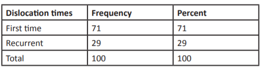
Table 2: The frequency of the dislocation side.
The radiography results confirmed shoulder dislocation in all patients. In addition, pre-radiography ultrasonography indicated that all patients had a shoulder dislocation. The results of radiography also confirmed the accuracy of reduction. Based on the study results, positive and negative predictive value, sensitivity, feature, and accuracy of ultrasonography were equal to 100%.
DISCUSSION
Nowadays, the use of state-of-the-art technologies at the patient bedside has increased the accuracy of medical diagnoses and reduced the need for some methods such as radiography that emit ionizing radiation. Hence, the significance of determining the diagnostic accuracy and value of these new methods to replace traditional ones necessitates such studies [12, 13]. In the present study, the diagnostic value of the clinical use of ultrasonography for the diagnosis of joint dislocation and confirmation of its reduction was evaluated. According to the results, the ultrasonography performed by a trained resident confirmed all cases of dislocation diagnosis and reduction confirmation by radiography. Until 2009, shoulder reduction used to be confirmed by radiography. The most important drawback of this method was the need for the elimination of the effects of anesthetics. In case of failure in the reduction, there was a need for re-anesthesia. Therefore, there was a need for an alternative clinical method for the confirmation of reduction. Halberg et al. (2009) proposed the possibility of using ultrasonography for the confirmation of reduction [14]. In a posterior dislocation, AP radiograph is not helpful in 50% of cases. In case of doubt about dislocation and final confirmation, it is necessary to perform CT Scan. In terms of cost-benefit and exposure to radiation, no remarkable result has been reported in previous studies. Clinical ultrasonography in the emergency department is the alternative and available modality. In a case report, David et al. used ultrasonography for the diagnosis of dislocation but their results cannot be generalized to other populations due to lack of imaging studies and the small number of patients [15]. Today, ultrasonography is increasingly being used in the examination of shoulder pains [16].
In a report published by Blakeley et al., all the 5 patients were successfully examined using ultrasonography and it was concluded that ultrasonography is an appropriate alternative modality for the confirmation of joint reduction [17]. Davies (2004) successfully used ultrasonography in the diagnosis of the shoulder anterior dislocation. Referring to the fact that ultrasonography is commonly used in the diagnosis of articular lesions, such as effusion, and musculoskeletal disorders, such as rotator cuff injuries, he proposes the use of this modality for the diagnosis of anterior dislocation as a quick and accessible way [18]. Abbasi et al. (2013) reported that the sensitivity of ultrasonography in the diagnosis of shoulder dislocation and confirmation of its reduction is equal to 100% [19]. This is consistent with the findings of the present study. Flinders et al. (2016) stated that ultrasonography is helpful in the diagnosis of shoulder dislocation and confirmation of its reduction. They also claimed that, along with the examination of hematoma and soft tissues injuries, this method makes it possible for the emergency physicians to easily diagnose the shoulder joint dislocation in patients, especially those with decreased consciousness, in the fast track section of the emergency department [20]. Lahham et al. reported the sensitivity and specificity of ultrasonography in the diagnosis of shoulder dislocation to be 93.3% and 98.5%, respectively. Therefore, it is an appropriate alternative for the diagnosis of dislocation [21]. In another study, Lahham et al. reported that the sensitivity, specificity, and the positive and negative predictive value of ultrasonography (from the back view) are equal to 100%. Compared to the study of Abbasi et al., they used only one back view, instead of two back and lateral views, but the sensitivity and specificity of the test did not decrease [22]. In a review study, Ahmadi et al. evaluated the replacement of radiography with ultrasonography. Based on this report, ultrasonography is an appropriate alternative for the diagnosis of dislocation. In addition, the use of ultrasonography along with clinical examination and asking the patient some questions increases the certainty of diagnosis and eliminates the need for radiography [23]. In this study, none of the patients had fractures or traumatic lesions following the shoulder dislocation. Hence, it was not possible to evaluate the diagnostic power of ultrasonography in relation to complications of shoulder dislocation. In addition, since all of the patients had the anterior dislocation shoulder, the power of ultrasonography in the diagnosis of posterior and lower dislocations. CONCLUSION The results of this cross-sectional study showed that ultrasonography is an appropriate alternative to radiography for the diagnosis of anterior dislocations, and trained physicians can definitely diagnose by getting the patient’s status, clinical examination, and ultrasonography. In addition, after the reduction and removal of anesthesia effects, ultrasonography can be used to confirm the accuracy of reduction and the need for re-sedation can be eliminated in the case of failure. Finally, given the above-mentioned limitations, it is necessary to conduct more studies on larger samples and different types of dislocation.
LIMITATIONS In this study, none of the patients had fractures or traumatic lesions following the shoulder dislocation. Hence, it was not possible to evaluate the diagnostic power of ultrasonography in relation to complications of shoulder dislocation. In addition, since all of the patients had the anterior dislocation shoulder, the power of ultrasonography in the diagnosis of posterior and lower dislocations. Therefore, we recommend that a prospective study with a wider types of the shoulder dislocation will be constructed to examine the precision of the ultrasonography in diagnosis the several types of shoulder dislocations. Also, due to the importance of determining the accuracy of ultrasonography in detecting the comorbid fractures, it is essential to design a study about the patients with both dislocation and fractures in the shoulder joint.
REFRENCESE 1. Gottlieb, Michael and Frances Russell. (2017). Diagnostic Accuracy of Ultrasound for Identifying Shoulder Dislocations and Reductions: A Systematic Review of the Literature.” Western Journal of Emergency Medicine. 18(5): 937-942. 2. Rockwood and Green’s. (2001). Fractures in Adults, 5th ed. Lippincott Williams and Wilkins. 18(6): 1110 3. Campbell’s. (1998). Operative orthopedics, 9th ed. Mosby. 3: 2397. 4. Rockwood and Green’s. (2001). Fractures in Adults, 5th ed. Lippincott Williams and Wilkins. 2: 1112. 5. Robinson CM, Seah M and Akhtar MA. (2011). The Epidemiology, risk of recurrence, and functional outcome after an acute traumatic posterior dislocation of the shoulder. J Bone Joint Surg. 93(17): 1605-13. 6. Campbell’s. (1998). Operative orthopedics, 9th ed. Mosby. 3: 2399. 7. Chong M, Karataglis D and Learmonth D. (2006). Survey of the management of acute traumatic first-time anterior shoulder dislocation among trauma clinicians in the UK. Ann R Coll Surg Engl. 88(5): 454-458. 8. Itoi E1, Hatakeyama Y, Kido T, Sato T, et al. (2003). A new method of immobilization after traumatic anterior dislocation of the shoulder. A preliminary study. J Shoulder Elbow Surgery. 12(5): 413-415. 9. Hendey GW and Kinlaw K. (1996). Clinically significant abnormalities in postreduction radiographs after anterior shoulder dislocation. Ann Emerg Med. 28(4): 399-402. 10. Ahmadi K, Hashemian AM and Sineh Sepehr K. (2015). Sonography as a new modality in the management of shoulder dislocation. Reviews in Clinical Medicine. 2(2): 100-102. 11. Yuen CK, Mok KL, Kan PG and Wong YT. (2009). Ultrasound diagnosis of anterior shoulder dislocation. Hong Kong J Emerg Med. 16(1): 29-34. 12. Akyol C, Gungor F, Akyol AJ, Kesapli M, et al. (2016). Pointof-care ultrasonography for the management of shoulder dislocation in ED. The American journal of emergency medicine. 34(5): 866-370. 13. McBride T and Kalogrianitis S. (2012). Dislocations of the shoulder joint, Trauma. 14(1): 47-56. 14. Halberg MJ, Sweeney TW and Owens WB. (2009). Bedside ultrasound for verification of shoulder reduction. The American journal of emergency medicine. 27(1): 134e5. 15. Mackenzie DC and Liebmann O. (2013). Point-of-care ultrasound facilitates diagnosing a posteriorr shoulder dislocation. The Journal of emergency medicine. 44(5): 976-978. 16. Levine BD, Motamedi K and Seeger LL. (2012). Imaging of the shoulder: a comparison of MRI and ultrasound. Current sports medicine reports. 11(5): 239-243. 17. Blakeley C, Spencer O, Newman-Saunders T and Hashemi K. (2009). A novel use of portable ultrasound in the management of shoulder dislocation. Emergency Medicine Journal. 26(9): 662-663. 18. Bize P, Pugliese F, Bacigalupo L and Bianchi S. (2004). Unrecognized bilateral posterior shoulder dislocation diagnosed by ultrasound (2003: 11b). European radiology. 14(2): 350-352. 19. Abbasi S, Molaie H, Hafezimoghadam P, Zare MA, Abbasi M, et al. (2013). Diagnostic accuracy of ultrasonographic examination in the management of shoulder dislocation in the emergency department. Ann Emerg Med. 62(2): 170-e5. 20. Flinders A and Seif D. (2016). Point-of-Care Ultrasound in Diagnosis and Treatment of Luxatio Erecta (Inferior Shoulder Dislocation). Journal of Medical Ultrasound. 24(2): 70-73. 21. Lahham S, Lane N, Trinh A and Fox JC. (2015). Evaluation of Shoulder Injury in The Emergency Department: Utility Of Bedside Ultrasound in The Diagnosis of Acute Shoulder Dislocation. Academic Emergency Medicine. 22: S303. 22. Lahham S, Becker B, Chiem A, Joseph LM, et al. (2016). Pilot Study to Determine Accuracy of Posterior Approach Ultrasound for Shoulder Dislocation by Novice Sonographers. Western Journal of Emergency Medicine. 17(3): 377-82. 23. Ahmadi K, Hashemian AM and Sineh Sepehr K. (2015). Sonography as a new modality in the management of shoulder dislocation. Reviews in Clinical Medicine. 2(1): 100-102.