Information Links
Related Conferences
Current Issue Volume 10, Issue 2 - 2026
Comprehensive Prosthetic Management of Acquired Maxillary Defects: A Narrative Review
Alapati Sowmya, Pavan kumar T, Kalamalla A SaranBabu*, Harsha Priya G, Vineela Reddy Katasani, Madireddygari Rithika
Department of Prosthodontics, Narayana Dental College & Hospital, Nellore, Andhra Pradesh, India
*Corresponding Author: Dr. Kalamalla A SaranBabu, Associate Professor, Department of Prosthodontics, Narayana Dental College and Hospital, Nellore, Andhrapradesh, India, Tel: 8886078222, Email: [email protected]
Received Date: May 27, 2026
Published Date: June 13, 2026
Citation: Sowmya A, et al. (2026). Comprehensive Prosthetic Management of Acquired Maxillary Defects: A Narrative Review. Mathews J Dentistry. 10(2):68.
Copyrights: Sowmya A, et al. © (2026).
ABSTRACT
Acquired maxillary defects are complex anatomical deformities resulting from surgical resection, trauma, infection, or pathological conditions affecting the maxilla and adjacent structures. These defects produce significant impairment in speech, mastication, swallowing, esthetics, and psychosocial well-being. Prosthodontic rehabilitation using obturator prostheses remains one of the most predictable and widely accepted treatment modalities for restoring oral function and facial form in such patients. Obturators re-establish the separation between the oral and nasal cavities, improve phonetics, aid deglutition, and enhance patient confidence. Depending on the stage of healing and treatment requirements, obturators may be classified as surgical, interim, or definitive prostheses. Support, retention, stability, defect classification, tissue state, and residual dentition must all be carefully taken into account while designing a prosthesis. Recent advances including hollow bulb obturators, flexible silicone prostheses, CAD/CAM technology, and implant-supported obturators have significantly improved rehabilitation outcomes. This narrative review discusses the etiology, classification, objectives, types, design principles, impression techniques, hollow bulb prostheses, recent technological advancements, and clinical considerations involved in the prosthodontic management of acquired maxillary defects.
Keywords: Acquired Maxillary Defects, Obturator Prosthesis, Maxillectomy, Hollow Bulb Obturator, Maxillofacial Prosthodontics, CAD/CAM Obturator.
INTRODUCTION
Maxillofacial prosthodontics is a specialized branch of prosthodontics concerned with the rehabilitation of patients with congenital or acquired defects involving the stomatognathic and craniofacial structures. Acquired maxillary defects are among the most common intraoral defects encountered in maxillofacial rehabilitation and frequently result from surgical management of benign or malignant neoplasms, traumatic injuries, infections, or radiation-induced tissue destruction [1,2]. These defects compromise the normal separation between the oral and nasal cavities, thereby affecting speech, swallowing, mastication, esthetics, and psychological well-being [2,3].
The primary objective of prosthodontic rehabilitation in such patients is to restore function and improve quality of life through the fabrication of obturator prostheses [1,4]. The term “obturator” is derived from the Latin word obturare, meaning “to close or stop up.” Obturators are prostheses designed to close congenital or acquired tissue openings of the palate and associated structures. They play a crucial role in restoring oral competence, reducing nasal regurgitation, improving speech intelligibility, and supporting facial contours [3].
Depending upon the stage of healing, obturator prostheses may be categorized into surgical obturators, interim obturators, and definitive obturators. Advances in biomaterials, digital technology, and implant dentistry have considerably enhanced the prognosis and acceptance of obturator prostheses. This review discusses the current concepts and prosthodontic principles involved in the rehabilitation of acquired maxillary defects.
Acquired defect:
Acquired maxillofacial defects are those that are treated by other than congenital / developmental influences. They are in effect man-made defects. These defects are more often related to surgical intervention for the elimination of disease process / to trauma resulting in the loss / significant alteration of the normal anatomic features of the oral and facial structures.
Etiology of Acquired Maxillary Defects
Acquired maxillary defects may arise due to several causes, including:
Surgical resection of malignant or benign tumors
- Traumatic injuries
- Osteoradionecrosis
- Fungal infections such as mucormycosis
- Congenital defect corrections
- Radiation burns
- Pathological bone destruction
Among these, surgical maxillectomy performed for management of oral and maxillofacial malignancies is the most common cause [2,5]. Depending on the extent of resection, patients may experience varying degrees of impairment involving speech, swallowing, mastication, salivary control, and facial esthetics [4,6].
Classification of Maxillary Defects
Proper classification of maxillary defects is essential for treatment planning and framework design [2,3]. Several classifications have been proposed; however, for partially edentulous maxillary deformities, the Aramany classification is still the most commonly used method.
Aramany Classification
Class I, II, III, IV, V, VI Maxillary Defect Classification
Mohamed Aramany classified acquired maxillary defects into six categories based on the relationship between the defect and the remaining teeth [7,8]. This classification assists in designing obturator frameworks with appropriate support, retention, and stability [9].
As the class number increases, the complexity of the defect and the prosthesis design also increases.
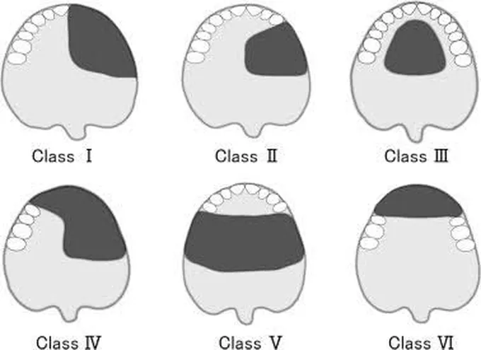
Class I
Midline resection with one side of the teeth left intact.
Class II
Unilateral defect posterior to remaining anterior teeth.
Class III
Central palatal defect with preservation of teeth bilaterally.
Class IV
Bilateral defect crossing the midline with limited remaining teeth.
Class V
Posterior bilateral defect with anterior teeth remaining.
Class VI
Anterior defect with bilateral posterior teeth remaining.
Table 1. Prosthodontic considerations of Aramany classes
|
Aramany class |
Prosthodontic Considerations |
|
I |
· Simple framework design · Clasping of remaining teeth · Good support and stability from residual structures |
|
II |
· Rests placed on anterior and posterior abutments · Indirect retention required · Cross-arch stabilization important |
|
III |
· Rests provided on both sides of the arch · Double reciprocal retention may be used · Broad palatal coverage improves stability |
|
IV |
· Complex framework design required · Rests on both anterior and posterior teeth · Indirect retention is crucial · Cross-arch stabilization needed |
|
V |
· Anterior teeth contribute significantly to retention · Palatal strap design is most commonly used · Indirect retention improves stability |
|
VI |
· Rests primarily placed on posterior teeth · Indirect retention from posterior abutments · Broad palatal coverage enhances stability and support |
The Brown classification is another system commonly used in oncologic reconstruction as it considers both vertical and horizontal components of the defect [5,10].
Objectives of Prosthodontic Rehabilitation:
The primary objectives of rehabilitation include:
- Separation of oral and nasal cavities
- Restoration of mastication and swallowing
- Improvement in speech intelligibility
- Enhancement of facial esthetics
- Preservation of residual structures
- Psychological rehabilitation
- Reduction of nasal regurgitation
- Improvement in patient comfort and confidence [1,2]
Obturator prostheses also aid in wound healing, support surgical dressings, reduce contamination of the surgical site, and help maintain tissue contours during healing [2,3].
Types of Obturators:
Surgical Obturator
A surgical obturator is a temporary prosthesis inserted during or immediately after surgery. It acts as a matrix for surgical packing and assists in healing.
Immediate Surgical Obturator
The immediate surgical obturator is fabricated before surgery and inserted immediately following resection. It provides immediate separation between the oral and nasal cavities and allows early restoration of speech and swallowing [11,12].
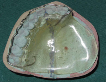
Figure 1. Immediate surgical obturator.
Advantages [2,11] include:
- Reduced postoperative contamination
- Enhanced healing
- Improved patient morale
- Maintenance of facial contour
- Reduced scar contracture
However, retention can be difficult in extensive defects, particularly in edentulous patients.
Delayed Surgical Obturator
Delayed surgical obturators are inserted approximately 7–10 days after surgery following removal of surgical packing. These prostheses are particularly useful in edentulous patients and allow better adaptation to healing tissues [2].
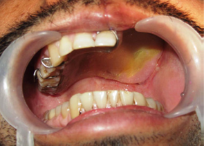
Figure 2. Intra oral view of delayed surgical obturator.
Interim Obturator
An interim obturator is fabricated several weeks after surgery during the healing phase. It replaces the surgical obturator and may include replacement teeth in the defect region.
The interim obturator is modified periodically using tissue conditioners or relining materials to accommodate rapid tissue changes occurring during healing [2,4]. Functional impression techniques are frequently employed to improve peripheral seal and speech outcomes [4].
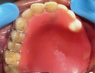
Figure 3. Intraoral View of Interim Obturator.
Definitive Obturator
A definitive obturator is fabricated after complete healing and tissue stabilization, usually 3–6 months following surgery.5 These prostheses are designed to provide long-term restoration of function and esthetics.
Definitive obturators may be fabricated using acrylic resin, cast metal frameworks, flexible materials, or implant-assisted designs depending on the clinical scenario [4,9].
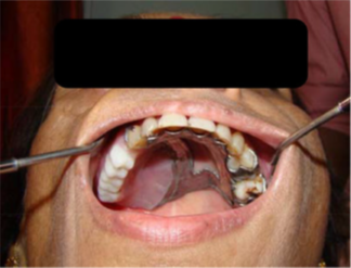
Figure 4. Intra oral view of definitive obtutaror.
Important considerations include:
- Defect size and contour
- Remaining dentition
- Availability of undercuts
- Mouth opening
- Soft tissue mobility
- Patient dexterity and hygiene
Design Principles of Obturator Prostheses
Successful obturator rehabilitation depends on three major prosthodontic principles support, retention and stability [1,9]
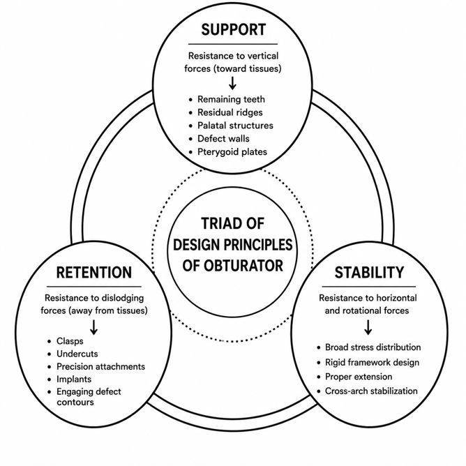
Figure 5. pictorial representation of triad of design principles of obturator.
Support
Support refers to resistance against movement toward the tissues [2,9]. Support may be derived from:
- Remaining teeth
- Residual alveolar ridge
- Hard palate
- Defect walls
- Nasal septum
- Pterygoid plates
Retention
Retention prevents dislodgement of the prosthesis [2]. It may be obtained from:
- Clasps
- Tissue undercuts
- Precision attachments
- Engaging defect contours
- Dental implants
Stability
Stability refers to resistance against horizontal or rotational forces. Broad stress distribution and appropriate framework design improve prosthesis stability.
Hollow Bulb Obturators
In large maxillary defects, hollow bulb obturators are frequently employed to lower prosthesis weight while preserving sufficient extension into the defect [4,13].
Advantages [4,13]
Reduced prosthesis weight
- Improved retention
- Better speech resonance
- Enhanced patient comfort
- Reduced stress on supporting tissues
Open Hollow Bulb
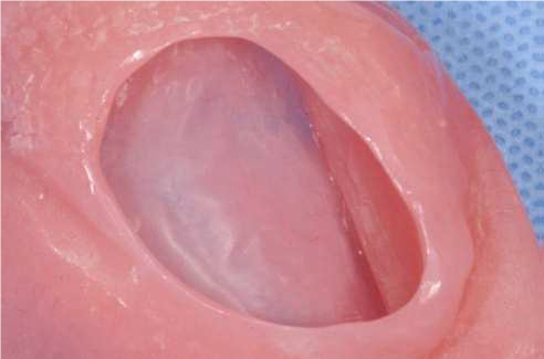
Figure 6. Open lid obturator.
Advantages:
- Easier cleaning
- Reduced weight
- Simpler fabrication
Disadvantages:
- Accumulation of nasal secretions
- Difficulty in polishing internal surfaces
Closed Hollow Bulb:
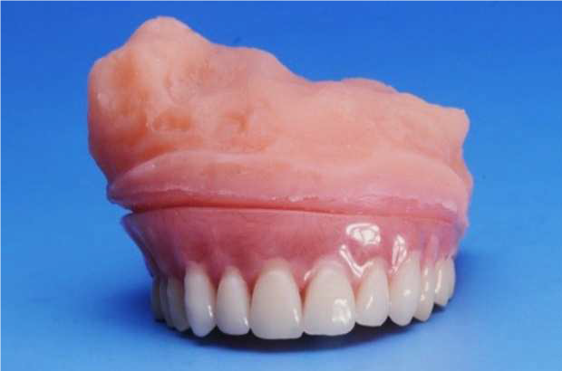
Figure 7. Closed hollow bulb obturator.
Advantages:
- Reduced fluid accumulation
- Improved hygiene barrier
Disadvantages:
More difficult fabrication
Potential microbial contamination if leakage occurs
Several techniques have been described for fabrication of hollow obturators, including [14,15]:
- Salt technique
- Sugar technique
- Ice technique
- Putty index method
- Double flask technique
Table 2. Comparative evaluation of open and closed hollow bulb obturator prostheses
|
Parameters |
Closed Hollow Bulb Obturator |
Open Hollow Bulb Obturator |
|
Basic Design |
Bulb portion is completely enclosed without any opening |
Bulb contains an opening communicating with the defect cavity |
|
Internal Configuration |
Sealed hollow cavity |
Hollow cavity with accessible internal surface |
|
Weight Reduction |
Provides reduction in prosthesis weight |
Also reduces overall prosthesis weight effectively |
|
Hygiene Maintenance |
Internal surface is difficult to access for cleaning |
Easier cleaning due to direct accessibility |
|
Fluid Accumulation |
Lesser accumulation of food debris and nasal secretions |
Greater tendency for accumulation of nasal secretions |
|
Fabrication Complexity |
More technique-sensitive and time-consuming |
Comparatively simpler fabrication procedure |
|
Risk of Microbial Growth |
May occur if leakage develops within sealed cavity |
Increased risk due to continuous exposure to secretions |
|
Patient Comfort |
Comfortable in patients with excessive nasal discharge |
Comfortable in patients capable of maintaining hygiene |
|
Maintenance Requirements |
Requires periodic evaluation for seal integrity |
Requires frequent cleaning and irrigation |
|
Speech Resonance |
Improved resonance due to enclosed air chamber |
Adequate resonance with lighter prosthesis design |
|
Indications |
Suitable for patients requiring improved hygiene barrier |
Preferred in patients with good manual dexterity and hygiene maintenance |
|
Main Advantage |
Prevents direct contamination of internal hollow space |
Easy to clean and lightweight |
|
Main Limitation |
Difficult internal cleaning and repair |
Accumulation of secretions and odor formation |
Impression Procedures
Accurate impressions are critical for successful obturator fabrication [2].
Preliminary Impression
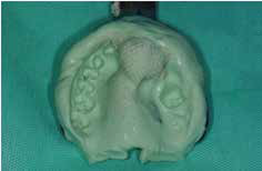
Figure 8. Preliminary impression made with alginate.
Preliminary impressions are generally made using irreversible hydrocolloid with undercuts blocked using gauze or cotton tied with floss [2].
Final Impression
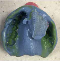
Figure 9. Elastomeric impression.
Custom trays extending into the defect are fabricated. Border molding and functional movements are used to record dynamic tissue contours. Elastic impression materials such as polyvinyl siloxane are preferred for final impressions due to their accuracy and dimensional stability [2].
Functional impression techniques help achieve improved peripheral seal and speech outcomes [4].
Speech and Functional Rehabilitation
Speech impairment, especially hypernasality, is common in maxillectomy patients due to communication between the oral and nasal cavities [6].
Obturator prostheses improve [4,6]:
- Speech intelligibility
- Swallowing
- Mastication
- Salivary control
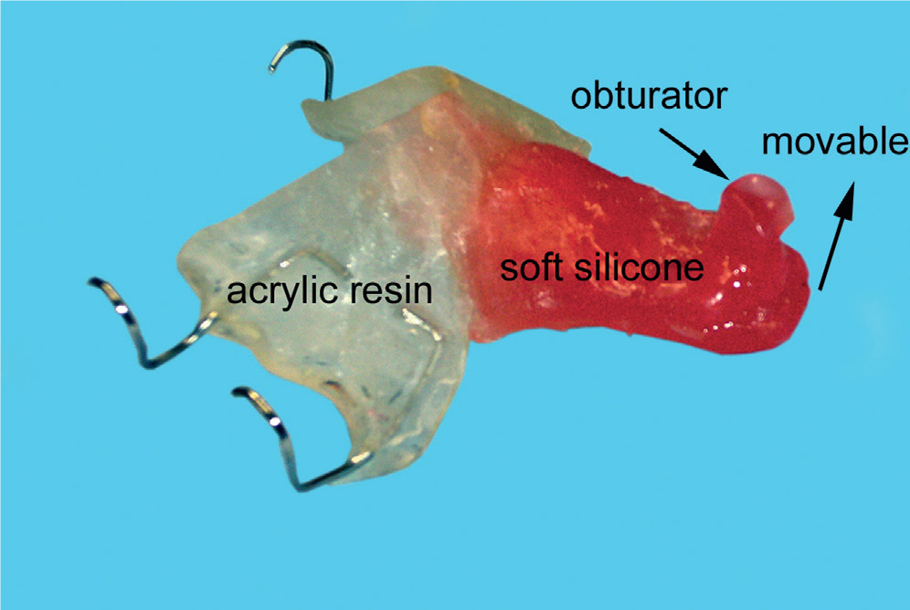
Patients with extensive soft palate defects may require speech aid prostheses such as:
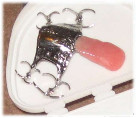
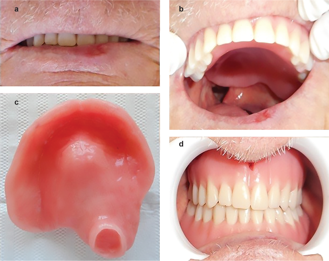
(1) (2) (3)
Figure 10. (1), (2): Palatal lift prosthesis; (3): Meatal obturator.
- Palatal lift prosthesis
- Speech bulb obturator
- Meatal obturator [2]
Speech evaluation should include assessment of nasality, articulation, and air escape during phonation.
Implant-Supported Obturators
Conventional obturators may exhibit compromised retention in extensive defects. Implant-supported obturators significantly improve prosthesis stability and patient satisfaction [16].
Common implant options include:
- Zygomatic implants
- Pterygoid implants
- Conventional implants
- Magnetic attachments
- Bar-supported systems
Advantages include:
- Improved retention
- Enhanced chewing efficiency
- Better speech outcomes
- Reduced prosthesis movement
However, implant placement may be limited by radiation therapy, bone availability, and systemic conditions.
Digital and CAD/CAM Technologies
Recent advancements in digital prosthodontics have transformed maxillofacial rehabilitation [17,18].
Applications [18] include:
Intraoral scanning
- CAD/CAM obturators
- 3D printed prostheses
- Surgical planning
- Virtual reconstruction
Advantages [17,18] include:
- Reduced chairside time
- Improved precision
- Better patient acceptance
- Reproducibility
- Enhanced esthetics
Digital workflows are particularly useful in complex defects and implant-assisted rehabilitation.
Psychological Considerations
Maxillary defects frequently result in emotional distress, social withdrawal, and reduced self-esteem [6,19]. Early prosthodontic intervention helps improve patient confidence and facilitates reintegration into social life [19].
Counseling and multidisciplinary management involving surgeons, prosthodontists, speech therapists, and psychologists significantly enhance rehabilitation outcomes.
CONCLUSION
Acquired maxillary defects present significant functional and psychological challenges requiring comprehensive prosthodontic rehabilitation. Obturator prostheses remain the cornerstone of treatment due to their ability to restore oral function, speech, esthetics, and patient confidence [1,9]. Appropriate classifications, careful framework design, accurate impression procedures, and consideration of support, retention, and stability [1,9] are essential for successful outcomes. Hollow bulb prostheses, implant-assisted rehabilitation, and CAD/CAM technologies have further improved treatment predictability and patient comfort [16,18]. A multidisciplinary approach combined with recent technological advancements can significantly enhance the quality of life of patients with acquired maxillary defects [20].
REFERENCES
- Desjardins RP. (1978). Obturator prosthesis design for acquired maxillary defects. J Prosthet Dent. 39(4):424-435.
- Beumer J, Curtis TA, Marunick MT. (1996). Maxillofacial rehabilitation: prosthodontic and surgical considerations. St Louis: Ishiyaku EuroAmerica.
- Chalian VA, Drane JB, Standish SM. (1971). Maxillofacial prosthetics: multidisciplinary practice. Baltimore: Williams & Wilkins.
- Keyf F. (2001). Obturator prostheses for hemimaxillectomy patients. J Oral Rehabil. 28(9):821-829.
- Brown JS, Shaw RJ. (2010). Reconstruction of the maxilla and midface. Br J Oral Maxillofac Surg. 48(5):307-315.
- Tayfun N, Kendi TK, Yildiz C, Findik Y. (2017). Evaluation of quality of life in patients with obturator prosthesis. Niger J Clin Pract. 20(9):1076-1082.
- Aramany MA. (1978). Basic principles of obturator design for partially edentulous patients. Part I: Classification. J Prosthet Dent. 40(5):554-557.
- Aramany MA. (1978). Basic principles of obturator design for partially edentulous patients. Part II: Design principles. J Prosthet Dent. 40(6):656-662.
- Parr GR, Gardner LK, Rahn AO. (1989). Prosthodontic principles in framework design of maxillary obturator prostheses. J Prosthet Dent. 62(2):205-212.
- Bidra AS, Jacob RF, Taylor TD. (2012). Classification of maxillectomy defects: a systematic review. J Prosthet Dent. 107(2):121-129.
- Appadurai R, Lingeshwar D, Dilshad B, Maria S. (2019). Surgical retention for immediate obturator in maxillectomy patients. Indian J Dent Res. 30(1):133-136.
- Farias A, Hegde C, Krishnaprasad D. (2013). Simplified technique to make an immediate surgical obturator for maxillectomy patient. J Interdiscip Dent. 3(2):125-128.
- Wu YL, Schaaf NG. (1989). Comparison of weight reduction in different designs of solid and hollow obturator prostheses. J Prosthet Dent. 62(2):214-217.
- Habib BH, Driscoll CF. (2004). Fabrication of a closed hollow obturator. J Prosthet Dent. 91(4):383-385.
- Rilo B, Dasilva JL, Ferros I, Mora MJ, Santana U. (2005). A hollow-bulb interim obturator for maxillary resection. J Prosthet Dent. 93(4):383-385.
- Roumanas ED, Nishimura RD, Beumer J. (1997). Clinical evaluation of implants retaining edentulous maxillary obturator prostheses. J Prosthet Dent. 77(2):184-190.
- Hatamleh MM, Watson J. (2013). Construction of obturator prosthesis using rapid prototyping. Int J Prosthodont. 26(5):497-500.
- Jiao T, Zhu C, Dong X, Gu X. (2021). Rehabilitation of maxillary defects with CAD/CAM obturator prostheses. J Prosthet Dent. 125(4):675-682.
- Goiato MC, Pesqueira AA, Ramos da Silva C, Gennari Filho H, Micheline Dos Santos D. (2009). Patient satisfaction with maxillofacial prosthesis. Literature review. J Plast Reconstr Aesthet Surg. 62(2):175-180.
- Singh M, Bhushan A, Kumar N, Chand S. (2019). Obturator prosthesis rehabilitation after maxillectomy: a review. Int J Appl Dent Sci. 5(4):255-260.
