Information Links
Related Conferences
Previous Issues Volume 7, Issue 1 - 2023
Artificial Intelligence in Maxillofacial Radiology: A Bibliometric Study
Gulay Altan Salli1, Elifhan Alagoz2,*, Nilufer Gursoy3, Irfan Sarica2
1Assistant Professor, Department of Oral and Maxillofacial Radiology, Faculty of Dentistry, University of Beykent, Buyukcekmece, Istanbul, Turkey
2Assistant Professor, Department of Oral and Maxillofacial Radiology, Faculty of Dentistry, Bezmialem Vakif University, Fatih, Istanbul, Turkey
3Research Assistant, Department of Oral and Maxillofacial Radiology, Faculty of Dentistry, Bezmialem Vakif University, Fatih, Istanbul, Turkey
*Corresponding Author: Elifhan ALAGOZ, Department of Oral and Maxillofacial Radiology, Bezmialem Vakif University, Vatan Street, Adnan Menderes Avenue, 34093, Fatih, Istanbul, Turkey; Tel No: +90 542 342 59 82; e-mail: [email protected]; [email protected]; [email protected]
Received Date: January 17, 2023
Publication Date: January 31, 2023
Citation: Salli GA, et al. (2023). Artificial Intelligence in Maxillofacial Radiology: A Bibliometric Study. Mathews J Dentistry. 7(1):33.
Copyright: Salli GA, et al. © (2023)
ABSTRACT
Purpose: The aim of this study is to evaluate the global trend in Artificial Intelligence (AI) research involving maxillofacial radiology and query a large database for a comprehensive analysis for those wishing to do more research in this area. Materials & Methods: All publication searches were performed using the PubMed databases. From January 1989 to March 2022, all AI-related publications were selected using following search terms: “artificial intelligence dental radiology”, “deep learning dental radiology”, “machine learning dental radiology”, “Convolutional Neural Network dental radiology”, “neural network dental radiology”. Totally, 971 articles were found, 732 articles were excluded, 239 articles were included and analyzed for the specified bibliometric criterias. Then including publications were categorized by country of origin, institution, type of article, journal name, impact factor of journal, subspecialties, study design, publication year, number of citation, AI tecnic and imaging modality. Statistical analysis was performed using IBM SPSS Statistics version 28.0(IBM, Chicago, IL). Results: According to results, an increase was observed in the number of publications over the years, the most publications were made in 2021(100) and the most publications from Korea (48). The institution that conducts the most studies on AI is Charité-Universitätsmedizin (8.44%) and the journal in which the studies are published the most is Dentomaxillofacial radiology (24%). The most cited publication was the Korean study published in 2018, with 283 citations. Panoramic radiograph (75) was the most used imaging technique, and CNN (99) was used from AI techniques. Conclusion: This analysis provides researchers with a comprehensive overview of AI-related research in maxillofacial radiology, providing guidance for future studies.
Keywords: Bibliometric analysis, Artificial Intelligence in maxillofacial radiology, CNN, Deep learning, Machine learning
INTRODUCTİON
AI is a highly developed branch of computer science that can mimic the functioning of the human brain. The term AI was first used by John McCarthy in 1956 [1]. The various techniques of AI which are being applied in various sectors include machine learning (ML), deep learning (DL), Artificial neural networks (ANNS), convolutional neural networks (CNNs), etc [1]. ML is a subfield of AI that “learns “ internal statistical patterns in data to eventually make predictions on unseen data. DL is a ML technique that uses multi-layered mathematical operations to learn and draw conclusions from complex data such as images [2]. ANNs are AI methods that aim to provide machines with skills such as predicting by modeling the learning function, which is the most basic feature of the human brain, and extracting new information. CNNs is a class of DL methods which has become dominant in various computer vision tasks and is attracting interest across a variety of domains, including radiology. Accurate diagnosis is a prerequisite for successful clinical practice and treatment. In this regard, trained neural networks can be very useful for diagnosticians, especially in cases with multifactorial etiology [1,3].
AI applications have helped doctors, dentists and other medical professionals in countless fields. Areas such as health information database, epidemic and syndromic surveillance, predictive models, decision support, medical imaging and provide geographical coordinates corresponding to a location of health data [1,4]. In recent years, there has been a significant increase in the number of AI research, especially in engineering, biotechnology and healthcare [5]. There are extensive articles in the literature about the benefits of AI applications in medicine and dentistry too. It also emphasized the importance of this technology for diagnostic accuracy, effectiveness of treatment, and facilitation of the overall clinical process, dental education, manufacturing of dental prostheses, patient management, oral disease, orthodontics, pain management, forensic dentistry, prediction of disease risk, detection of abnormalities/pathologies, diagnosis of disease, and evaluation of prognosis and dental radiology [1,6]. Following this, the same increasing trend is observed in the field of dental radiology, as it is suitable for artificial intelligence research due to large digital datasets. AI seems to be more adaptable to the medical industry especially radiology because of the ability of radiology to produce digitally coded images that can be more easily translated into computer language [6]. Despite the growing exponentially nature of the field, little is known about the global trend in dental radiology AI research [5].
Bibliometric analysis is a field of research that quantitatively analyzes information in a particular field by combining scientific data with statistical mathematical methods. Bibliometric analysis provides an overview of AI progress in its field and provides publication trends in dental radiology [7]. To the best of the present authors’ knowledge this is the first study to assess a bibliometric analysis of AI on dental radiology. The aim of this study was to conduct a comprehensive database search to assess the global trend in AI research in dental radiology.
METHODS
Search strategy criteria were used to construct a bibliometric research. All publication searches were performed using the PubMed databases. From January 1989 to March 2022, all AI-related publications were selected using the following search terms: “artificial intelligence dental radiology”, “deep learning dental radiology”, “machine learning dental radiology”, “CNN dental radiology”, “neural network dental radiology”, “computer learning dental radiology”.
The inclusion criteria were that the study was performed in the maxillofacial region and that the imaging technique was used in the study. Studies conducted outside the maxillofacial region, publications that do not refer to any imaging technique, congress-symposium papers, editorial articles, and publications whose full text cannot be accessed are excluded.
Using the keyword “artificial intelligence dental radiology”, 339 publications were found. When full-text or abstract reading, inclusion and exclusion criteria were applied, 186 publications were included in the study. 153 articles were excluded because they were not using dental imaging and suitable for this bibliometric study. Using the keyword “deep learning dental radiology”, 180 articles were found. However, only 26 articles were included in the study, as most of the articles matched the previous keyword. Using the keyword “machine learning dental radiology”, 180 articles were found. However, only 13 articles were included in the study, as most of the articles matched the previous keywords. Using the keyword “CNN dental radiology”, 75 articles were found. However, only 3 articles were included and as most of the articles matched the previous keywords. And finally using the keyword “neural network dental radiology”, 197 articles were found and 11 articles were included. Totally, 971 articles were found in the databases Pubmed, using keywords. 732 articles were excluded in the study because of they were irrelevant, dental imaging not used, proceedings papers, meeting abstracts duplicate or editorial articles. Finally, 239 articles were included and analyzed for the specified bibliometric criterias. (Diagram 1).
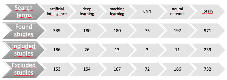
Diagram 1: Number of publications on studies found, included, or excluded when search criteria are used.
The resulting publication database was then categorized by country of origin, institution (first author), type of article (review, research etc.), journal name, impact factor of journal (-for last two years), subspecialties (main topic of study), study design (review, survey, comparative, retrospective, qualitative, etc.) publication year, number of citation, AI tecnic (CNN, NN, etc.) and imaging modality (panoramic radiography, periapical radiography, cone beam computed tomography (CBCT), etc.).
STATISTICAL ANALYSIS
Statistical analysis was performed using IBM SPSS Statistics version 28.0 (IBM, Chicago, IL). Frequency analysis was used to show the observation frequency and percentage distribution of the data. Statistical significance was evaluated by using crosstab and pearson chi-square test in the comparison of independent variables with each other.
RESULTS
According to the statistical results, there are a total of 239 AI studies using dental imaging in the last 32 years. Among these studies, the most original research has been done, and reviews are in the second place. Comparative study, preliminary study, survey study, phantom study and systematic reviews are among the study designs made. When AI studies are evaluated in terms of country, it is observed that publications are made from 36 different countries. Most studies, including 48 publications (20,1%), were from Korea. The second place is followed by China with 25 publications (10,5%). It is noteworthy that Turkey ranks 6th in this ranking with 13 publications (5,4%).
The top three institutions with the highest number of researches are Charité - Universitätsmedizin (Germany) with 13 (8.44%) studies, Seoul National University (Korea) with 11 (7.14%) studies, and Yonsei University (Korea) with 11 (7.14%) studies. The first six journals in which current studies are published most frequently are Dentomaxillofacial radiology with 24 (24%) publications, Scientific reports with 23 (23%) publications, Journal of clinical medicine and Journal of dentistry with 9 (9%) publications, Oral surgery, oral medicine, oral pathology and oral radiology and Imaging science in dentistry with 7% publications, respectively. When the impact factor of the journals is evaluated, this value varies between 0.360 and 9.730. The mean value is 2,91927, with a total of 110 studies published in journals above the mean value. There are 2 publications (0,8%) in the journal with the highest impact factor. One of these publications is from the USA and the other one is from Canada. When the citations to the publications are examined, it is seen that it varies between 0 and 283 and the mean value is 19.67. The most cited work was done in Korea, published in 2018, and received 283 citations. The impact factor of the journal in which it was published is 3.242.
When we evaluate the studies made in dental radiology related to AI by years, it is seen that the most publications, a total of 100 studies (41,8%), were made in 2021 and 67 publications (28%) were made in 2020. There was an increase in direct proportion between the number of publications and the years. (Graphic 1) When the studies in dental radiology related to AI are evaluated in terms of the imaging technique used, it is seen that the maxillofacial imaging technique was used in a total of 205 studies (85,8%). In 34 studies (14,2%), it was noticed that no imaging technique was used. These are reviews, systematic reviews and survey studies. It is observed that panoramic radiographs are mostly used among the imaging techniques, a total of 75 publications (31,4%). This is followed by the CBCT with 34 publications (14,2%). On the other hand, periapical radiographs, are in third place, used in a total of 22 publications (9,2%) (Graphic 2).
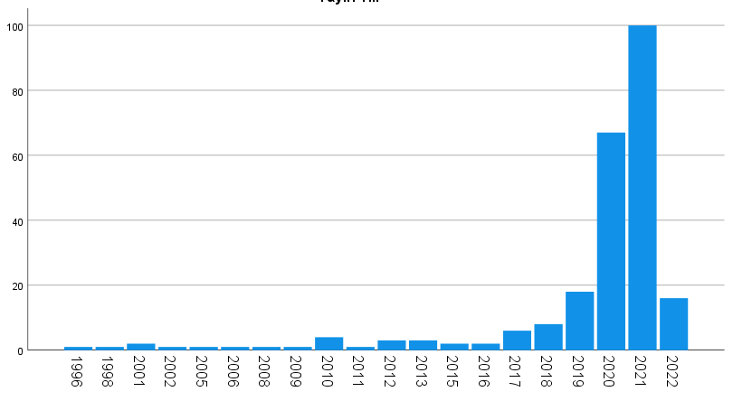
Graphic 1: Number of publications on AI in dentomaxillofacial by years.
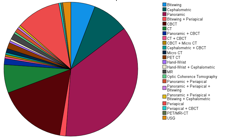
Graphic 2: Distribution of imaging techniques used in publications on AI in the diagram.
Among the AI techniques, CNN was used in 99 (41.4%) studies, both generative adversarial networks (GAN) and CNN in 2 (0.8%) studies, NN in 14 (5.9%) studies, and U-Net in 12 (5%) studies. In 21 (8.8%) studies, AI was mentioned in general (survey, review, systematic review). In 12 (5%) studies, ML was used in general and DL was used in 10 (4.2%) studies without giving any information about the sublayers. Bayesian CNN was used in 10 (4.2%) studies, D-CNN in 10 (4.2%) studies, R-CNN in 10 (4.2%) studies, and ANN in 10 (4.2%) studies.
DISCUSSION
In this study, research on AI in maxillofacial radiology was analyzed by examining the amount of increase in publications by years, the AI techniques used, the distribution by countries, imaging techniques, the number of citations and the impact factor of the journals in which they were published, using bibliometric data.
Wang, et al. [8] examined trends in the application of DL networks in medical image analysis in a bibliometric study in which they examined a total of 2685 original articles between 2012 and 2020. They found that the number of publications from 2017 to 2020 increased substantially, accounting for 97.2% of all included articles. In this study, results were found parallel to that study. According to the current results, an increase has been observed in the number of publications over the years, and a jump has been observed in studies related to AI in the last 5 years, similar to the results of Wang L, et al [8]. However, it has been observed that there are more publications in 2021 than in 2022 in the current study. This is the result of searching only the first three months of 2022, while searching for all months in 2021. The increasing number of publications shows that AI plays a major role in diagnosis, treatment planning and prognosis evaluation in maxillofacial radiology, dentistry and medical medicine.
West E, et al. analyzed 8813 AI-based publications in Radiology between 2000-2018 [5]. According to the results of this study, they reported that the 12 most productive countries were the United States, China, Germany, the United Kingdom, Canada, Japan, the Netherlands, France, India, Italy, South Korea and Australia, respectively. While the USA accounted for approximately 35-50% of the total publication between 2000 and 2018, China was stated to be the second most productive country in the world, contributing 18% of the total production in 2018. Contrary to these results in our study, the 6 most productive countries are Korea, China, Japan, USA, Germany and Turkey, respectively. The reason for this difference is multifactorial. There may be exclusion factors (not using imaging technique, proceedings papers, meeting abstracts). While the study carried out research until 2018, the current study also included 2022 and the great leap in the number of major publications has been made in the last 5 years. In addition, while the related study examined publications in medical radiology, the current study examined publications specific to the maxillofacial region. In general, in the study of West , et al. [5], Turkey could not enter any ranking, while it is in the 6th place in the current study. This can be explained by the fact that the state-sponsored budget allocated to scientific research is higher in the relevant countries (United States (16.5%), China (3.6%)) and the socio-economic status of the countries is good [5].
According to the results of West E, et al. [5].'s study, the most common publication types were articles (53.7%), proceedings papers (38.0%) and meeting abstracts (6.8%). The most frequently published journals were NeuroImage (16.1%), IEEE Transactions on Medical Imaging (6.9%), and Medical Physics (6.9%). In our analysis, 183 (76.6%) of 239 publications were original studies and were the most frequently identified studies. This is followed by reviews with 25 (10.5%) studies. The journals in which current studies are published most frequently are Dentomaxillofacial radiology with 24 (24%) publications, Scientific reports with 23 (23%) publications, Journal of clinical medicine and Journal of dentistry with 9 (9%) publications. Differences between studies can be attributed to exclusion factor differences, differences in the number of publications evaluated, and the budget allocated to scientific research.
Wang L, et al. [8] reported that magnetic resonance imaging (MRI) (24.4%), computed tomography (CT) (22.0%) were mostly used as imaging methods in their study on DL networks in medical image analysis between 2012 and 2020. In our study, panoramic radiographs were used most frequently, including 75 (31.4%) studies. This is followed by CBCT with 34 (14.2%) publications and periapical radiographs with 22 (9.2%) publications. In the current study, MRI was used in 3 (1.3%) publication, CT was used in 14 (5.9%) publications, and both CT and CBCT were used in 1 (0.4%) publication. The differences between studies can be attributed to the difference in the number of publications examined and the difference in the imaging technique most commonly used in the maxillofacial region.
According to the statistical results of Wang L, et al. [8]'s study, CNN (n = 1626) including CNN-based and CNN-derived DL studies were the most frequently used network types, with U-Net (n = 639) being applied most frequently in image segmentation (n = 349). Similarly with that study, CNN-based (101) and CNN-derived DL (36) studies are the most frequently used network type in current study.
PubMed is a very dynamic database, but it is a platform where more publications are viewed in a shorter time. Even if the same keywords are searched, different search results may appear at different times. However, browsing data in Pubmed requires sufficient time and careful work. In bibliometric studies, databases such as Web of Science and Scopus are used for ease of use and analysis. Pubmed database was used in our study. This is the limitation of our study.
As a result, the use of different databases will not make a difference, as details such as imaging technique or AI models used require full-text evaluation when scanning in all databases used.
CONCLUSION
AI holds the power to change all the usual patterns in the field of health and dentistry. Recently, there has been a great interest in AI studies. Dentomaxillofacial radiology is a very suitable branch for these applications because it is very convenient in terms of image data. The proliferation of studies in the field of AI will also contribute to dentomaxillofacial radiology. Studies in this field have shown a huge increase and successful results, especially in the last 5 years. More research is needed for dentists to start using AI in their daily practice and to make their work easier.
REFERENCES
- Sharma S. (2019). Artificial intelligence in dentistry: the current concepts and a peek into the future. Int J Contemp Med Res. 6(12):5-9.
- Schwendicke F, Golla T, Dreher M, Krois J. (2019). Convolutional neural networks for dental image diagnostics: A scoping review. J Dent. 91:103226.
- Kalappanavar A, Sneha S, Annigeri RG. (2018). Artificial intelligence: A dentist's perspective. J Med Radiol Path Sur. 5(2):2-4.
- Pakdemirli E, Wegner U. (2020). Artificial Intelligence in Various Medical Fields With Emphasis on Radiology: Statistical Evaluation of the Literature. Cureus. 12(10).
- West E, Mutasa S, Zhu Z, Ha R. (2019). Global Trend in Artificial Intelligence-Based Publications in Radiology From 2000 to 2018. AJR Am J Roentgenol. 213(6):1204-1206.
- Hung KF, Montalvao C, Tanaka R, Kawai T, Bornstein MM. (2020). The use and performance of artificial intelligence applications in dental and maxillofacial radiology: A systematic review. Dentomaxillofacial Radiol. 49(1):20190107.
- Aksoy S, Aksoy U, Orhan K. (2022). An overview of the 35 years of research in the oral radiology: a bibliometric analysis. Oral Radiol. 38(2):183-191.
- Wang L, Wang H, Huang Y, Yan B, Chang Z, Liu Z, et al. (2022). Trends in the application of deep learning networks in medical image analysis: Evolution between 2012 and 2020. Eu J Radiol. 146:110069.