Information Links
Related Conferences
Previous Issues Volume 7, Issue 2 - 2023
Aesthetic Rehabilitation of Peg-Shaped Maxillary Permanent Lateral Incisors: Ultraconservative Approach
Amira Kikly1,*, Sabra Jaâfoura2, Foued Brigui, Ameni Chadlia Belghith3, Wided Glii3, Neila Zokkar4, Nabiha Douki4
1Associate Professor, Department of Dentistry, University Hospital, Sahloul, Tunisia, Faculty of Dental Medicine, University of Monastir, Avicenna Street, 5000 Tunisia, Research Laboratory LR12ES11, University of Monastir, Tunisia
2Associate Professor, Department of Dental Biomaterials, Faculty of Dental Medicine, University of Monastir, Avicenna Street, 5000 Tunisia, Laboratory of Dento-Facial, Clinical and Biological Approach (ABCDF) LR12ES10, University of Monastir, Tunisia
3Department of Dentistry, University Hospital, Sahloul, Tunisia, Faculty of Dental Medicine, University of Monastir, Avicenna Street, 5000 Tunisia, Research Laboratory LR12ES11, University of Monastir, Tunisia
4Professor, Department of Dentistry, University Hospital, Sahloul, Tunisia, Faculty of Dental Medicine, University of Monastir, Avicenna Street, 5000 Tunisia, Research Laboratory LR12ES11, University of Monastir, Tunisia
*Corresponding Author: Amira Kikly, Associate professor, Department of Dentistry, university hospital, Sahloul, Tunisia, Faculty of Dental Medicine, University of Monastir, Avicenna Street, 5000 Tunisia; Email: [email protected]
Received Date: September 6, 2023
Publication Date: October 13, 2023
Citation: Kikly A, et al. (2023). Aesthetic Rehabilitation of Peg-Shaped Maxillary Permanent Lateral Incisors: Ultraconservative Approach. Mathews J Dentistry. 7(2):39.
Copyright: Kikly A, et al. © (2023)
ABSTRACT
Objective: The aim of this article is to develop a simplified clinical protocol for the management of a peg-shaped maxillary lateral incisor using a direct restoration technique. Materials and methods: A 14-year-old girl consulted us for aesthetic discomfort in the anterosuperior sector, due to the presence of a peg-shaped right maxillary lateral incisor. A direct composite resin restoration was used for the aesthetic rehabilitation of this case. Result: It is therefore possible to achieve aesthetic and reproducible results using the direct technique, provided a strict and rigorous protocol is followed. Clinical assessment of the case report was made using FDI criteria Discussion: Peg-shaped maxillary lateral incisors can have many aesthetic and functional consequences. Their aesthetic rehabilitation must respect the therapeutic gradient. At present, no exhaustive protocol has been adopted. A set of decision criteria must be established in order to indicate the most appropriate therapeutic solution. Direct restoration with composite resin is the most conservative solution, as it has the advantage of being reversible, unlike indirect restorations. Previewing the results of treatment with a wax-up illustrating the final result is essential and helps communication between practitioner and patient, as well as the patients' decision-making process.
Keywords: anterior esthetics, peg-shaped lateral maxillary teeth, Composites, direct restoration
INTRODUCTION
The peg-shaped maxillary lateral incisor is defined as a conical, undersized maxillary lateral incisor. This shape anomaly corresponds to a reduction in its mesio-distal diameter, and is most prevalent in late-series teeth such as maxillary lateral incisors. The prevalence of peg-shaped maxillary permanent lateral incisors was 1.8% overall. This prevalence has been shown to differ according to race, population type and gender. Prevalence rates were higher in orthodontic patients and in women. Although the prevalence of unilateral and bilateral lateral incisors was similar, the left side was twice as frequent as the right [1].
Its restoration, like any restoration, must respect the notion of therapeutic gradient. The treatment options are numerous: direct restoration with composite resin, composite or ceramic veneers, or corono-peripheral reconstructions. Composite resins are the most conservative solution, as they have a low tissue cost and, unlike indirect restorations, are reversible. In the event of failure, it remains possible to switch to more invasive solutions. Direct restoration is performed using a wax-up approach that illustrates the final result. This is followed by a silicone key to guide the restoration. This solution may be chosen for the short term, while awaiting a more permanent restoration. But it can also be chosen as a medium-term solution, provided the protocol is strictly adhered [1.2].
The aim of this work is to present, through a case study, the clinical steps involved in the aesthetic rehabilitation of a peg-shaped maxillary lateral incisor using a composite resin restoration technique.
CASE REPORT
This is a 14-year-old girl complaining about the unsightly appearance of her anterosuperior sector, which is due to the presence of a peg-shaped right maxillary lateral incisor (Figure 1). The patient wanted a solution that was aesthetically pleasing, economical and fast. After an aesthetic and functional analysis, an aesthetic rehabilitation using direct composite resin restoration was indicated. A wax-up approach was performed and a silicone guide was prepared (Figure 2). Color selection was carried out in a clean mouth and on hydrated teeth. A shade guide specific to the composite used was employed for this step.
Once the composite masses had been selected, small samples could be placed on the tooth to better visualize their integration. The dental dam was then placed (Figure 3) and the silicone guide was fitted (Figure 4). After surfacing the enamel with a diamond-tipped cylindrical burr (Figure 5) and applying an adhesive system with prior two-stage etching and rinsing (Figures 6,7&8), the palatal wall was built up with enamel composite, using the silicone key as a layering support (Figures 9&10). A second layer is then applied, reconstituting most of the cavity with dentin composite (Figure 11). This is the body of the tooth. It is this dentine mass that will give the restoration its shade. Next, enamel composite is placed on top of the dentin layer to build up the proximal walls and vestibular face (Figure 12). The transparent matrix was placed and held in place during light-curing. Finally, the finishing step was carried out to ensure proper aesthetic and functional integration of the final restoration (Figures 13&14).
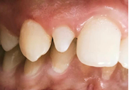
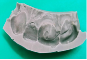
Figure 1: Preoperative clinical view. Figure 2: Wax up + silicone guide.
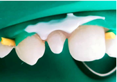
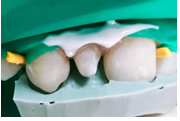
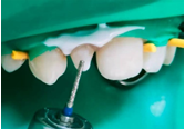
Figure 3: Rubber dam isolation. Figure 4: Testing the silicone guide. Figure 5: Enamel surfacing.
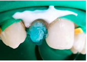
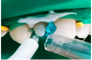
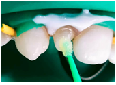
Figure 6: Etching. Figure 7: Rinsing and drying. Figure 8: Adhesive application.
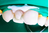
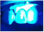
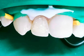
Figure 9: Edification of palatal wall and proximal walls with A2 enamel shade. Figure 10: Photopolymerization. Figure 11: D2 dentin resin layer and light-curing.
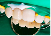
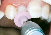
Figure 12: Vestibular enamel layer and light-curing. Figure 13: Polishing the restoration.
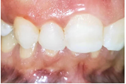
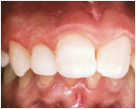
Figure 14: Final result. Figure 15. 6-month follow-up.
A follow-up was made at six months (Figure 15) and the clinical assessment of the case report was made using FDI criteria (Table 1)
Table 1: Description of selected FDI criteria.
|
Selected FDI criteria |
Surface finishing |
Color and translucency stability |
Anatomical shape |
Material fracture and retention |
Proximal anatomical shape |
Post-operative sensitivity and pulpal vitality |
|
Score |
1 |
2 |
1 |
1 |
1 |
1 |
DISCUSSION
The peg-shaped maxillary lateral incisor is defined as a conical, undersized maxillary lateral incisor. This shape anomaly corresponds to a reduction in its mesio-distal diameter, and preferentially concerns teeth at the end of a series, such as maxillary lateral incisors and wisdom teeth. Microdontia is often associated with other anomalies, notably agenesis.
Restoration of the conoid incisor, like all restorations, must respect the notion of a therapeutic gradient (Thirlet and Attal 2009). Indeed, there are many treatment options: direct restoration with composite resin, placement of composite or ceramic veneers, or peripheral coronal reconstructions. But it's important to remember that the treatment used to restore the aesthetics of the smile should always be as minimally invasive as possible. In line with this philosophy, composite restorations are one of the most conservative therapies for healthy dental tissue; and therefore, this concept needs to be understood and mastered to enable a complete and informed choice of the options that are available to patients. Direct composite resin restorations are the most conservative solution, as they are low in tissue cost, and have the advantage of being reversible, unlike indirect restorations. In fact, this solution can be chosen in the short term to satisfy an aesthetic demand while awaiting a more permanent, more invasive restoration [3].
In our case, an impression is essential to record the anatomy of the tooth to be restored, as well as the adjacent and antagonistic teeth. Based on the impression, the dental technician will create a wax-up/wax-out model to illustrate the final result. This is followed by a silicone key to be used as a guide for the restoration.
This step is essential for our patient's acceptance of the treatment plan. The patient can easily project his or her own image and give his or her opinion.
Setting up the surgical field is essential to ensure safety, salivary isolation and working comfort. The split dam technique may be indicated [4].
This technique ensures correct, optimal repositioning of the silicone key and correct salivary isolation. Nonetheless, it is less watertight than conventional dam placement. In fact, this technique can also be considered, provided we can correctly reposition our layering guide and have sufficient clearance [5].
The composite resin injection restoration technique is a feasible option for the aesthetic rehabilitation of our case. It enables a wax-up produced on a plaster model or virtual composite model to be reliably transposed into the mouth via a transparent gutter.
In this situation, the use of highly filled flowable resins means that this technique can be used for transitional restorations in young patients awaiting bone growth [6-8].
So, when the patient is looking for a rapid aesthetic and functional result with limited financial resources, injectable resins can be a good solution [9].
Nano-filled composites are currently the preferred choice, as they offer a wide range of shades for improved aesthetics, good strength, and low wear and polish ability. A better understanding of the behavior and optical properties of dental tissues also means that direct restorations of peg-shaped incisor can be performed more naturally. Unlike indirect restorations, these types of restoration do not require excessive tooth preparation. Preparations are limited to the treatment surfaces that will receive the composite material. In this respect, composite restorations are among the most conservative therapies available.
Natural teeth are polychromatic, presenting a wide variety of colors and shades. Shade selection is therefore an essential step in identifying the details that nuance the tooth. This step must be carried out on a clean tooth, in natural light and with the contralateral tooth intact as a reference. Shading can be carried out using a custom-made shade guide, with several thicknesses of enamel and dentin materials.
Alternatively, small increments of composite can be positioned and cured at the junction of the middle and cervical thirds of the tooth in order to select the dentin shade, and at the free edge to select the enamel shade. Once the shade has been selected, the composite dots can be easily removed.
It is important to understand the optical behavior of tooth structures in order to identify composite resins that will reproduce the characteristics of the tooth. Indeed, a successful restoration begins with the correct choice of material. However, there is no such thing as a "miracle" material, and the final result depends on the practitioner's manual skills and ability to choose techniques that deliver reproducible results. As a result, it's possible to use composite resin from one brand, opalescent and translucent from another, and a final one for brightness, for the same restoration. However, it is always preferable to use the same brand to simplify the procedure.
After enamel surfacing, etching and adhesive application, the restoration was made following the principles of L. Vanini's three-layer layering technique. This technique ensures the natural integration of aesthetic restorations [10.11].
The palatal surface and proximal ridges are restored with an enamel-shaded composite. Next, dentin-shaded composites of decreasing opacity are layered obliquely over the dentin tissue medially and distally. The number of layers may differ according to the size of the restoration. Finally, the last layer of enamel composite covers the dentin masses. Intensives can be placed between the dentin lobes and the superficial enamel layer to recreate areas of high opalescence or specific dyschromia.
For the finishing step, a cylindrical-conical diamond bur was used to remove excess material and re-contour the restoration, leading to a natural final appearance. In addition, a smooth composite surface facilitates the subsequent polishing step. Finally, polishing can be conventionally carried out using abrasive strips, siliconized discs or tips with abrasive paste. This step results in a more natural finish.
It is therefore possible to achieve aesthetic and reproducible results using the direct technique, provided a strict and rigorous protocol is followed [12].
a follow-up must be set up to ensure the clinical evaluation of our restoration. Indeed, it has been found that the FDI criteria should not only be reserved for clinical trials, but should also be used in our daily practice in the dental office for the evaluation of restorations using the direct technique. By using these criteria, clinicians can evaluate their own work and develop a critical approach, thus providing them with a guide as to when to act again [13].
CONCLUSION
Once the patient's request has been formulated, the best restorative solution must be proposed, respecting the notion of therapeutic guardianship. All decision-making criteria must be collected. An interview with the patient is essential to understand his or her expectations. Direct composite resin restorations may be chosen as a first-line treatment, particularly in cases of incomplete growth or unilateral anomalies. The bonding protocol must be followed to ensure the durability of the restoration.
REFERENCES
- Hua F, He H, Ngan P, Bouzid W. (2013). Prevalence of peg-shaped maxillary permanent lateral incisors: a meta-analysis. Am J Orthod Dentofacial Orthop. 144(1):97-109.
- Andrei OC, Farcaşiu C, Bisoc A, Tărlungeanu DL, Tănăsescu LA, Dina MN, et al. (2021). Unilateral agenesis of permanent superior canine in familial peg-shaped lateral incisors: rare case report and literature review. Rom J Morphol Embryol. 62(4):1045-1050.
- Izgi AD, Emrah Ayna E. (2005). Direct restorative treatment of peg-shaped maxillary lateral incisors with resin composite: a clinical report. J Prosthet Dent. 93(6):526-529.
- Devictor A, Beyly T. (2021). Résines injectées : les nouvelles approches pour réaliser des restaurations antérieures simples et durables. Info Dent. 32(2):140-147.
- Hosaka K, Tichy A, Motoyama Y, Mizutani K, Lai WJ, Kanno Z, et al. (2020). Post-orthodontic recontouring of anterior teeth using composite injection technique with a digital workflow. J Esthet Restor Dent. 32(7):638-644.
- Terry DA, Powers JM. (2015). Résine composite injectable : une nouvelle technique - Part 1. DT Study 3:26-31.
- Pomperski M. (2021). Technique d’injection de composite dans le secteur antérieur. Info Dent. 103(9):31-35.
- Geštakovski D. (2019). The injectable composite resin technique: minimally invasive reconstruction of esthetics and function. Clinical case report with 2-year follow-up. Quintessence Int. 50(9):712-719.
- Ammannato R, Ferraris F, Marchesi G. (2015). The index technique » in worn dentition : a new and conservative approach. Int J Esthet Dent. 10(1):68-99.
- Vanini L. (1996). Light and color in anterior composite restorations. Practical Periodontology and Aesthetic Dentistery. 8(7):673-682.
- Omeish N, Nassif A, Feghali S, Vi-Fane B, Bosco J. (2022). Esthetic and functional rehabilitation of peg-shaped maxillary lateral incisors: Practical recommendations. Clin Case Rep. 10(3):e05507.
- Vanini L. (2010). Conservative restorations that mimic nature: a step-by-step anatomical stratification technique. Journal of cosmetic dentistry. 26(3):80-98.
- Hickel R, Peschke A, Tyas M, Mjör I, Bayne S, Peters M, et al. (2010). FDI World Dental Federation - clinical criteria for the evaluation of direct and indirect restorations. Update and clinical examples. J Adhes Dent. 12(4):259-272.